
Amphinemura qiliana, Li & Teslenko & Yang, 2020
|
publication ID |
https://doi.org/10.11646/zootaxa.4743.3.7 |
|
publication LSID |
lsid:zoobank.org:pub:613D68E6-A60D-4ED8-BB4F-5403360F669A |
|
DOI |
https://doi.org/10.5281/zenodo.3691592 |
|
persistent identifier |
https://treatment.plazi.org/id/03A2CB24-9E2F-B13B-FF48-A083FF71FDAF |
|
treatment provided by |
Plazi |
|
scientific name |
Amphinemura qiliana |
| status |
sp. nov. |
Amphinemura qiliana View in CoL sp. nov.
( Figs. 11–19 View FIGURE 11 View FIGURES 12–15 View FIGURES 16–19 )
Examined material. Holotype male, China: Qinghai Province, Qilian County, Arou Temple, Daotang River , 3150 m.a.s.l., 13.VII.2011, coll. W. Li ( HIST) . Paratypes, 1 male, same locality and date as holotype ( HIST) , 1 male (cleared and mounted), same locality and date as holotype ( FSC EATB FEB) .
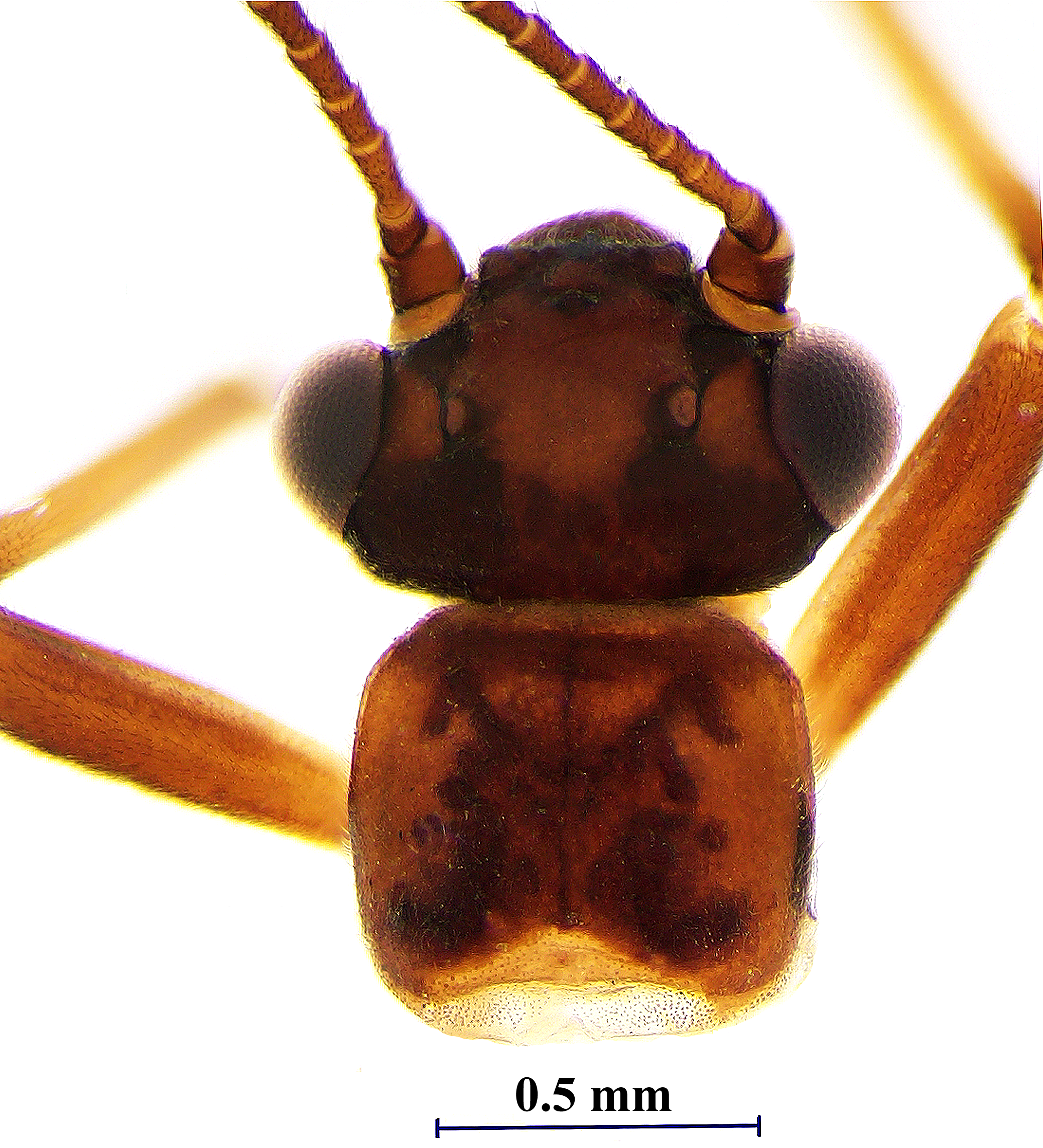
Description. Adult habitus. Body length from tip of the head to the apex of abdomen, males, 4.0-4.5 mm, forewing length, males, 5.1-5.8 mm; wingspan, males, 11.2-12.6 mm. Head brown with U-shaped black band anterior of the anterior ocellus extending onto clypeus, lateral angles of the clypeus with rounded light-brown spot; M-line black and distinct ( Fig. 11 View FIGURE 11 ). A small rectangular black spot present on each inner side at the top of the epicranial suture in the antennal area. Interocellar area with a large light brown spot rounded posteriorly. A small semi-oval light brown spot present medially between posterior ocellus and the compound eye. Occiput covered by small black spots along the epicranial stem and behind each eye, extending to posterior ocelli. Pronotum light brown with rounded angles and X-shaped pattern consisting of small black spots which form a figure with sharp anterior angles and rounded posterior branches, posterior part of the pattern more pronounced ( Fig. 11 View FIGURE 11 ).
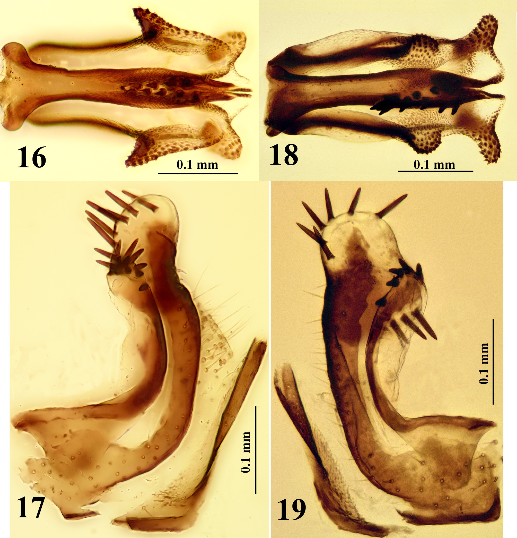
Male. Posterior margin of tergum 9 with notch and interrupted medially by a scattered fringe of long inwardly curved setae ( Fig. 12 View FIGURES 12–15 ). Subgenital plate of sternum 9 elongate, trapezoid in the first half anteriorly, with an obtuse angle in the middle of the outer edge, narrowed distally and terminating in a long rounded tongue-shaped extension; vesicle elongated, not reaching ½ length of the subgenital plate ( Fig. 13 View FIGURES 12–15 ). Tergum 10 medially forming a triangular sclerite plate with pointed anterior angles; two paramedial elongated leaf-shaped sclerites extend from the base of the epiproct to the triangular sclerite plate ( Figs. 12, 15 View FIGURES 12–15 ). Sternum 10 occupied by the broad bases of the paraprocts. Cerci membranous, oval, covered by hairs ( Figs. 12‒14 View FIGURES 12–15 ). Paraprocts complex in ventral view: the inner paraproctal lobe elongated, less sclerotized than the other lobes, blade-like along medial edge ( Figs. 13 View FIGURES 12–15 , 17 View FIGURES 16–19 ); middle lobe large, elongate, and transverse at the base, mostly sclerotized at the inner edge and distally, extending first backward and then forward and up, curved dorsally; basally and along the inner edge with hairs ( Figs. 13 View FIGURES 12–15 , 17 View FIGURES 16–19 ). Dorsal face of the middle lobe apex a membranous rounded knob ( Fig. 17 View FIGURES 16–19 ), covered with a fringe of 6-8 long blunt black spines along the upper and rear face. Outer lobe is a narrow sclerite which bends outward and curves medially. Posterior branch of the outer lobe scoop shaped and slightly widened, its tip lies in the loop of the sclerite of the middle lobe, outer margin with 9-12 pointed black spines of the different size which are generally arranged in two rows ( Fig. 17 View FIGURES 16–19 ).
Epiproct in dorsal view lyre-shaped, narrowed at the base, widened and rounded anteriorly, with two anterolateral heavily sclerotized finger-shaped projections curved downward and densely covered with stout spicules ( Fig. 15a View FIGURES 12–15 ); tip of the epiproct bifurcated ( Fig. 12 View FIGURES 12–15 ), consisting of thin tips of two ventral sclerites covered by fine spicules ( Fig. 15b View FIGURES 12–15 ). Dorsum of the epiproct densely covered with fine spicules, surface divided by fine suture that anteriorly forms a shallow cleft separating two short membranous rounded lobes ( Fig. 15c View FIGURES 12–15 ). Lateral arms ( Fig. 15d View FIGURES 12–15 ) of the dorsal sclerite are long, narrow and darkly sclerotized, each lateral arm forks ( Fig. 14 View FIGURES 12–15 ) in the second half of length, the dorsal branch of the lateral arm extending to the base of anterolateral heavily sclerotized finger-shaped projection ( Fig. 15a View FIGURES 12–15 ); the ventral branch of the lateral arm supporting lateral wing-shaped membranous lobe with an acute triangular apex directed obliquely downward, and covered with stout spicules especially densely on the posterior edge of the acute apex ( Figs. 14, 15f View FIGURES 12–15 , 16 View FIGURES 16–19 ). In ventral view, the paired ventral sclerite heavily sclerotized, broad at the base, converge in the first ½ of their length, branched and narrowed towards the apex ( Figs. 16 View FIGURES 16–19 , 15b View FIGURES 12–15 ). In lateral view, the ventral sclerite form keel-shaped ridges; each ridge bears 5 pairs of heavily sclerotized stout spines ( Figs. 14 View FIGURES 12–15 , 16 View FIGURES 16–19 ). Basal sclerites represented by two triangular sclerotized patches located at the basolateral margins of the epiproct ( Figs. 12, 15 View FIGURES 12–15 ).
Female. Unknown.
Remarks. Amphinemura qiliana sp. n. appears to be closely related to A. zwicki Teslenko, 2015 ( Teslenko 2015) . Both species share the remarkable pair of anterolateral projections of the epiproct and the keel-shaped ventral sclerite with five pairs of stout spines. However, the anterolateral projection of A. qiliana is finger-like, longer than that of A. zwicki and curved dorsally downward; A. zwicki has bulbous projections directed towards each side of the bifurcated tip of the epiproct. Amphinemura qiliana can be also separated from A. zwicki by the shape of the lateral lobes of the epiproct which are wing-shaped with triangular apex directed obliquely downward and originating from the anterolateral apex of epiproct, longer than one third of epiproct. The lateral lobes of A. zwicki are bulbous, rounded, and close to or covering partially the keel-shaped ridge of ventral sclerite ( Fig. 18 View FIGURES 16–19 ). Spines of the ventral sclerite of the epiproct of A. qiliana are short as compared to A. zwicki , which has long and pointed spines, especially at the base of keel ( Fig. 18 View FIGURES 16–19 ). Additional differences are the shape and number or location of the spines on the middle and outer paraproctal lobes: the middle paraproctal lobe of A. zwicki distally enlarged with spines on dorsal and ventral edges ( Fig. 19 View FIGURES 16–19 ) whereas the middle paraproctal lobe of A. qiliana is narrowed, shorter, and the spines located on dorsal edge. The outer lobe of A. zwicki has at most nine spines along the outer edge and A. qiliana bears 9-12 spines arranged in two rows ( Fig. 17 View FIGURES 16–19 ).
Distribution. The new species is known from the Daotang River, Qilian Mountains at 3,150 m.a.s.l. in northwestern of China, straddling the border between Gansu and Qinghai provinces.
Etymology. The specific name Amphinemura qiliana refers to the type locality Qilian Mountains or Qilian County.

| FSC |
Fredericton Stock Culture Collection |
No known copyright restrictions apply. See Agosti, D., Egloff, W., 2009. Taxonomic information exchange and copyright: the Plazi approach. BMC Research Notes 2009, 2:53 for further explanation.
|
Kingdom |
|
|
Phylum |
|
|
Class |
|
|
Order |
|
|
Family |
|
|
Genus |