
Conura leucotela (Walker, 1862)
|
publication ID |
https://doi.org/ 10.11646/zootaxa.4942.3.5 |
|
publication LSID |
lsid:zoobank.org:pub:FF2841C8-D952-44E0-83AC-75C3FA5953EC |
|
DOI |
https://doi.org/10.5281/zenodo.4619705 |
|
persistent identifier |
https://treatment.plazi.org/id/03A3AD23-6257-C660-FF29-7C6EFBC22B99 |
|
treatment provided by |
Plazi |
|
scientific name |
Conura leucotela |
| status |
|
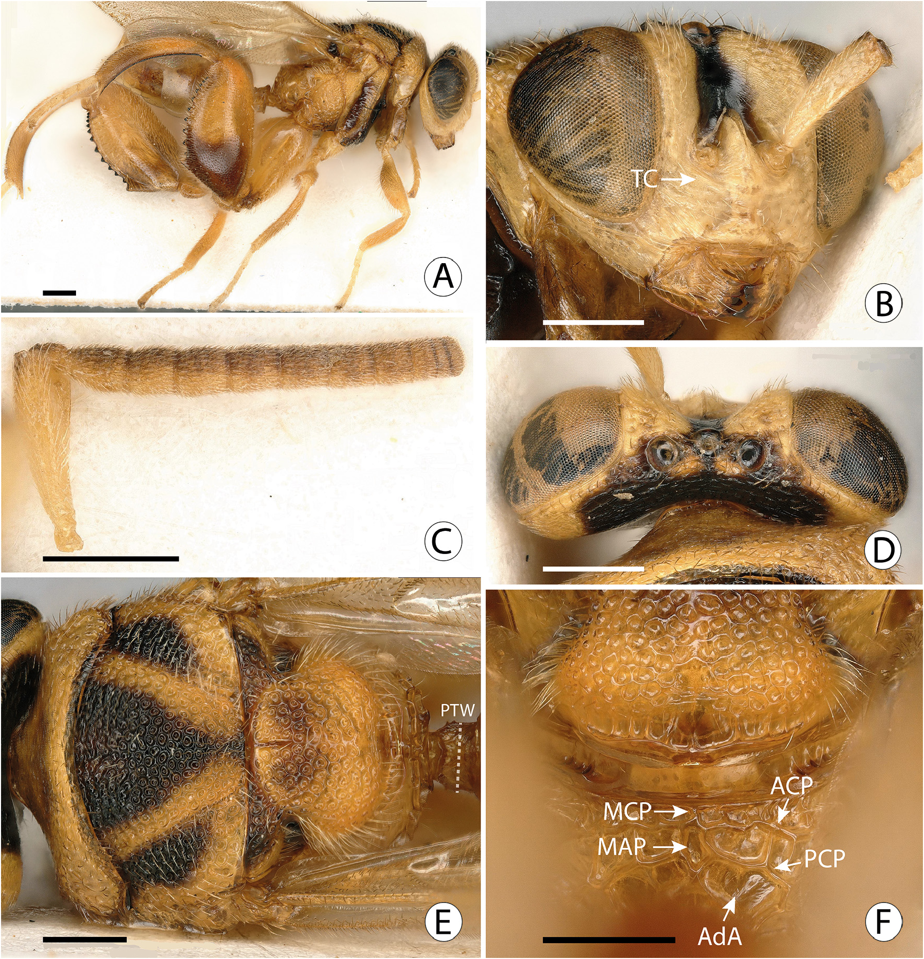
Conura leucotela group sp. 1
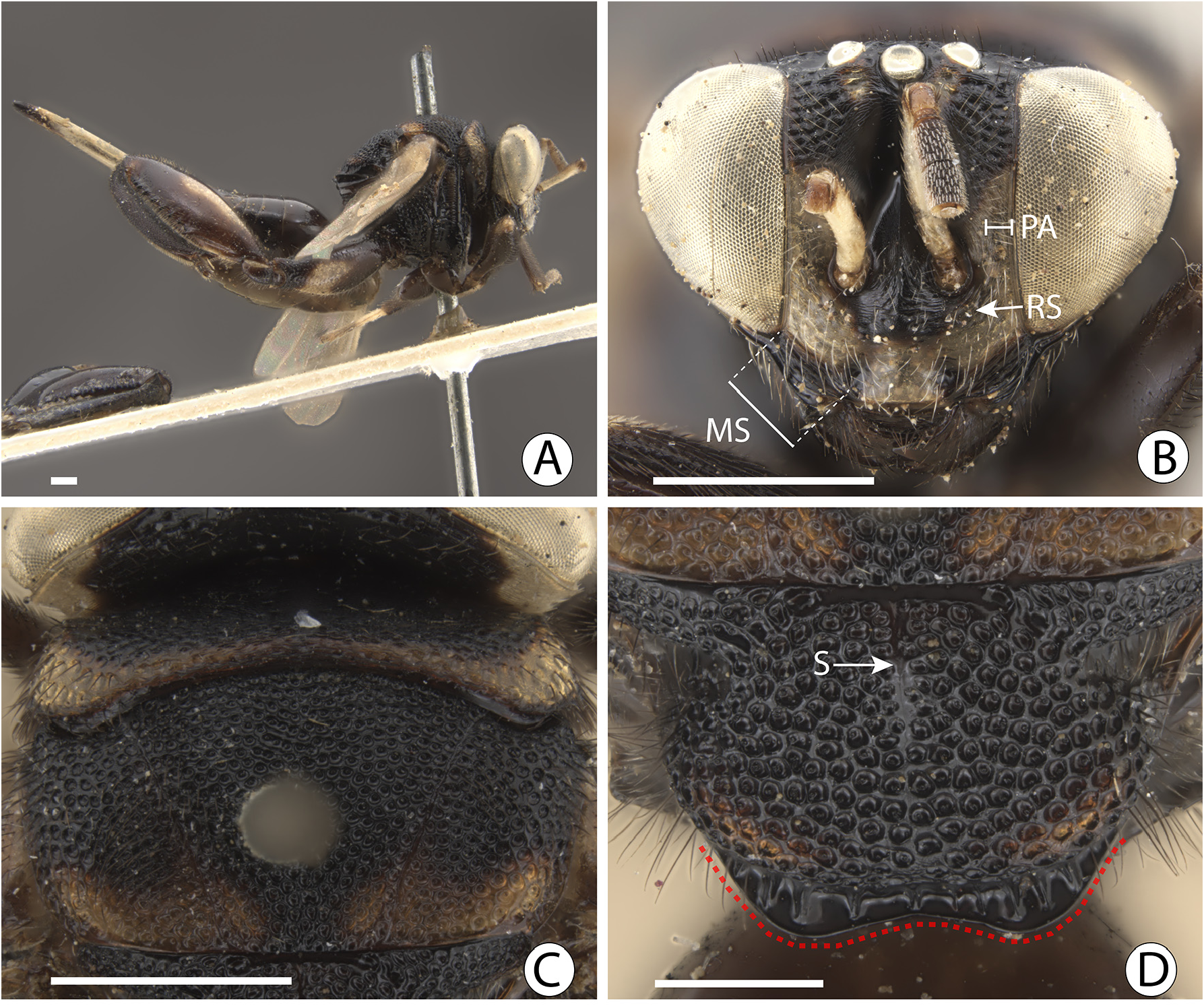
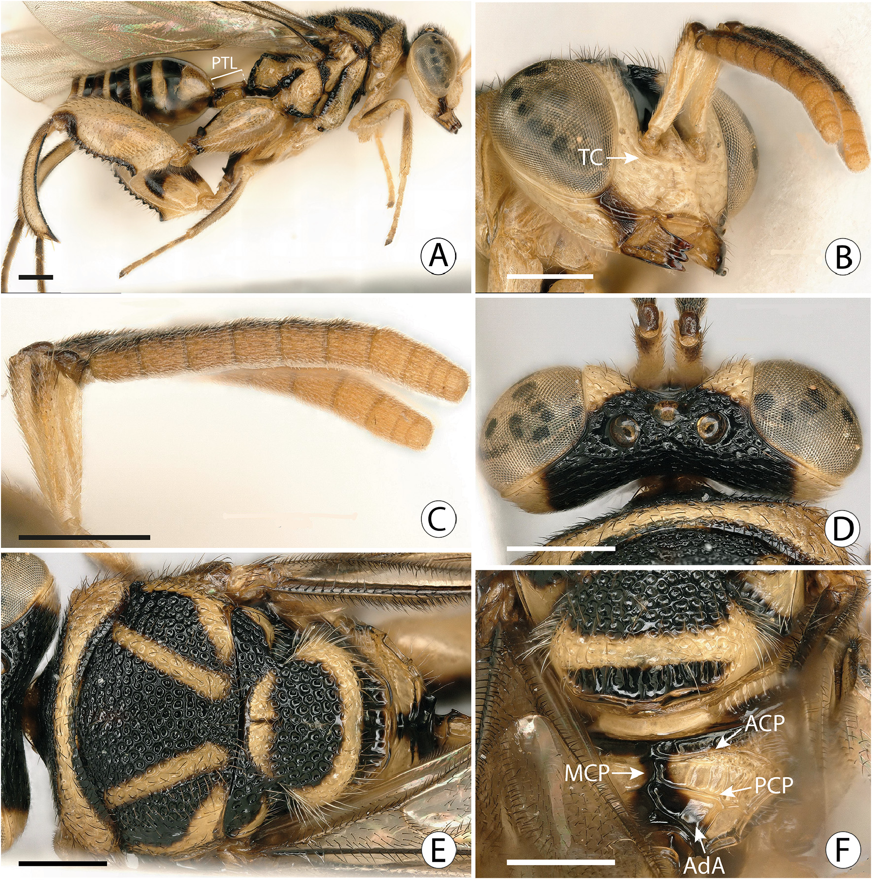
Figs 7 View FIGURE 7 A–F
Description. MALE. Length 5.75 mm
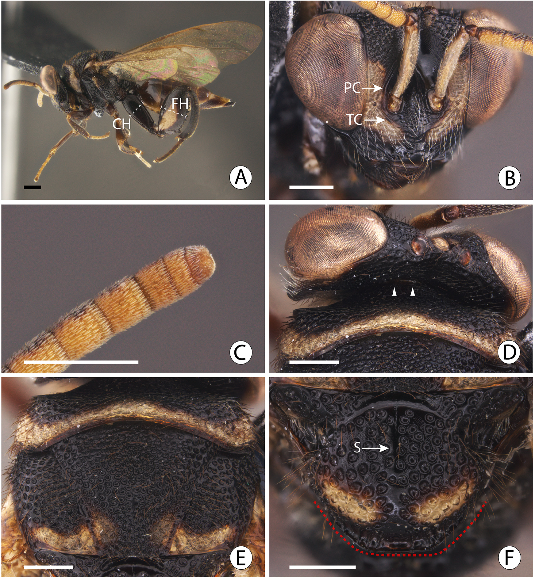
Color. Body mainly yellow ( Fig. 7A View FIGURE 7 ), but venter of Fu1–Fu6, Fu7 and clava dark yellow ( Figs 7B, C View FIGURE 7 ), with dorsal apex of antennal scape ( Figs 7B, C View FIGURE 7 ), basal two thirds of mandible ( Fig. 7B View FIGURE 7 ), dorsolateral spot on pronotum ( Fig. 7A View FIGURE 7 ), and lateral side of mesotibia ( Fig. 7A View FIGURE 7 ) brown, and the following black: pedicel, anellus, dorsum of Fu1–Fu6 ( Figs 7B, C View FIGURE 7 ), antennal scrobe ( Fig. 7B View FIGURE 7 ), strip on malar area ( Fig. 7B View FIGURE 7 ), ventral and dorsal margin of mandible, mandibular teeth ( Fig. 7B View FIGURE 7 ), vertex ( Figs 7D, E View FIGURE 7 ); median area of occiput ( Fig. 7D View FIGURE 7 ), pronotal neck, posterodorsal margin of pronotal collar, inverted triangle on median lobe of mesoscutum ( Fig. 7E View FIGURE 7 ), internal two thirds of lateral lobe of mesoscutum ( Fig. 7E View FIGURE 7 ), dorsum of axilla ( Fig. 7E View FIGURE 7 ), axillula ( Fig. 7E View FIGURE 7 ), disc and apex of mesoscutellum ( Figs 7E, F View FIGURE 7 ), posterolateral margin of metanotum ( Fig. 7F View FIGURE 7 ), median strip and almost the whole perimeter of propodeum ( Figs 7A, 7F View FIGURE 7 ), spot on anterior face of mesopectus ( Fig. 7A View FIGURE 7 ), strip along the mesofemoral depression ( Fig. 7A View FIGURE 7 ), carina of mesocoxal foramen, spot on dorsum of acropleuron ( Fig. 7A View FIGURE 7 ), posterior margin and spot on venter of mesepimeron ( Fig. 7A View FIGURE 7 ), anterior and ventral margins of metapleuron ( Fig. 7A View FIGURE 7 ), spot on anterior face of profemur ( Fig. 7A View FIGURE 7 ), posterior face of mesotibia, spot on internal face and apex of metacoxa ( Fig. 7A View FIGURE 7 ), venter of metafemur, spot on internal face of metafemur ( Fig. 7A View FIGURE 7 ), margins of metatibia ( Fig. 7A View FIGURE 7 ), metatarsus, base and apex of petiole, and transverse band on gastral tergites ( Fig. 7A View FIGURE 7 ). Wings slightly smoky, veins dark brown ( Figs 7A, 7E, F View FIGURE 7 ). Setae mainly black ( Figs 7A, 7E View FIGURE 7 ), but following with white setae: lower half of parascrobal area, lower face ( Fig. 7B View FIGURE 7 ), antennae ( Figs 7B, C View FIGURE 7 ), gena, front and middle legs (with the exception of some black setae on pro- and meso- femora and tibiae), venter of mesopleuron, mesepimeron, metapleuron, venter of metacoxa, internal and external face of metafemur (with some black scattered black setae), external face of metatibia (with some scattered black setae), metatarsus, callus ( Fig.7A View FIGURE 7 ), and laterally on mesoscutellum and propodeum ( Figs 7A, 7E View FIGURE 7 ).
Head. Clava tapering toward apex ( Fig. 7C View FIGURE 7 ); lower face with inconspicuous transverse carina below antennal foramen ( Fig. 7B View FIGURE 7 ).
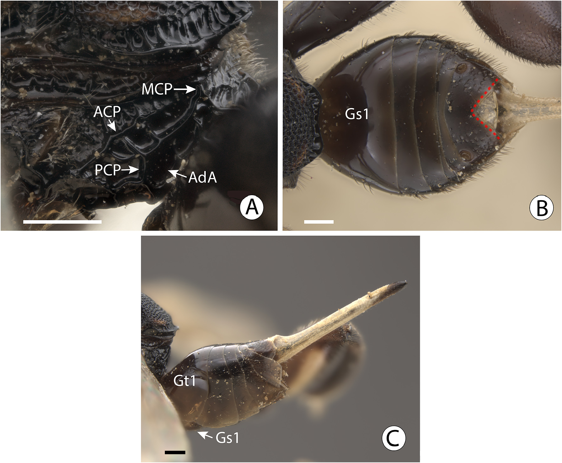
Mesosoma. Median area of mesoscutum with regular umbilicate fovea, interstices not so narrow, giving sculpture a reticulate appearance ( Fig. 7E View FIGURE 7 ); mesoscutellum without basal smooth and shiny transverse band ( Fig. 7E View FIGURE 7 ), median unfoveate strip restricted to basal quarter ( Fig. 7E View FIGURE 7 ), frenal carina forming sublateral lobes ( Fig. 7E View FIGURE 7 ); metascutellum convex, smooth and shiny, without median fovea or impression ( Fig. 7F View FIGURE 7 ); propodeum oblique ( Fig. 7A View FIGURE 7 ), anterior costula conspicuous throughout ( Fig. 7F View FIGURE 7 : ACP), median carina continuous, sinuous and extending virtually from anterior to posterior margin ( Fig. 7F View FIGURE 7 : MCP), portion anterior to posterior costulae about 0.5× median length of propodeum, posterior costula conspicuous and limited to posterior half of propodeum ( Fig. 7F View FIGURE 7 : PCP), adpetiolar area without submedian carinae ( Fig. 7F View FIGURE 7 : AdA); metafemur with 13 small teeth, basal tooth the largest, followed by a minute tooth and then by medium sized teeth ( Fig. 7A View FIGURE 7 ).
Metasoma. Petiole visible dorsally, about 1.5× as long as wide, basal lamina present dorsally and ventrally, median dorsal carina present over anterior third, and with strong and complete dorsolateral carina delimiting a lateral sulcus ( Fig. 7A View FIGURE 7 ).
FEMALE. Unknown.
Host. Unknown.
Distribution. French Guiana (Arrondissement of Cayenne).
Remarks. Conura sp. 1 and Conura sp. 2, both based on males, differ from the females of C. leucotela , C. paraleucotela and C. pseudoleucotela by having the body mainly yellow with black or brown markings ( Fig. 7A View FIGURE 7 ), whereas the females are mainly black or dark with yellow markings ( Figs 1A View FIGURE 1 , 3A View FIGURE 3 , 5A View FIGURE 5 ). Although this difference in body color is conspicuous between the sexes, all have the pattern of coloration on the dorsum of the mesosoma somewhat similar (here the differences are on the extension of the black markings), including sharing a transverse yellow strip on the pronotum, an external yellow strip on the lateral lobe of the mesoscutum ( Figs 1C View FIGURE 1 , 3E View FIGURE 3 , 5E View FIGURE 5 , 7E View FIGURE 7 ), and a posterior yellow strip on the mesoscutellum ( Figs 2B View FIGURE 2 , 3F View FIGURE 3 , 5F View FIGURE 5 , 7E View FIGURE 7 ). We have seen some hundreds of species of Conura , many of them with associated males and females, and the opposite sexes are quite similar in color, not as different as between the males and females species of the leucotela group above. Conura sp. 1 and Conura sp. 2 also differ from described females of leucotela species by their longer petioles, which is distinctly longer in Conura sp. 1 ( Fig. 7A View FIGURE 7 ) and Conura sp. 2 ( Fig. 8A View FIGURE 8 ), though this difference may reflect sexual dimorphism. In species of Conura with similar mesosoma-metasoma articulation systems to that of C. leucotela and C. pseudoleucotela (in Conura fusiformis (Ashmead) and other species of fusiformis species group, see item Discussion below), male petioles are distinctly longer than for females. Conura sp. 1 differs from the other known leucotela group species by having the antennal clava tapering apically ( Fig. 7A View FIGURE 7 ) (slightly tapering in C. pseudoleucotela , , not tapering in the remaining species), propodeum with a continuous median carina that extends virtually between the anterior and posterior margins ( Fig. 7F View FIGURE 7 : MCP) (median carina interrupted and not extending along the propodeal length in other species), petiole about 1.5× as long as wide, with median and two dorsolateral carinae ( Figs 7A, 7E View FIGURE 7 ) (at most 0.6× as long as wide, without median carina and with at most one dorsolateral carina in other species).
Material Examined. Male labeled ‘ French Guiana, [ Arrondissement of Cayenne , Sinnamary], camp ORSTOM , piste de Saint-Elie PK 17, on Solanum sp. 03.xi.1989 (G. Delvare)’ ( CIRAD) .
| CIRAD |
Centre de Cooperation Internationale en Recherche Agronomique pour le Developpement |
No known copyright restrictions apply. See Agosti, D., Egloff, W., 2009. Taxonomic information exchange and copyright: the Plazi approach. BMC Research Notes 2009, 2:53 for further explanation.