
Pachycynodon cf. filholi Schlosser, 1888
|
publication ID |
https://doi.org/10.5252/geodiversitas2019v41a15 |
|
publication LSID |
urn:lsid:zoobank.org:pub:9DD3CC29-3AEA-44B8-8E8F-6AD882DF5B1C |
|
DOI |
https://doi.org/10.5281/zenodo.3703509 |
|
persistent identifier |
https://treatment.plazi.org/id/03A48799-1A69-FF95-FF40-FB586D6AFD1C |
|
treatment provided by |
Valdenar |
|
scientific name |
Pachycynodon cf. filholi Schlosser, 1888 |
| status |
|
Pachycynodon cf. filholi Schlosser, 1888
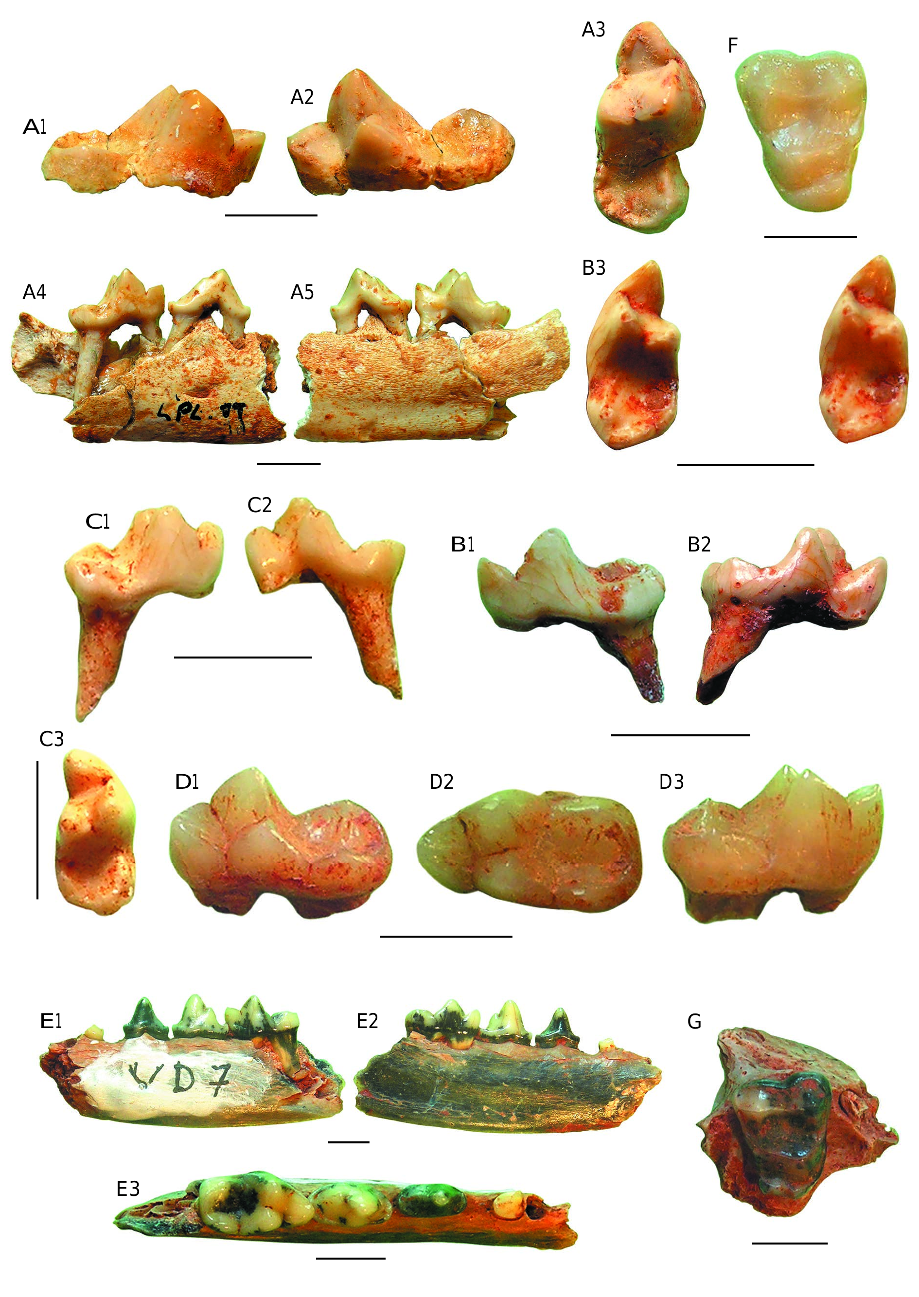
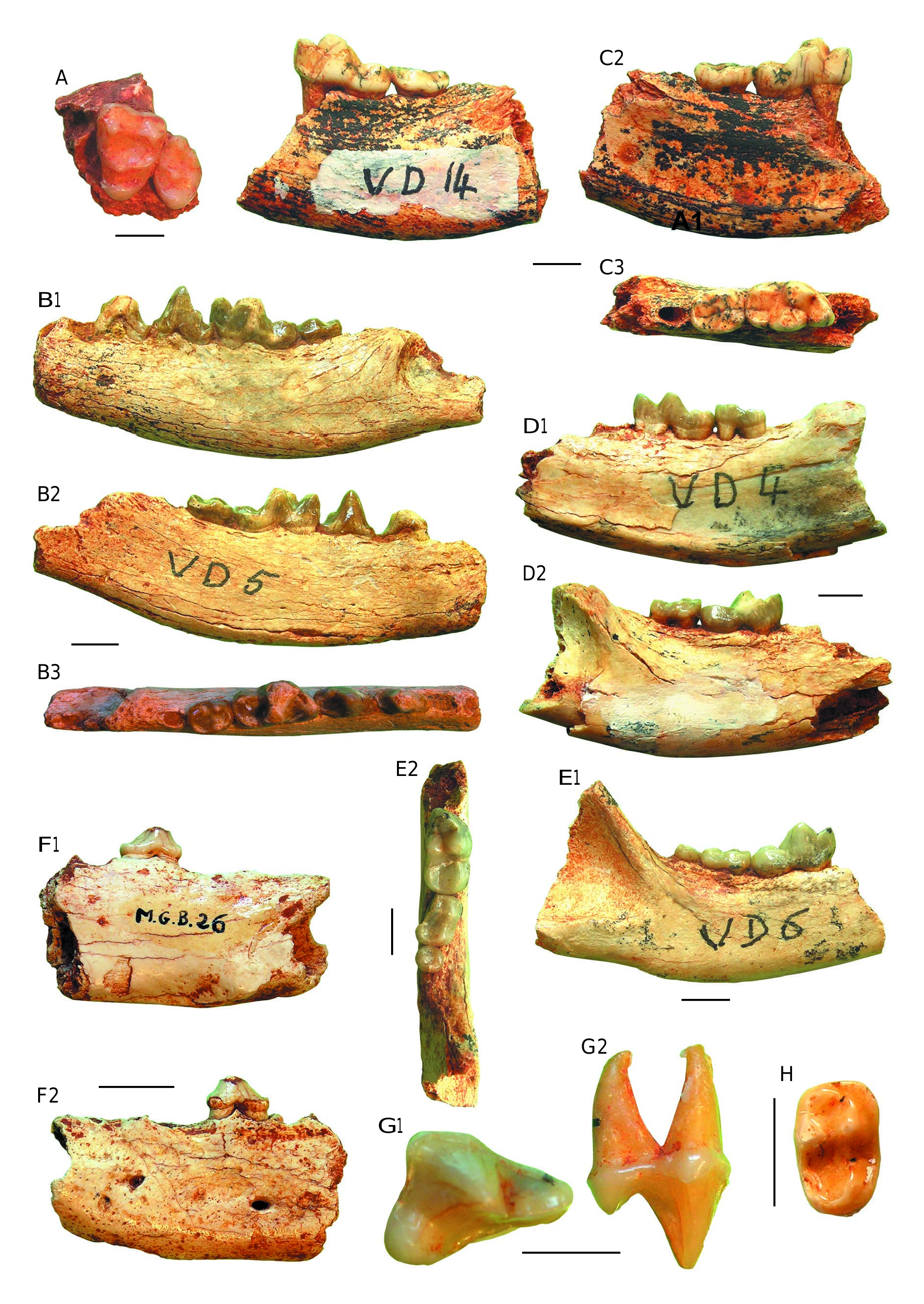
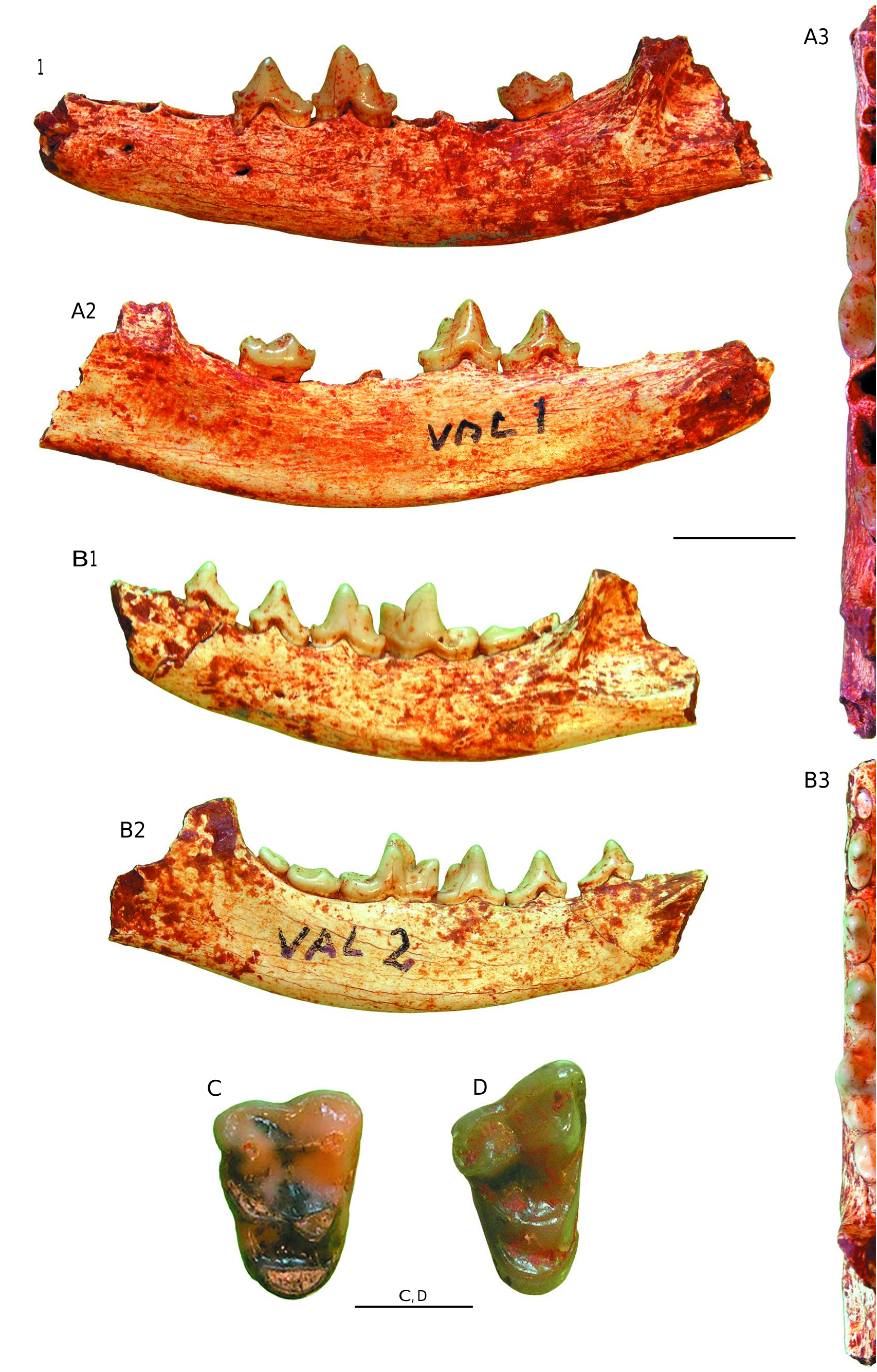
( Figs 2E View FIG ; 3B, H View FIG )
TYPE MATERIAL. — Lectotype by designation of Teilhard ( Teilhard 1915: 38): fragment of left hemi-mandible with broken p2, p3-m1 ( Schlosser 1888: 30, pl. IX, figs 1, 3, 5), “Phosphorites du Quercy” without any biostratigraphic information.
NEW MATERIAL. — UM VD 7 left hemi-mandible with p3-m1; UM VD 8 left hemi-mandible; UM VD 10 small fragment of mandible with m2; UP LPL 14 m 2; UM VD 5 hemi-mandible with p3-m2.
REMARKS
Schlosser (1888) established P. filholi on a fragment of mandible with p3-m1 from the “Phosphorites du Quercy” and he added a fragment of edentulous maxilla ( Schlosser 1888: pl. IX, fig. 16) from the bulk of the Quercy specimens without any special link to the mandible.
Among the material from Valbro, some mandibular specimens seem to be close to this species. A hemi-mandible (UM VD7) with mesial alveolus and distal root of p2, and fresh p3-m1 is quite similar to a cast of the type of P. filholi ( Fig. 2E View FIG 1-E View FIG 3 View FIG ). The almost symmetrical and pointed p3 displays small mesial and distal cingulids with a small, pointed mesial spur; its mesial and distal edges are sharp and pointed, with a profile that is slightly concave. The p4 is clearly higher than p3 and the protoconid of m1, its mesial and distal cingulids are more developed than in p3 and the distal cingulid forms a true small talonid. A pacd is situated at mid-height of the distal cristid of p4. The low m1 has a small cingulid running along the base of the paraconid-protoconid blade, the former appears smaller than the metaconid in lingual view. The paraconid–metaconid contact at mid-height closes the trigonid valley. The latter is slightly shifted distally and can clearly be seen in buccal view behind of the protoconid, which is separated from the hypoconid by a large sinus. The hypoconid is quite large and slopes gently lingually to the base of the entoconid, which is fused into a slender entocristid slightly higher than the hypoconid. Medially, the entoconid is separated by a small notch from the metaconid and distally joins the hypocristid to close the talonid basin. The p2 and p3 ( Teilhard 1915: pl. IV, figs 7-9) are sharp.
There is no m 2 in the type specimen, but there is an m 2 in the hemi-mandible VD5 that we identify as P. cf. filholi ( Fig. 3B View FIG 1-B View FIG 3 View FIG ). The p3 of VD5 is partly broken off but was smaller than the thin and pointed p4 that displays a small pacd and a minute talonid. The m1 has a paraconid that is less oblique than that of P. dubius and thus the paraconidprotoconid shearing blade is more prominent. In the talonid of m1, the entoconid is partly broken off. In m2, which is rectangular, the trigonid occupies half of the surface of the crown; the protoconid and metaconid are subequal in size, both having a small wear facet on the dentine at the tip. In front of the protoconid and metaconid, a mesial cristid closes a mesial fovea. The protoconid is separated by a sinus from the hypoconid, which, in turn, is followed by a cristid running to the metaconid and closing a bowlshaped basin.
An isolated rectangular and elongate m2 from the site of La Plante2 (UP LPL14) is too small to fit P. boriei or P. curvirostris ( Fig. 3H View FIG ). It displays a complete trigonid, a faint cingulid is present along the base of a small but clear paraconid displaying a small pit of dentine at its tip. The paraconid cristids close the minute trigonid basin mesially. Both protoconid and metaconid display a wear facet at the tip. This facet is larger in the latter. The proto- and metalophids separate the trigonid and talonid basins, while distally a post-hypocristid and entocristid close the bowlshaped talonid basin.
The corpus mandibulae of UM VD8 is shallow and tapering rostrally. There are two small mental foramina, one below p2, and another below p3. The m1, in which part of the trigonid is missing, has a morphology similar to that of UM VD7. Some isolated m1s could belong to the same taxon.
We hesitantly add to these remains a fragment of mandible (UM VD10) with m2. Its crown is partially worn but we can see the large wear pit of the protoconid, the less marked metaconid and, mesially, the small well with dentine that would may correspond to the paraconid. There is a sinus between the protoconid and the hypoconid. The hypoconid and the entoconid are worn with large appearance of dentine. The talonid tapers distally and the basin is narrow.
| NEW |
University of Newcastle |
| UM |
University of Marburg |
| UP |
University of Papua and New Guinea |
No known copyright restrictions apply. See Agosti, D., Egloff, W., 2009. Taxonomic information exchange and copyright: the Plazi approach. BMC Research Notes 2009, 2:53 for further explanation.