
Synergus ramoni Nieves-Aldrey & Medianero
|
publication ID |
https://doi.org/10.5281/zenodo.276876 |
|
DOI |
https://doi.org/10.5281/zenodo.6189360 |
|
persistent identifier |
https://treatment.plazi.org/id/03A487F3-FFF5-C603-FF2A-6215FAEBEC13 |
|
treatment provided by |
Plazi |
|
scientific name |
Synergus ramoni Nieves-Aldrey & Medianero |
| status |
sp. nov. |
Synergus ramoni Nieves-Aldrey & Medianero sp. nov.
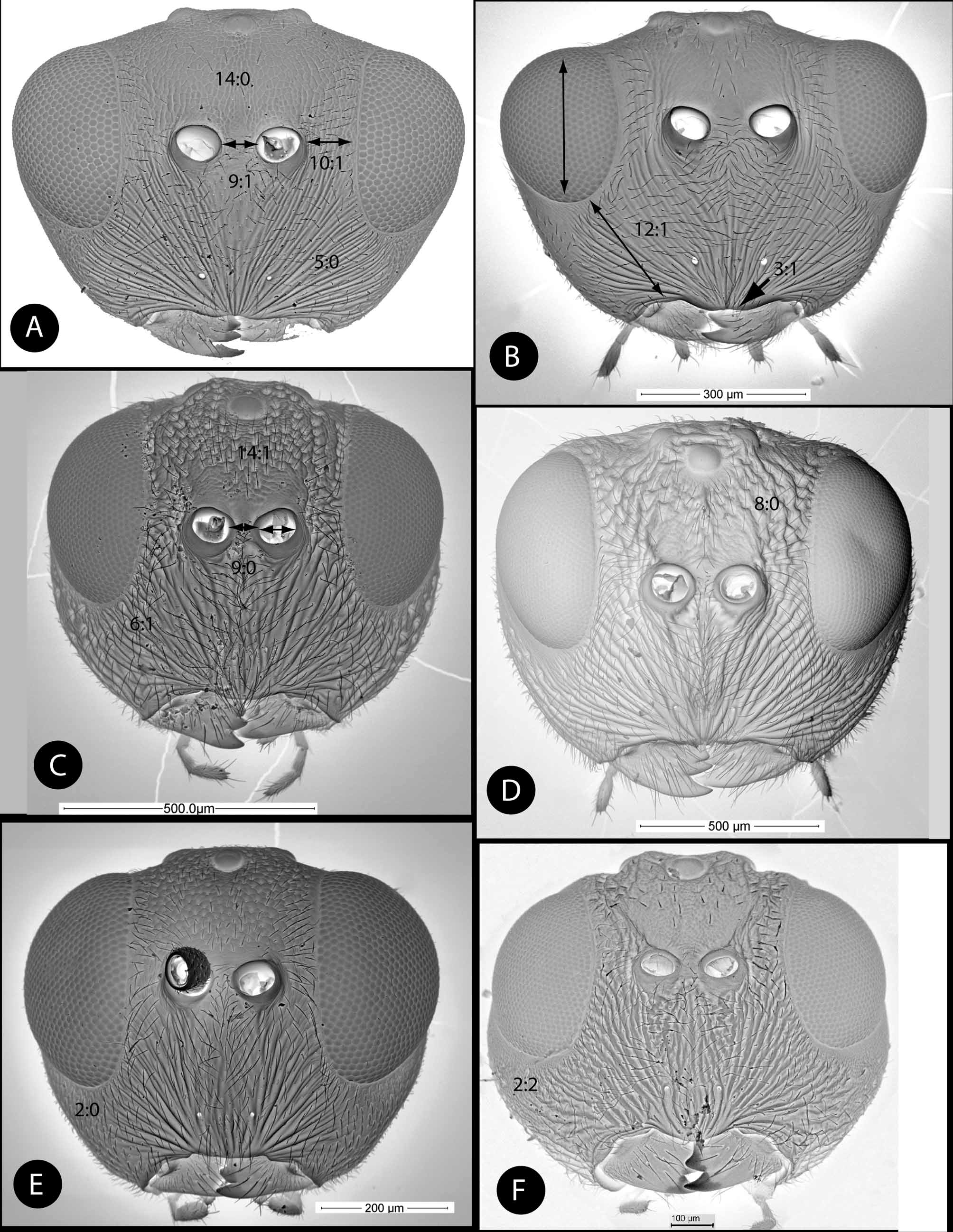
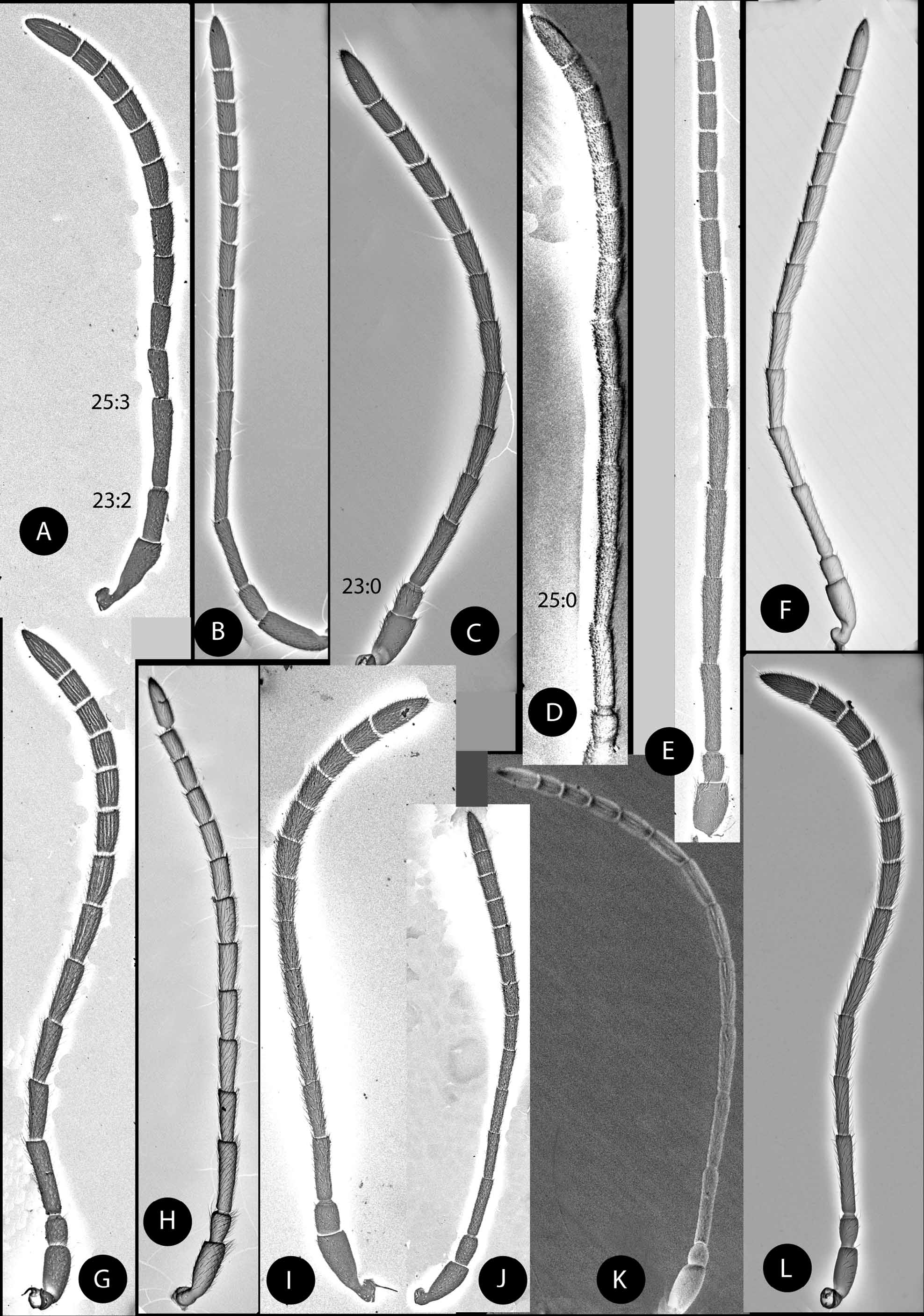
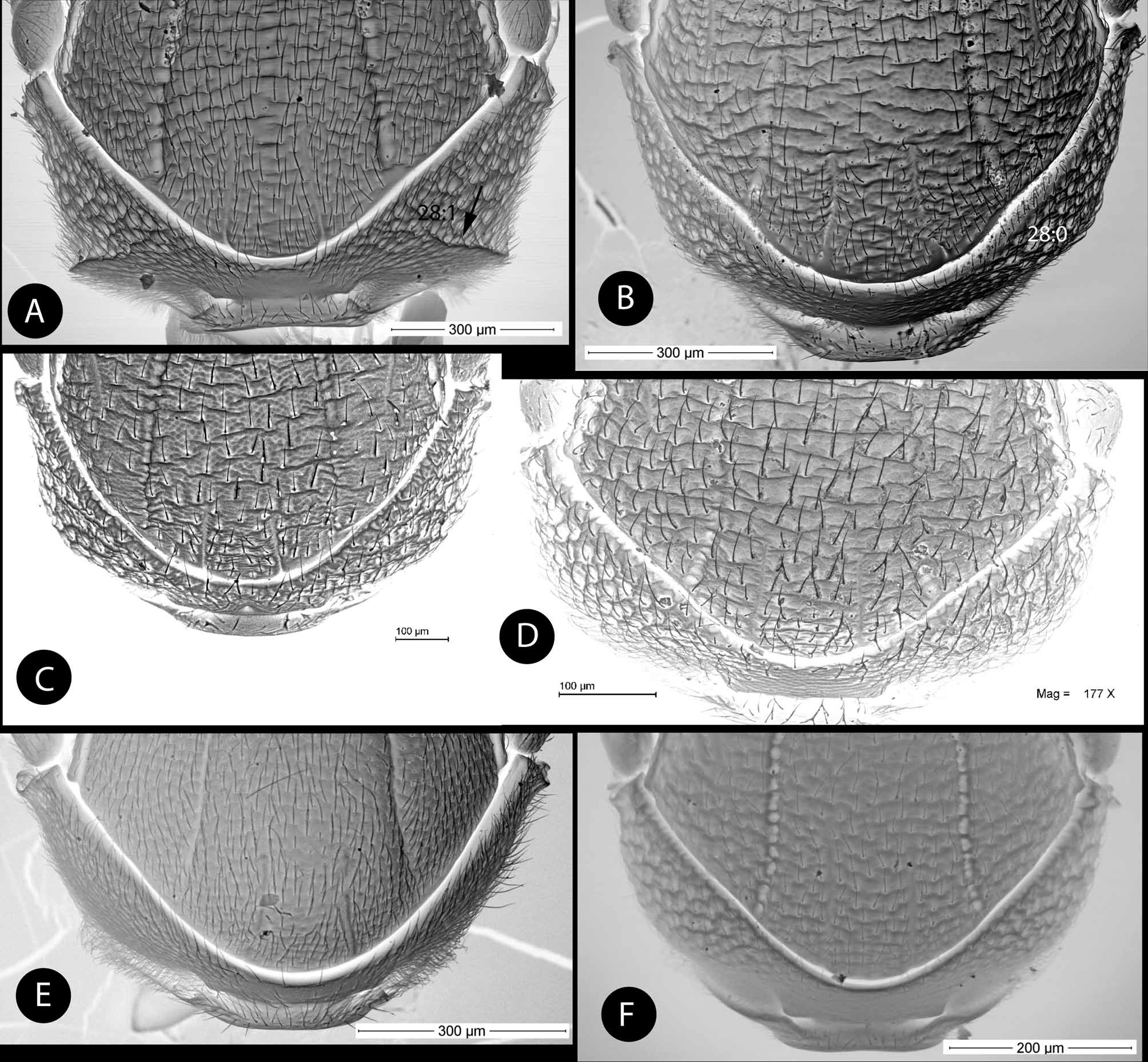
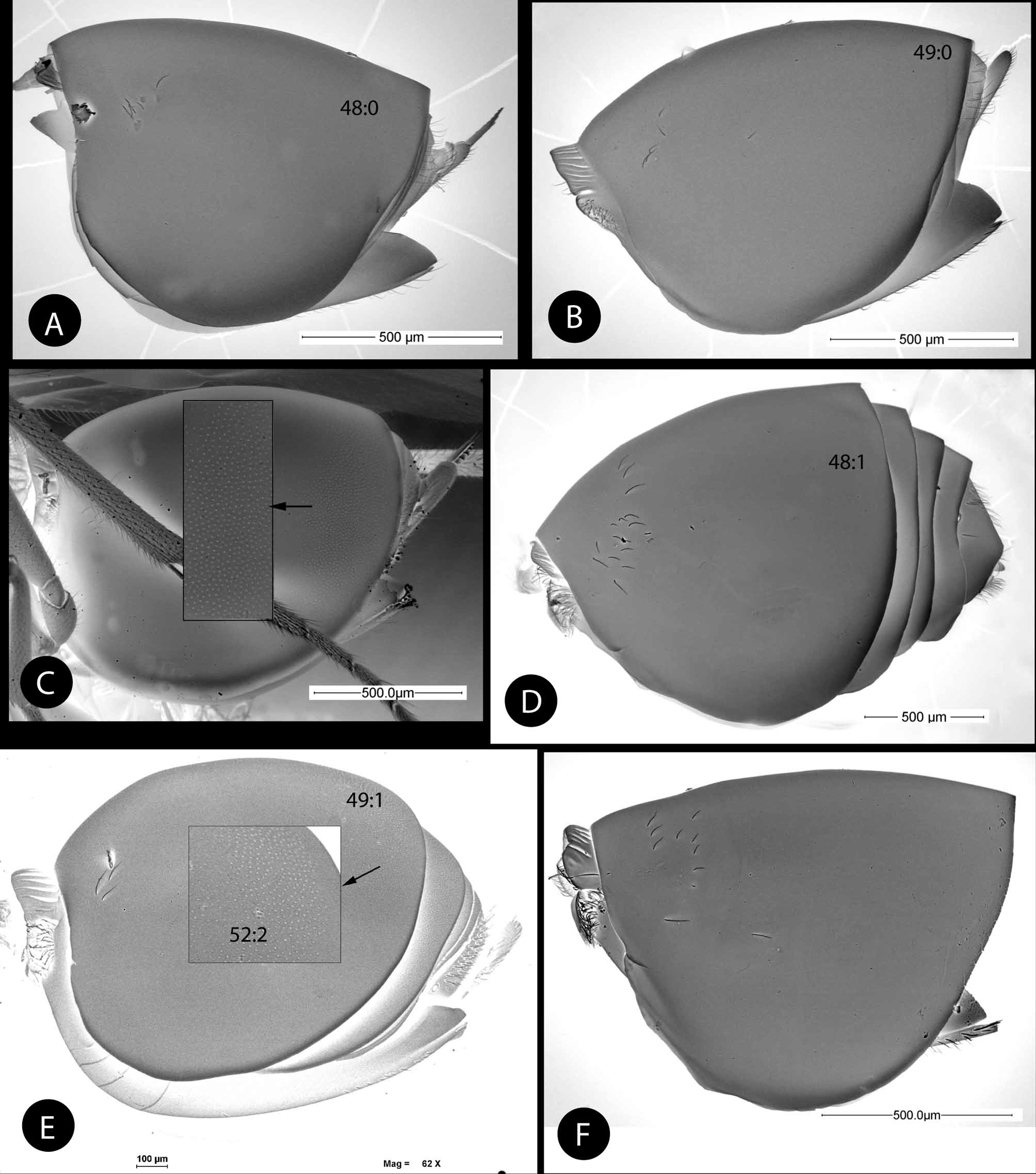
( Figs. 1 View FIGURE 1 C, 3F, 5C, 6G, 7B, 8C, 10C, 12D, 13F, 16B, 17C, 18E–F)
Type material. Holotype ̩ ( Fig. 18 View FIGURE 18 F) (in Museo Nacional de Ciencias Naturales, Madrid, Spain ( MNCN), cardmounted. Cat. nº 2097). PANAMA, Chiriquí, Boquete, El Salto, 8º 47' 32.08” N, 82º 27' 37.09” W, 1,431 m; ex gall of an unidentified cynipid genus on twigs of Quercus salicifolia Née (Fagaceae) ( Fig. 22 View FIGURE 22 A), gall collected 27.xi.2008, E. Medianero leg. Paratypes: 13, 1̩, same data as holotype; 13,1̩, Boquete, Road to Volcancito, 1,404 m, 22.x.2008. E. Medianero leg. In the MNCN. Additionally, 1̩ paratype of the type series was dissected for SEM observation (in the MNCN).
Etymology. Named after Ramón Hernández, dear friend of the first author, in acknowledgment of his invaluable help in the first collecting trip of oak gall wasps of Panama, which led to this study.
Diagnosis and comments. This species is closely related to S. mesoamericanus , being similar in a majority of their diagnostic morphological characters. It differs from that species with respect to its black coloration, narrower head, larger ocellar triangle and the posterior ocelli being closest to the margin of eyes. Furthermore, the frons is more closely punctate, and the frontal carinae are less marked in S. ramoni ; the sculpture of mesopleuron is weaker; the speculum is partially smooth; and the radial cell is 2.8 times as long as wide (less than 2.5 in S. mesoamericanus ).
Description. Body length 2.75 mm (range 2.69–2.77; N = 3) for females; 2.41 (range 2.21–2.61; N = 2) for males.
Head, mesosoma and metasoma predominantly black; mouthparts and sides of frons near the toruli yellowish; metasoma ventrally red brown; antennae blackish yellow; distal half less dark. Legs mainly black, apex of femora and tibiae and tarsi dark yellowish. Forewing hyaline, slightly darkened; veins brown. The male differ in having entirely the face, genae, and the antennal flagellum yellowish.
Female. Head in dorsal view ( Fig. 3 View FIGURE 3 F) 1.9 times as wide as long. Gena not expanded behind compound eye. POL 1.6 times longer than OOL, posterior ocellus separated from inner orbit of eye by 1.3 times its diameter. Head in anterior view ( Fig. 1 View FIGURE 1 C) narrow, only 1.1 times wider than high, genae very slightly expanded, strongly punctate. Face moderately pubescent, with marked, irregular, irradiating carinae from clypeus, reaching ventral margin of eye and ventral margin of toruli; branched near compound eyes. Ventral margin of clypeus straight not projected over mandibles. Malar space 0.6 as long as height of compound eye. Toruli situated slightly below mid-height of compound eye; distance between toruli short, shorter than diameter of a torulus; distance between mesal margin of compound eye and lateral margin of a torulus shorter than diameter of a torulus, including rim. Frons closely and conspicuously punctate, frontal carinae present but obscured by the punctate sculpture ( Figs. 1 View FIGURE 1 C, 3F). Vertex and occiput rugose-punctate.
Female antenna with 14 segments ( Fig. 5 View FIGURE 5 C); flagellum filiform. Relative lengths of antennal segments: 19:11:27:18:19:19:19:17:15:15:14:13:13:22; Pedicel 1.4 as long as wide; F1 1.5 as long as F2. Ultimate flagellomere 1.7 times longer as long as F11. Placodeal sensillae on F8–F11 disposed in one single row of 2–4 visible sensillae in each flagellomere. Male antenna with 15 segments; F1 excaved medially and moderately expanded apically ( Fig. 6 View FIGURE 6 G).
Mesosoma. Pronotum without a lateral pronotal carina ( Fig. 7 View FIGURE 7 B). Lateral surface of pronotum with rugosepunctate sculpture. Mesoscutum ( Fig. 8 View FIGURE 8 C) with marked, undulate transverse rugae, the interspaces with coriarious sculpture. Notauli percurrent, well impressed, wider posteriorly, wider than transscutal fissure; moderately converging posteriorly. Median mesoscutal impression visible but shallowly impressed. Anteroadmedian signa well marked. Scutellar foveae ellipsoidal, with some rugose sculpture, anterior margins widely divergent from the fissure transscutal, posterior margins indistinct. Scutellum with rugose sculpture; not margined laterally and posterodorsally ( Figs. 8 View FIGURE 8 C, 10C). Mesopleuron ( Fig. 10 View FIGURE 10 C) medially with weak longitudinal striae, the interspaces with coriarious sculpture, the striae being weak and hardly visible dorsolaterally in the speculum ( Fig. 10 View FIGURE 10 C).
Lateral propodeal carinae distinct, broad, subparallel. Median propodeal area pubescent, with some vertical rugae prolonged into the nucha. Nucha strongly sulcate, dorsally and laterally.
Legs. Tarsal claw ( Fig. 16 View FIGURE 16 B) with base produced into a small secondary acute tooth measuring about 1/4 of length of apical tooth.
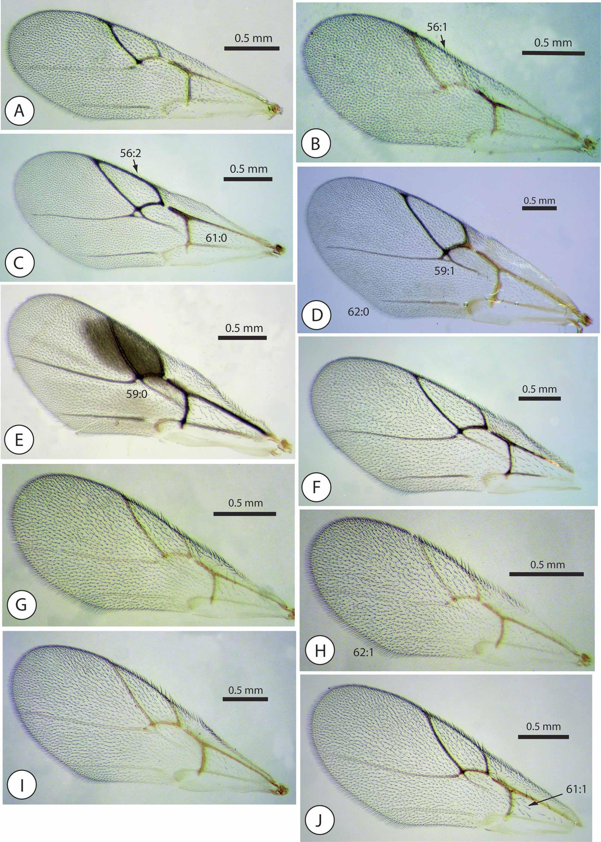
Forewing ( Fig. 17 View FIGURE 17 C). Radial cell 2.8 times longer than wide. All the veins well pigmented, R1 well visible along margin of radial cell, the radial cell appearing unambiguously closed ( Fig. 17 View FIGURE 17 C; areolet distinct; vein Rs+M visible. Basal cell with dense, closely spaced setae. Apical margin of wing with a short fringe of setae.
Metasoma ( Fig. 13 View FIGURE 13 F). As long as head plus mesosoma. First metasomal tergum longitudinally sulcate dorsally. Metasomal tergum T2+3 fused, smooth and shining, covering almost the entire metasoma; without micropunctures; anteromedian area only with a group of about 14 setae. Projecting part of hypopygial spine slightly extended beyond attachment of lateral flap; apical setae not projected beyond apex spine.
Distribution. Known only from the type locality near Boquete and Volcán Barú (Chiriqui, Panama).
Biology. Synergus ramoni inhabits twig galls on Quercus salicifolia ( Quercus, Lobata section). The host gall is irregularly spherical and develops on twigs ( Fig. 22 View FIGURE 22 A). The host cynipid was not reared, and it is unknown.


| MNCN |
Museo Nacional de Ciencias Naturales |
No known copyright restrictions apply. See Agosti, D., Egloff, W., 2009. Taxonomic information exchange and copyright: the Plazi approach. BMC Research Notes 2009, 2:53 for further explanation.
|
Kingdom |
|
|
Phylum |
|
|
Class |
|
|
Order |
|
|
Family |
|
|
Genus |