
Synergus nicaraguensis Diaz & Gallardo, 1998
|
publication ID |
https://doi.org/10.5281/zenodo.276876 |
|
DOI |
https://doi.org/10.5281/zenodo.6189362 |
|
persistent identifier |
https://treatment.plazi.org/id/03A487F3-FFFA-C602-FF2A-615BFC38EF87 |
|
treatment provided by |
Plazi |
|
scientific name |
Synergus nicaraguensis Diaz & Gallardo, 1998 |
| status |
|
Synergus nicaraguensis Diaz & Gallardo, 1998
New record from Panama
Studied material. 1 3, 5Ƥ, Chiriquí, Renacimiento, 1,270 m, ex gall Disholcaspis sp. on Quercus lancifolia , 24.xi.2008. E. Medianero leg.
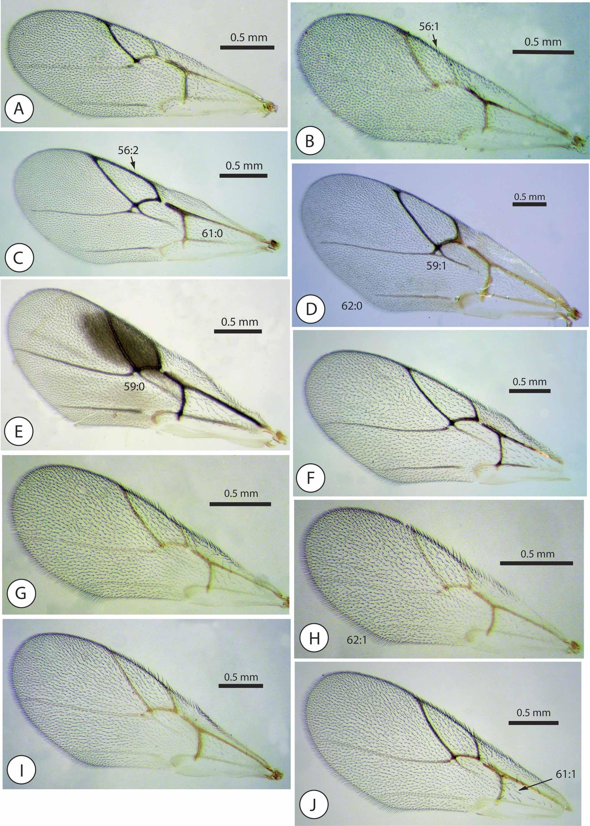
Diagnosis and comments. This species is easily recognizable and can be distinguished from all of the remaining Neotropical Synergus species by having a forewing with a shaded infuscate area over the radial cell extended beyond the Rs vein. Other diagnostic features of this species are: radial cell short and wide, less than 2.5 times as long as wide. F1 3 as long as the pedicel. Metasomal T2+3 posteriorly closely and distinctly punctate, with the punctures extended over about 1/3 of the total length of the segment.
Until now, knowledge of this species was limited to the materials of the type locality (Jinotega, Nicaragua) ( Diaz & Gallardo 1998). Our record from Panama is the second for this species in Central America, considerably increasing the size of its known distribution area. We examined the type material of S. nicaraguensis and compared it with our materials from Panama. The Panamanian insects differ slightly from the type material in their coloration, especially with respect to their almost entirely yellow legs, which are darker in the type specimens.
Additional descriptive data. Some descriptive date and illustrations of morphological characters which were omitted in the original description are here given.
Female (habitus, Fig. 19 View FIGURE 19 A). Head in dorsal view ( Fig. 4 View FIGURE 4 B) 2 times as wide as long; genae not expanded behind compound eye. POL 1.5 as OOL, posterior ocellus separated from inner orbit of eye by about its diameter. Head in anterior view ( Fig. 2 View FIGURE 2 E) 1.3 times wider than high. Facial carinae regular, deep, reaching ventral margin of eye and ventral margin of toruli; not branched near margin compound eyes. Ventral margin of clypeus straight. Frons with wide, slightly branched frontal carinae, reaching ocelli, weak piliferous punctures present.
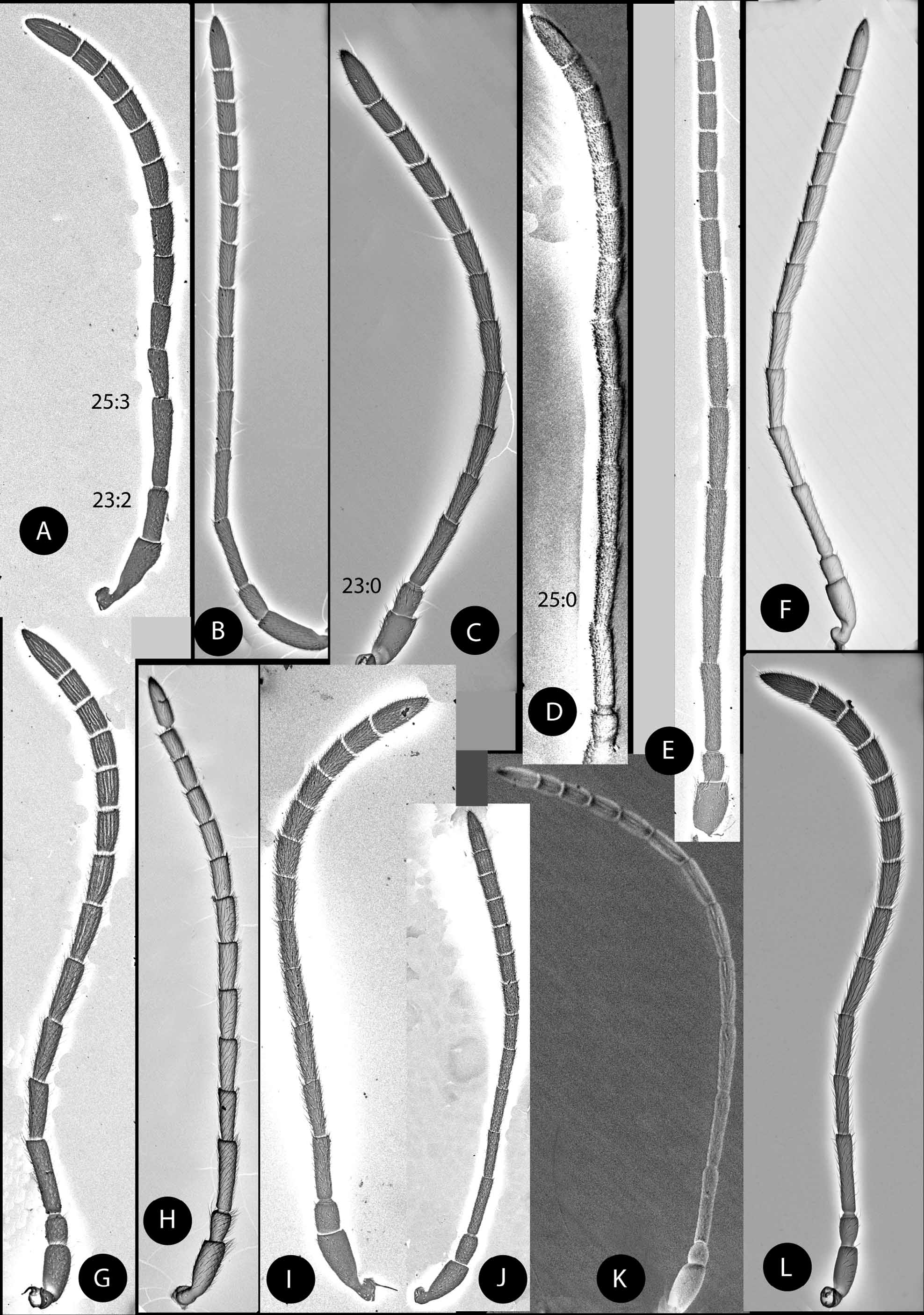
Female antenna with 14 segments ( Fig. 5 View FIGURE 5 E). Pedicel 1. 2 as long as wide; F1 as long as F2. Ultimate flagellomere 1.8 times as long as F11. F1 of male curved in de middle and moderately expanded apically.
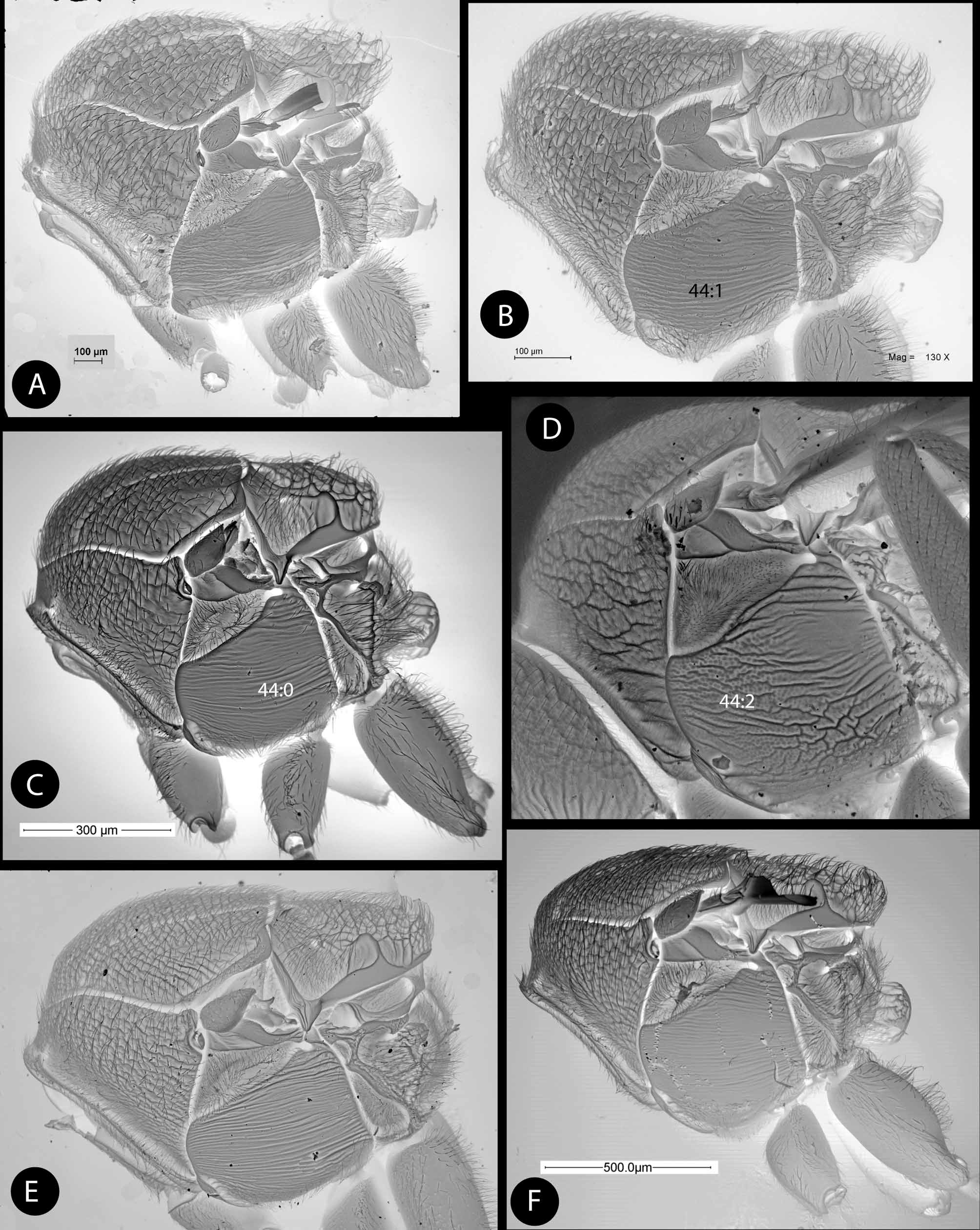
Pronotum without a lateral pronotal carina. Lateral surface of pronotum with rugose sculpture. Mesoscutum ( Fig. 8 View FIGURE 8 F) with wide, transverse, undulate, interrupted rugae, the interspaces with coriarious sculpture. Notauli percurrent, convergent posteriorly, wider than transscutal fissure; narrowly separated at meeting with the transscutal fissure. Median mesoscutal impression visible in posterior three quarts of mesoscutum but shallow. Scutellar foveae large, ellipsoidal, smooth, anterior margins widely divergent from the trasscutal fissure, posterior margins distinct. Scutellum not margined laterally, with strongly rugose sculpture. Mesopleuron ( Fig. 11 View FIGURE 11 A) with strong, irregular, longitudinal striae, extended into the entire surface of mesopleuron; coriarious sculpture visible in the interspaces. Lateral propodeal carinae distinct, broad, subparallel ( Fig. 12 View FIGURE 12 C).
Tarsal claw ( Fig. 16 View FIGURE 16 C) with base produced into a large secondary acute tooth measuring about 1/2 of length of apical tooth.
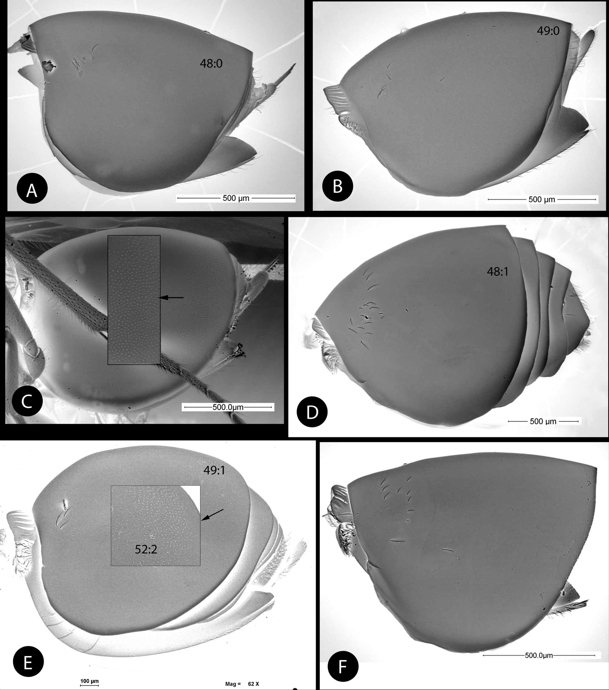
Forewing ( Fig. 17 View FIGURE 17 E). Present a shaded infuscate area over the radial cell and extended beyond the Rs vein; radial cell short and wide, less than 2.5 as long as wide; vein Rs+M visible. Basal cell with dense, closely spaced setae. Apical margin of wing with a short fringe of setae.
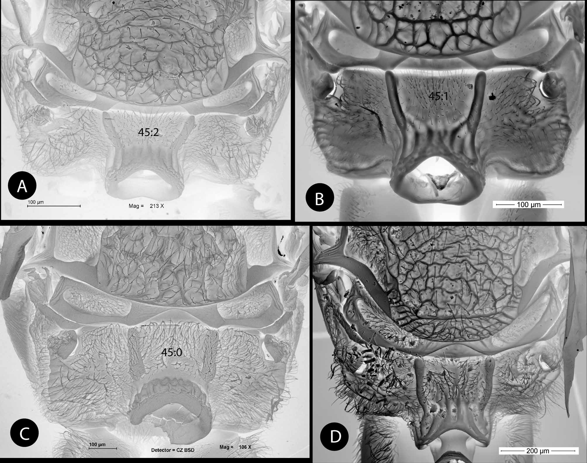
Metasoma ( Fig. 13 View FIGURE 13 E). Metasomal tergum T2+3 fused, smooth and shining, with close and distinct micropunctures in posterior half of metasoma; anteromedian area with a small group of 3–4 setae. Projecting part of hypopygial spine quite extended beyond attachment of lateral flap ( Fig. 15 View FIGURE 15 E); hypopigial setae arranged in two rows; apical setae not projected beyond apex spine.
Distribution. Recorded from Nicaragua and now from Panama (Chiriquí, Renacimiento).
Biology. The species was originally recorded as an inquiline in an unidentified stem gall on Quercus oleoides that was similar to galls of Xanthoteras quercusforticorne ( Diaz & Gallardo 1998) . We have reared this inquiline from galls of an undescribed Disholcaspis species (Medianero & Nieves-Aldrey, unpublished). The host galls are gregarious and are formed in stems of Quercus lancifolia ( Fig. 21 View FIGURE 21 H).


No known copyright restrictions apply. See Agosti, D., Egloff, W., 2009. Taxonomic information exchange and copyright: the Plazi approach. BMC Research Notes 2009, 2:53 for further explanation.
|
Kingdom |
|
|
Phylum |
|
|
Class |
|
|
Order |
|
|
Family |
|
|
Genus |