
Labyrinthus dunkeri ( Pfeiffer, 1852 )
|
publication ID |
https://doi.org/ 10.5281/zenodo.172563 |
|
DOI |
https://doi.org/10.5281/zenodo.6261405 |
|
persistent identifier |
https://treatment.plazi.org/id/03A4A163-FFC5-E36E-BA40-478D9D684FC9 |
|
treatment provided by |
Plazi |
|
scientific name |
Labyrinthus dunkeri ( Pfeiffer, 1852 ) |
| status |
|
Labyrinthus dunkeri ( Pfeiffer, 1852) View in CoL
Figures 1–14 View FIGURES 1 – 4 View FIGURES 5 – 8 View FIGURES 9 – 12 View FIGURES 13 – 16
Helix dunkeri Pfeiffer, 1852: 156 .
Description: Shell: ( Fig. 1 View FIGURES 1 – 4 ) As described by Solem (1966).
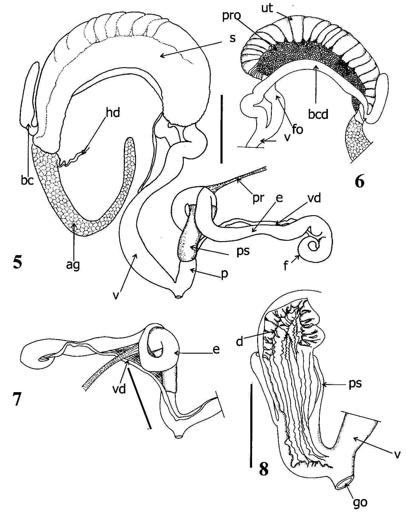
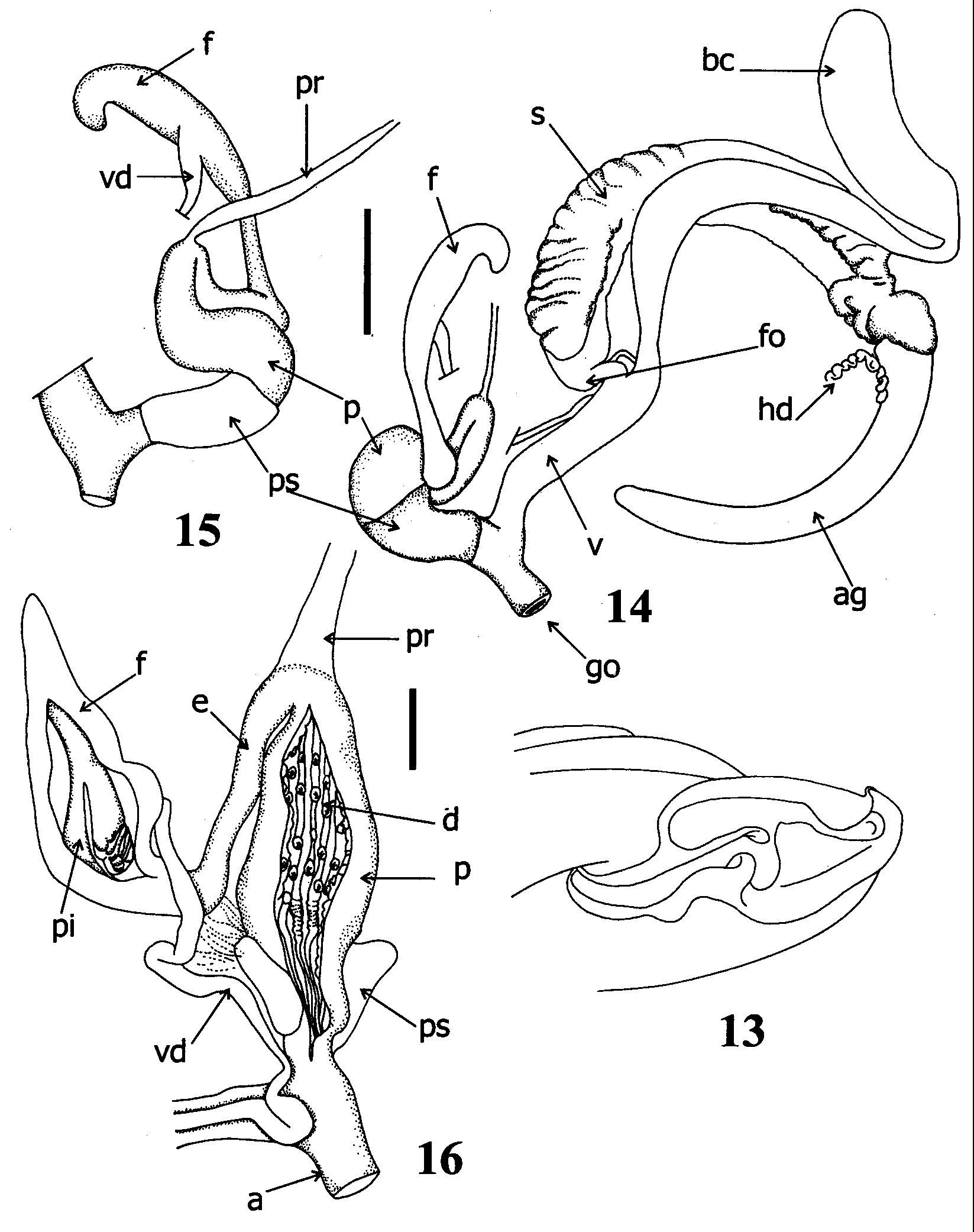
Pallial Organs: ( Figs 2, 3 View FIGURES 1 – 4 ) Pulmonary roof with few veins becoming more abundant, ramified near mantle collar ( Figs 2, 3 View FIGURES 1 – 4 ). Proximal portion of pulmonary roof not extending beyond top of kidney. Dark pigmented spots more abundant in distal portion of pulmonary roof ( Fig. 3 View FIGURES 1 – 4 ). Main pulmonary vein splitting into two main branches before reaching mantle collar. Many other minor veins running transversally to main veins. Kidney narrow, long, extending about 60% of pulmonary roof; secondary ureter closed from top of pulmonary roof to mantle collar, running parallel to rectum, ureteric interramus triangular, deeply excavated.
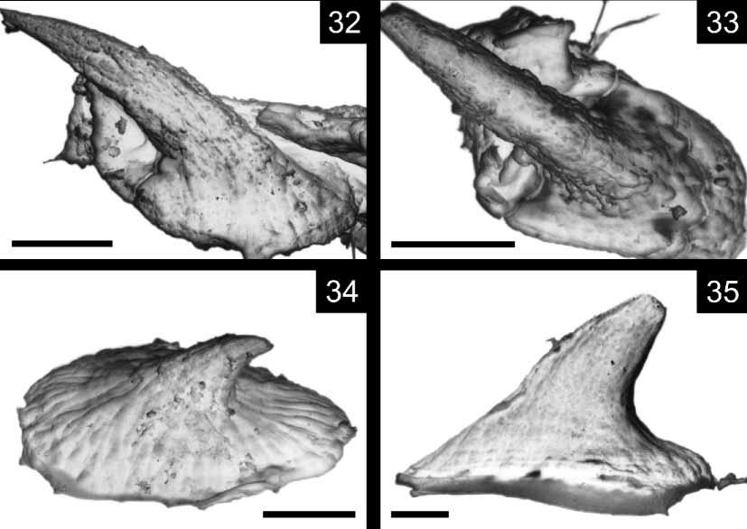
Reproductive system ( Figs 4–8 View FIGURES 1 – 4 View FIGURES 5 – 8 ; 32–33): Ovotestis consisting of single group of small digitiform acini located between second and third whorls, embedded in digestive gland ( Fig. 4 View FIGURES 1 – 4 ); hermaphroditic duct thin, long, convoluted, forming in middle portion, swollen vesicula seminalis. Fertilization pouchspermathecal complex (FPSC) not evident outside albumen gland, hermaphroditic duct folded over itself, penetrating at distal portion of albumen gland, beanshaped, continuous with spermoviduct ( Figs 4 View FIGURES 1 – 4 , 5 View FIGURES 5 – 8 ). Oviducal portion of spermoviduct organized into transversal folds over entire length. Prostatic zone continuing distally into long, vas deferens, descending parallel to vagina towards penioviducal angle. Vas deferens attached by connective tissue to junction of penis with oviduct. Ascending branch of vas deferens running parallel to penial complex, inserting in proximal epiphallus. Free oviduct a quarter of length of bursa copulatrix duct ( Fig. 6 View FIGURES 5 – 8 ). Natural position of free oviduct forming angle with vagina, bursa copulatrix duct continuous with free oviduct. Bursa copulatrix running parallel to spermoviduct until distal portion of albumen gland ( Fig. 5 View FIGURES 5 – 8 ). Bursa copulatrix and spermoviduct wrapped together by thin connective tissue. Bursa copulatrix sac elongated, oval, naturally reflexed over duct ( Figs 5, 6 View FIGURES 5 – 8 ). Inner structure of vagina consisting of thin parallel longitudinal folds, becoming thicker, deeper, with zigzag pattern. Distal portion of vagina more swollen than proximal part ( Fig. 5 View FIGURES 5 – 8 ). Vagina without hooked denticles. Penial complex composed by flagellum, epiphallus, penis ( Figs 5, 7 View FIGURES 5 – 8 ). Flagellum short, diameter decreasing towards proximal portion. Internally, flagellar pilaster with dorsal groove ending level where vas deferens enters epiphallus through papilla. Second longitudinal pilaster continuous within epiphallus. Epiphallus cylindrical, elongated with thick muscular walls. Internal structure consisting of thick medial pilaster with wrinkled surface without pustules. Internal pilasters running through entire epiphallusflagellum length. Vas deferens inserting in proximal epiphallus below flagellumepiphallus junction. Penis thick, reflexed over epiphallus, tightly packed with muscular fibers ( Fig. 7 View FIGURES 5 – 8 ). Penial sheath muscular, cylindrical, with upper proximal, lower distal borders well delimited ( Figs 5, 7 View FIGURES 5 – 8 ). Penis retractor becoming thicker before inserting at penisepiphallus junction. Internal structure of penis consisting of longitudinal, parallel ridges running through entire penis length. Ridges thicker with zigzag pattern at proximal portion, overlapped in part by penial sheath ( Fig. 8 View FIGURES 5 – 8 ). White small, fragile denticles with basal oval plate of about 300 m in length and long hook ( Figs 32, 33 View FIGURES 32 – 35. 32, 33 ). Denticles regularly distributed on top of zigzag ridges in proximal penis, absent in distal portion of penis ( Fig. 8 View FIGURES 5 – 8 ). Verge absent.
Digestive system ( Figs 9–12 View FIGURES 9 – 12 ): Jaw arcuate, apparently smooth without ribs. Thin transversal striae deeper than other perpendicular ones in jaw surface visible in SEM. Central tooth of radula unicuspid, lateral teeth unicuspid ( Fig. 9 View FIGURES 9 – 12 ) becoming bicuspids at level of N° 14, mesocone well developed ( Fig. 10 View FIGURES 9 – 12 ). First marginal teeth tricuspid ( Fig. 11 View FIGURES 9 – 12 ) close to margins of radula, marginal teeth become pluricuspis, wider ( Fig. 12 View FIGURES 9 – 12 ). Buccal mass muscular, spherical; esophagus cylindrical, open dorsally. Esophagus wall thick with inner longitudinal, parallel ridges. Two long salivary glands opening through thin ducts into dorsal portion of buccal mass at sides of esophageal opening. Esophageal crop walls thin, transparent without inner special sculpture. Stomach receiving anterior and posterior ducts of digestive gland; anterior opening into concavity between esophageal crop and intestine, posterior duct opening in parietal portion of stomach. Two internal typhlosoles arising from point of insertion of digestive gland ducts, one, longer, beginning at opening of anterior duct, running into proximal intestine. Intestine running along columelar side of visceral mass, turning down under anterior portion of gastric crop, then forming periaortic loop before finishing in rectum. Rectum closed along its entire length before opening into anus, running parallel to kidney, ending at mantle collar.
Material examined: COLOMBIA: FMNH 163709, Cesar Department, Sierra Perijá, El Roncon, 10–12 Km East of Becerril, 9°42'N, 73°11'W, 200– 300 m.; 16 September, 1969. Borys Malkin Coll. FMNH 173867 Cesar Department, Sierra de Perija, El Eocon, 10–12 km east of Becerril, 17 September 1969. B. Malkin Coll. AMNH 39705, Colombia.
Remarks: In species of Labyrinthus and Caracolus Montfort, 1810 , in which the jaw morphology was studied, it was reported as smooth or oxygnath ( Wurtz 1955). However, in L. dunkeri , the jaw showed incipient vertical ribs, and could not be consider as oxygnath. The morphology of the jaw in the rest of Labyrinthus species should be carefully examined to check if it really can be consider smooth and if this character can be used to diagnose the genus. The oxygnath condition is a character that has been used as unique of the American Belogona Euadenia (= Cepoliinae) ( Pilsbry 1894). However, this kind of jaw is also present in some Camaenidae and Sphincterochilidae among the Helicoidea ( Hausdorf 1998, Cuezzo 2003). The rest of the Helicoidean groups have an odontognath jaw ( Hausdorf 1998). The length of the hooked penis denticles and the absence of denticles in the vagina clearly separates L. dunkeri from the remaining species of Labyrinthus in which soft anatomy is known. The hooks on the dorsal portion of the denticles in L. dunkeri are more than twice the length than those in other species of Labyrinthus .


No known copyright restrictions apply. See Agosti, D., Egloff, W., 2009. Taxonomic information exchange and copyright: the Plazi approach. BMC Research Notes 2009, 2:53 for further explanation.
|
Kingdom |
|
|
Phylum |
|
|
Class |
|
|
Order |
|
|
Family |
|
|
Genus |
Labyrinthus dunkeri ( Pfeiffer, 1852 )
| Cuezzo, Maria Gabriela 2006 |
Helix dunkeri
| Pfeiffer 1852: 156 |