
Labyrinthus tarapotoensis ( Moricand, 1858 )
|
publication ID |
https://doi.org/ 10.5281/zenodo.172563 |
|
DOI |
https://doi.org/10.5281/zenodo.6261409 |
|
persistent identifier |
https://treatment.plazi.org/id/03A4A163-FFCD-E36B-BA40-47DD9DC448F2 |
|
treatment provided by |
Plazi |
|
scientific name |
Labyrinthus tarapotoensis ( Moricand, 1858 ) |
| status |
|
Labyrinthus tarapotoensis ( Moricand, 1858) View in CoL
Figures 23–31 View FIGURES 23 – 27 View FIGURES 28 – 31 ; 34–35
Helix tarapotoensis Moricand, 1858: 450 , pl.13, fig.2.
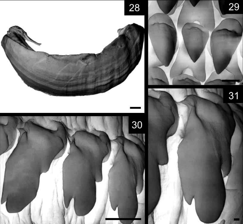
Description: Shell ( Fig. 23 View FIGURES 23 – 27 ): As described by Solem (1966: 80).
Pallial Organs ( Fig. 24 View FIGURES 23 – 27 ): Kidney narrow, long, occupying about 50–60% of pulmonary roof length. Both sides of main pulmonary vein with abundant minor veins running transversally respect to longitudinal axis of pulmonary roof. Main pulmonary vein running from pericardic cavity to mantle collar, not bifurcated. Pulmonary roof dark, with abundant black pigmentation bordering pulmonary veins. Secondary ureter closed from top of pulmonary roof to mantle collar. Ureteric interramus triangular, deeply excavated.
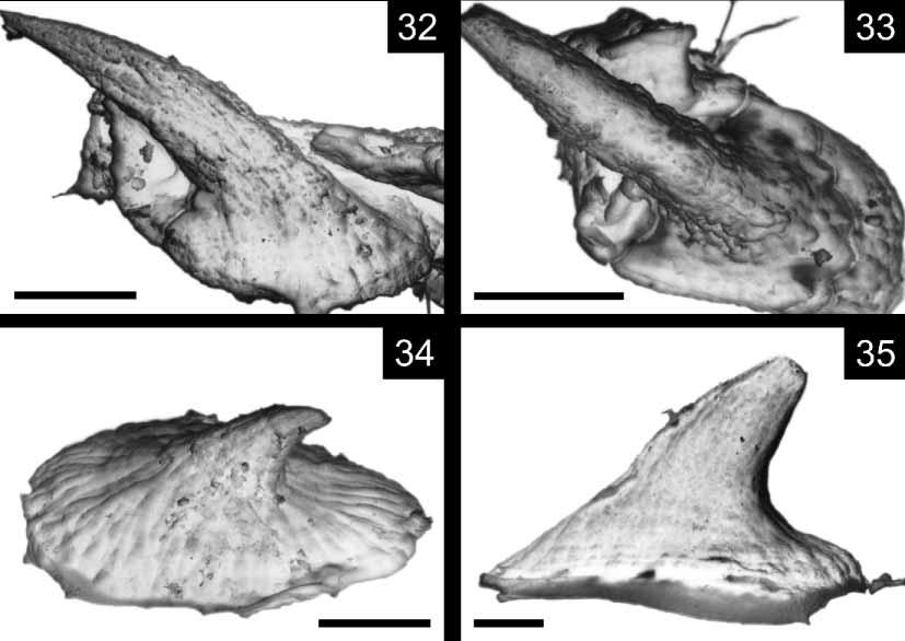
Reproductive system ( Figs 25–27 View FIGURES 23 – 27 , 32–33 View FIGURES 32 – 35. 32, 33 ): Ovotestis formed by single group of long nonbranched digitiform acini ( Fig. 26 View FIGURES 23 – 27 ). Albumen gland beanshaped, thin, long ( Fig. 25 View FIGURES 23 – 27 ). Hermaphroditic duct folded at distal portion inserting in albumen gland. FPSC externally undistinguishable. Proximal portion of spermoviduct voluminous, overlapping distal portion of albumen gland. Uterine portion of spermoviduct transversally folded. Prostatic portion of spermoviduct thin, prolonged into long vas deferens. Vas deferens running towards terminal genitalia surrounding vagina, wrapped by thin connective tissue. Free oviduct short, in natural position forming angle with vagina while duct of bursa copulatrix continuous with it ( Fig. 25 View FIGURES 23 – 27 ). Bursa copulatrix running parallel to spermoviduct up to distal portion of albumen gland. Duct of bursa copulatrix and spermoviduct wrapped together with thin connective tissue. Distal portion of bursa copulatrix sac folded over its duct. Distal portion of bursa copulatrix duct broadened respect to proximal portion. Vagina longer than phallic complex, proximal portion thicker. Inner structure of proximal vagina formed by longitudinal thick, deep cords, divided into rhomboidal portions, regular in shape. Each rhomboidal portion bearing white hooked denticles with oval bases. Denticles densely, regularly distributed in proximal vagina. Distal portion of vagina with longitudinal cords, not divided in rhomboids, lacking hooked denticles. Phallic complex formed by flagellum, epiphallus, penis. Flagellum thin, gradually tapering towards tip. Internally, longitudinal pilaster with dorsal groove ending at point of insertion of vas deferens or slightly lower on proximal epiphallus. Other two internal pilaster continuous within epiphallus. Epiphallus longer, thinner than penis. Penis thick, muscular, with distal portion ensheathed with part of epiphallus by penial sheath ( Figs 25, 27 View FIGURES 23 – 27 ). Penial reflexed over distal portion of epiphallus, attached to it by muscular strands. Penis retractor inserted into penialepiphallus junction. Vas deferens inserted in proximal epiphallus marking limit between epiphallus and flagellum. Epiphallus with internal fine longitudinal folds in zigzag pattern. Penis with thick pilasters folded, densely packed in proximal portion ( Fig. 27 View FIGURES 23 – 27 ). Numerous white hooked denticles with oval base of 360 m stuck to dorsal portion of pilasters ( Figs 32– 33 View FIGURES 32 – 35. 32, 33 ). Verge absent.
Digestive system: Jaw ( Fig. 28 View FIGURES 28 – 31 ) arched, without vertical ribs, only fine transversal grooves on surface. Upper and lower cutting edges even. Central tooth of radula monocuspid, sharp tip ( Fig. 29 View FIGURES 28 – 31 ). First lateral teeth unicupid, changing to bicuspidates. Marginal teeth tricuspid ( Figs 30, 31 View FIGURES 28 – 31 ) becoming wider, multicuspid close to edges of radula. Rest of digestive system as described for L. dunkeri .
Material examined: ECUADOR: FMNH 173038: Pastaza Department, Cusheme River, Cusheme. May 15, 1971. B. Malkin Coll. COLOMBIA: FML s/n: Nariño Department, Pto. Nariño. E. Dominguez leg.
Remarks: Hooked denticles in L. tarapotoensis are more abundant than in other species examined. These denticles are present not only on penis but also in vagina.
Labyrinthus tarapotoensis belongs to the L. raimondii species complex ( Solem 1966). Among the species included by Solem (1966) into this complex, L. raimondii (Philippi, 1867) is the most similar in shell size and morphology to L. tarapotoensis . However, L. tarapotoensis can be distinguished from L. raimondii by its white lip and less angulated periphery. Both species have similar shell diameter ( L. raimondii shell diameter: 38.6– 42.1 mm ( Solem 1966), L. tarapotoensis shell diameter: 41–42 mm). The anatomy of L. raimondii is not known so anatomical comparisons are not currently possible.

No known copyright restrictions apply. See Agosti, D., Egloff, W., 2009. Taxonomic information exchange and copyright: the Plazi approach. BMC Research Notes 2009, 2:53 for further explanation.
|
Kingdom |
|
|
Phylum |
|
|
Class |
|
|
Order |
|
|
Family |
|
|
Genus |
Labyrinthus tarapotoensis ( Moricand, 1858 )
| Cuezzo, Maria Gabriela 2006 |
Helix tarapotoensis
| Moricand 1858: 450 |