
Isomeria awa, Cuezzo, Maria Gabriela, 2006
|
publication ID |
https://doi.org/ 10.5281/zenodo.172563 |
|
DOI |
https://doi.org/10.5281/zenodo.6261415 |
|
persistent identifier |
https://treatment.plazi.org/id/03A4A163-FFD6-E373-BA40-40959B124F42 |
|
treatment provided by |
Plazi |
|
scientific name |
Isomeria awa |
| status |
sp. nov. |
Isomeria awa n. sp.
Figures 36–48 View FIGURES 36 – 38 View FIGURES 39 – 42 View FIGURES 43 – 48
Type material: Holotype: AvH unnumbered. Paratypes: FML 14796 A, from type locality.
Type Locality: COLOMBIA, Nariño department, Reserva Natural La Planada, Nariño, 1730 mts, N 1° 50' W 77° 43' Orejuella, J. E. & Cantillo, G. Coll.
Etymology: the species name refers to the indigenous Inkal Awa community that lives close to the Reserva Natural La Planada in the region of the Cordillera Occidental, Ricaurte County, Nariño Department in Colombia. The name Awa means "people of the mountain". Noun in apposition.
Diagnosis: shell body whorl with axial growth lines, small pustules all over surface. Irregular spots of dark brown coloration on dorsal shell surface. Parietal lamellae small, triangular, not merging with lip, small conical tooth on lower palatal peristomal lip. Umbilicus narrow, open. Vagina long with glandular pouch in distal portion. Presence of inner conical stimulatorlike structure in distal vagina.
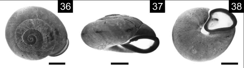
Description: Shell ( Figs 36–38 View FIGURES 36 – 38 ): Globose, 4 1/2 whorls, strong, body whorl periphery obtusely angulated; golden yellowish. Axial growth lines with small pustules all over shell surface. Irregular small spots of dark brown color on dorsal side of shell. Body whorl strongly descending behind aperture. Aperture subquadrate, chestnut coloration inside aperture. Peristomal lip white, strongly reflexed, parietal lip with prominent raised edge. Small, triangular parietal lamellae present, not merging with peristomal lip, lower palatal lip with small conical tooth. Suture shallow. Umbilicus narrow, open, with columnar groove noticeable, basal lip extension covering only a small portion.
Holotype shell measurements: D major= 39.7; D minor= 34.4; H= 21.5; D ap= 19.7; Hap= 16.3. Paratypes shell measurements (n= 3): D major= 38.8–39.4 (0 = 38.6); D minor= 33.2–34.2 (0 = 33.4); H= 21–21–5 (0 = 21.3); D ap= 19.3–20.1 (0 = 19.6): H ap= 16.2–16.9 (0 = 16.4).
Pallial Organs: Pulmonary roof black due to dense dark pigmentation. Thin grayish minor veins weakly marked, distinguishable mostly between rectum and kidney. Main pulmonary vein running from pericardial cavity to mantle collar, not branched. Kidney narrow, long, occupying about 70% of pulmonary roof length. Secondary ureter closes from top of pulmonary roof to mantle collar. Ureteric interramus triangular, deeply excavated, overlapped by mantle collar.
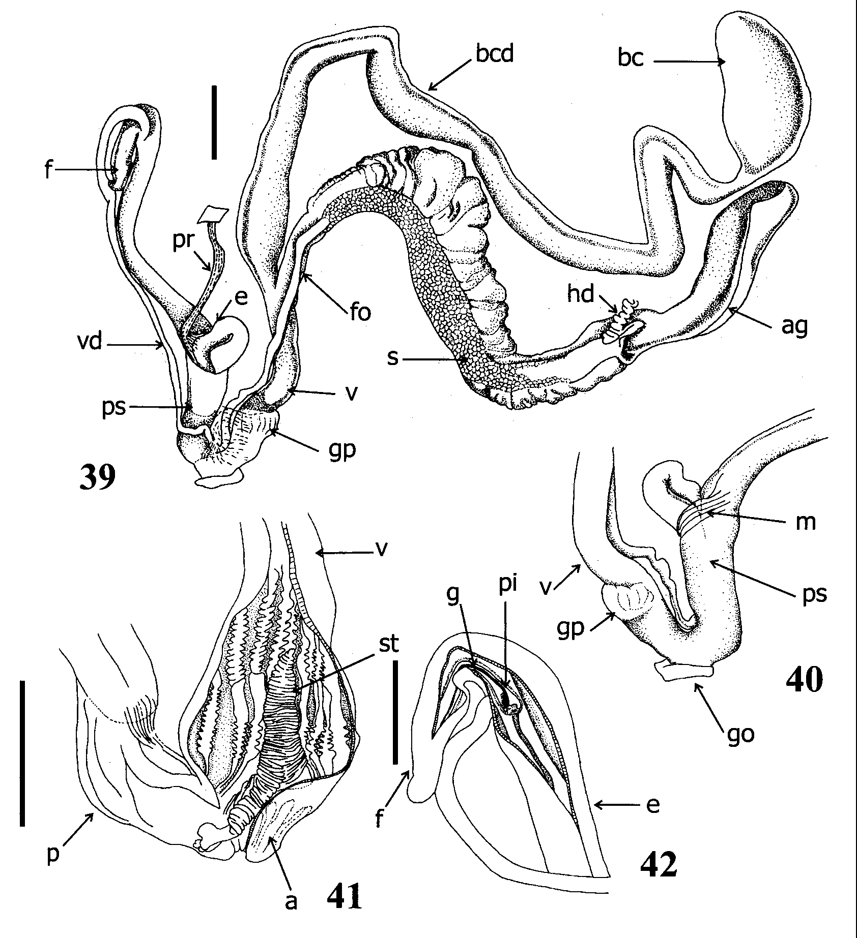
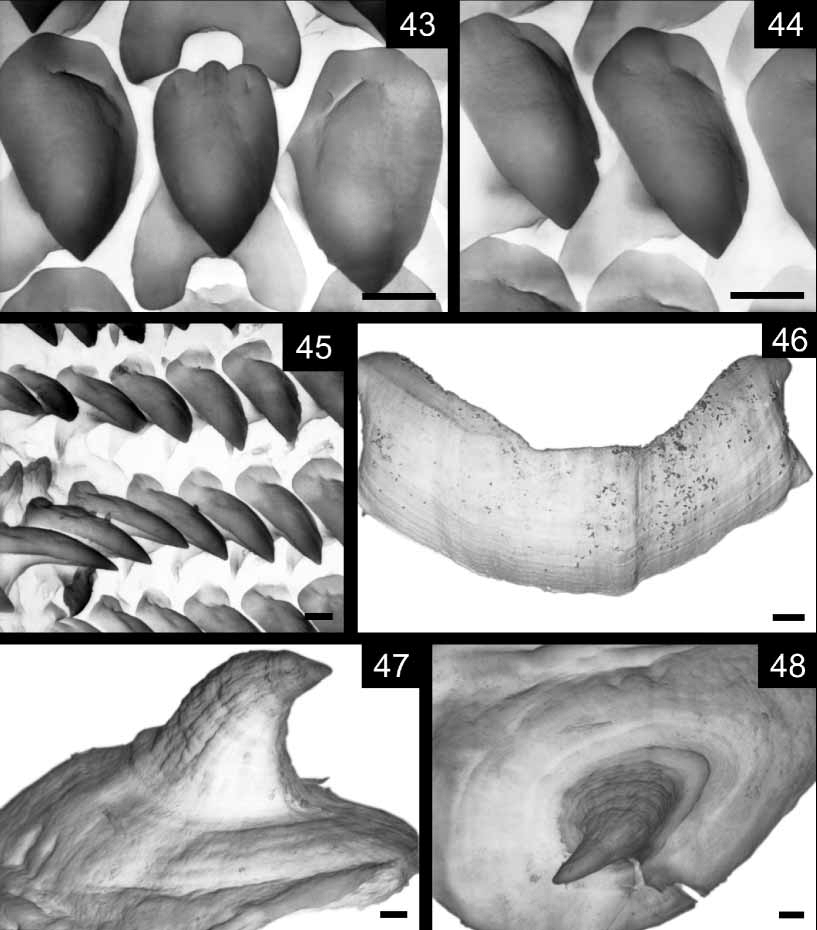
Reproductive system ( Figs 39–42 View FIGURES 39 – 42 , 47, 48 View FIGURES 43 – 48 ): Ovotestis formed by long, apically branched acini embedded in digestive gland. Hermaphroditic duct long, convoluted; distal portion of hermaphroditic duct reflexed over itself before inserting in distal portion of albumen gland. Fertilization pouchspermathecal complex (FPSC) not distinguishable. Duct of bursa copulatrix thicker than free oviduct, 1 1/2 times as long as spermoviduct length ( Fig. 39 View FIGURES 39 – 42 ). Sac oval, voluminous, not reflexed over its duct. Free oviduct short, slightly thinner than vagina. Vagina long, distal glandular pouch close to penioviducal angle ( Figs 39, 40 View FIGURES 39 – 42 ). Inner structure of proximal portion of vagina consisting of longitudinal, parallel, deep cords with scattered white hooked denticles. Conical stimulatorlike structure, present in distal portion of vagina ( Fig. 41 View FIGURES 39 – 42 ), attached through base to vagina's wall near vaginaatrium junction, raised free into vagina's lumen. Portion of stimulatorlike structure extending into atrium towards distal penis, ending into two fingerlike branches ( Fig. 41 View FIGURES 39 – 42 ). Penial complex formed by flagellum, epiphallus, penis ( Figs 39, 40 View FIGURES 39 – 42 ). Flagellum short, uniform diameter, tip folded. Internally, longitudinal pilaster with open dorsal groove running from top to junction with epiphallus, ending at point of insertion of vas deferens or slightly below ( Fig. 42 View FIGURES 39 – 42 ). Pilaster dorsal groove partially overlapped by extension of lateral wall, forming flap. Epiphallus longer, thinner than penis, distally reflexed over penis, overlapped by penis sheath. Penis long. Penial retractor inserting in distal epiphallus. Thick muscular strands attached penis to penis sheath ( Fig. 40 View FIGURES 39 – 42 ). Interior wall of proximal penis with thick, deep pilasters bearing hooked denticles regularly distributed, with oval bases of 180–200 m of length ( Figs 47, 48 View FIGURES 43 – 48 ). Denticles more abundant in penis than in vagina. Verge absent.
Digestive system ( Figs 43–46 View FIGURES 43 – 48 ): Jaw arched, incipient plaque in middle of jaw, fine transversal grooves more evident close to one edge ( Fig. 46 View FIGURES 43 – 48 ). Upper, lower cutting edges even. Central tooth of radula monocuspid ( Fig. 43 View FIGURES 43 – 48 ). First laterals monocuspid, changing into bicuspid ( Figs 44, 45 View FIGURES 43 – 48 ). Condition of marginal teeth unknown.
m.
Comparative Material examined: COLOMBIA: ZMZ 544602 Isomeria oreas (Koch, 1844) , G. Schneider, 1912; ZMZ 509673 Isomeria subelliptica (Mousson, 1869) , Holotype, Bagua, Amazonas, Wallis leg., 1869. ECUADOR: MUC s/n, Isomeria globosa , Pichincha Province. Puerto Quito, and 5/12/1983.M. Ituwalde. MUC s/n, Pichincha Prov., Puerto Quito, 16/1/1984. Leg. V. Zak.
Remarks: Isomeria awa belongs to the Isomeria oreas group of species ( Solem 1966). Isomeria awa is most similar in shell morphology to I. oreas , differing in being smaller (adult specimens of I. oreas , mean diameter = 57.3 mm; I. awa mean diameter= 39 mm); I. oreas has fine radial striae and malleations on the surface of the body whorl, while I. awa has axial growth lines with small pustules all over shell surface; the shell coloration in I. oreas is darker than in I. awa , without the characteristics irregular spots present in I. awa shells. The number of shell whorls is lower in I. awa (41/2 whorls) than in I. oreas (5– 5 1/2 whorls). Comparison based on anatomical characters is not possible because the anatomy of I. oreas is unknown. Isomeria subelliptica (Mousson, 1869) is similar to I. awa in that they both possess a small parietal tooth and open umbilicus, but I. awa has a larger shell [mean diameter of 39 mm, while I. subelliptica shell mean diameter is 30.9 mm ( Solem 1966)], the umbilicus is narrower than in I. subelliptica . Both species also differ in shell color, I. awa has a golden yellowish coloration with darker irregular spots while in I. subelliptica the shell possess an even dark brown coloration. As in the case of I. oreas , anatomical comparisons of I. awa with I. subelliptica are not possible due to the lack of information on the later. Isomeria globosa , whose anatomy was described by Solem (1966), differs from I. awa in not having the vagina's stimulator apparatus. Hooked denticles in vagina and penis, and a glandular pouch are present in I. globosa . A penial appendix is lacking in I. awa but it was described in I. globosa .
Two structures in the genitalia of I. awa probably represent unique characters within the genus, the presence of a stimulatorlike organ within the vagina and a glandular pouch externally visible in the distal portion of the vagina. This kind of stimulatorlike organ has never been reported before in any Camaenidae and resembles the ones described in Milacidae ( Wiktor 1987) . Other stimulatorlike appendages have been reported in certain Hygromiidae ( Hausdorf 1998) . However, several appendages have been called as "stimulators" ( Nordsieck 1987; Schileyko 1991; Hausdorf 1998) so that the homology of this kind of structures can only be tested by phylogenetic analyses. The glandular pouch found in I. awa is also present in I. globosa in which it has been described as a ring of low knobs around the vagina ( Solem 1966). A glandular pouch is characteristic of some species of Polydontes Montfort, 1810 , also member of the Camaenidae , however, this kind of structure appears in the penial complex but not in the vagina. Hooked denticles with oval bases are also present, both in vagina and penis, in some Labyrinthus , with similar morphology although they are smaller in I. awa . These denticles are usually more abundant in the penis than in the vagina in Labyrinthus as well as in Isomeria . Another character shared with Labyrinthus is the presence of an internal flagellar pilaster, which in I. awa , shows a distinct open dorsal groove. Marginal teeth were not observed in the radulas preparations because they were broken.
| FML |
Fundacion Miguel Lillo |
No known copyright restrictions apply. See Agosti, D., Egloff, W., 2009. Taxonomic information exchange and copyright: the Plazi approach. BMC Research Notes 2009, 2:53 for further explanation.