
Ubiquepuella telytokous Fernandes
|
publication ID |
https://doi.org/ 10.11646/zootaxa.4032.4.5 |
|
publication LSID |
lsid:zoobank.org:pub:84FFF17C-6737-42F5-8DB3-8F3325368E0A |
|
DOI |
https://doi.org/10.5281/zenodo.6105186 |
|
persistent identifier |
https://treatment.plazi.org/id/03A51067-FFB9-FF84-FF10-FCC5C5C6F8B9 |
|
treatment provided by |
Plazi |
|
scientific name |
Ubiquepuella telytokous Fernandes |
| status |
|
Ubiquepuella Fernandes View in CoL , n. gen.
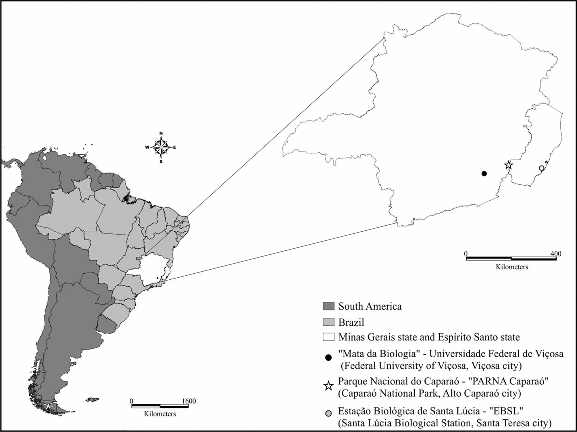
( Figs. 1–21 View FIGURES 1 – 5 View FIGURES 6 – 17 View FIGURE 21 )
" Ectecous sp. A" in Sperber 1999, pg 156.
" Ectecous " in Sperber et al. 2007, pg 3.
" Ectecous sp.1" in Szinwelski et al. 2013, pg 759. " Ectecous sp.1" in Szinwelski 2013: chapter 4, pg 53. Type Species. Ubiquepuella telytokous Fernandes , n. sp.
Etymology. The Ubiquepuella name is a combination of the Latin words "ubique" (everywhere, everyplace) and "puella" (girl, lady), because the type species is usually found in abundance along its distribution and has only females.
Diagnosis. Head with triangular shape in frontal view ( Fig. 8 View FIGURES 6 – 17 ); fastigium and vertex separated by transversal groove forming one marked step ( Fig. 6 View FIGURES 6 – 17 ); pronotum transverse, with anterior and posterior margins straight ( Figs. 1, 5 View FIGURES 1 – 5 ); posterior tibia with seven dorsal spurs, being four outer and three inner ( Fig. 10 View FIGURES 6 – 17 ); ovipositor smooth ( Figs. 14, 15 View FIGURES 6 – 17 ); copulatory papilla dorsoventrally flattened with lateral border sclerotized ( Fig. 4 View FIGURES 1 – 5 a, b, c); sclerotized area of papilla longer than wide, with internal concave surface and apex narrower than the base ( Fig. 4 View FIGURES 1 – 5 a, b, c).
Description. Size medium. Head as wide as pronotum ( Figs. 1, 5 View FIGURES 1 – 5 ), with triangular shape on frontal view ( Figs. 8 View FIGURES 6 – 17 ); transverse groove between vertex and fastigium forming one marked step ( Fig. 6 View FIGURES 6 – 17 ); fastigium wider than long with distal border sub-straight, on dorsal view ( Figs. 1, 5 View FIGURES 1 – 5 ); long semi-erected bristles in the central portion of fastigium; occiput and vertex covered with short and fine dark brown bristles; prominent black eyes ( Figs. 5 View FIGURES 1 – 5 , 6 View FIGURES 6 – 17 ); three ocelli with different sizes ( Fig. 8 View FIGURES 6 – 17 ); maxillary palps with fourth palpomere greater than first and second, but smaller than third and fifth ( Fig. 7 View FIGURES 6 – 17 ); last palpomere with rounded apex. Transverse pronotum, covered with short and fine dark brown bristles; anterior and posterior margins with row of long semi-erected bristles ( Figs. 1, 5 View FIGURES 1 – 5 , 6 View FIGURES 6 – 17 ); lateral lobes shorter posteriorly with antero-ventral angle of nearly 90 degrees and postero-ventral angle greater than 90 degrees ( Fig. 7 View FIGURES 6 – 17 ); forewings not reaching the third abdominal tergite, without specialized veins ( Figs. 1, 2 View FIGURES 1 – 5 ); hindwings absent; fore tibia with two apical spurs and one tympanum on the inner surface, which is absent in the outer surface; middle tibia with three apical spurs; hind tibia with seven dorsal spurs, being four outer and three inner ( Fig. 10 View FIGURES 6 – 17 ), two row of spines in the dorsal surface, three apical inner and outer spurs, being the median spurs the largest on both sides; basitarsus with two rows of spines on dorsal surface ( Fig. 11 View FIGURES 6 – 17 ). Abdominal tergites with many spots in shades of brown and yellow ( Fig. 1 View FIGURES 1 – 5 ). Laterally flattened ovipositor, apex smooth ( Fig. 15 View FIGURES 6 – 17 ). Reproductive system following the general insect pattern, with two ovaries opening to lateral oviducts that connect to a common oviduct, which opens to the spermatheca duct ( Fig. 14 View FIGURES 6 – 17 ); copulatory papilla flattened dorsoventrally, with sclerotized lateral margins and concave inner surface ( Fig. 4 View FIGURES 1 – 5 a,b,c); membranous spermatheca ( Fig. 3 View FIGURES 1 – 5 ).


No known copyright restrictions apply. See Agosti, D., Egloff, W., 2009. Taxonomic information exchange and copyright: the Plazi approach. BMC Research Notes 2009, 2:53 for further explanation.