
Hydropsyche trifora (Li & Tian 1990)
|
publication ID |
https://doi.org/ 10.11646/zootaxa.4374.1.1 |
|
publication LSID |
lsid:zoobank.org:pub:6DDFE314-126F-4C82-936F-33C1C28EAC6E |
|
DOI |
https://doi.org/10.5281/zenodo.5987722 |
|
persistent identifier |
https://treatment.plazi.org/id/03A5866E-C101-7E3F-D4E9-FDD238BEFF44 |
|
treatment provided by |
Plazi |
|
scientific name |
Hydropsyche trifora (Li & Tian 1990) |
| status |
|
Hydropsyche trifora (Li & Tian 1990) , Ceratopsyche
( Figs. 12–15 View FIGURE 12 View FIGURE 13 View FIGURE 14 View FIGURE 15 )
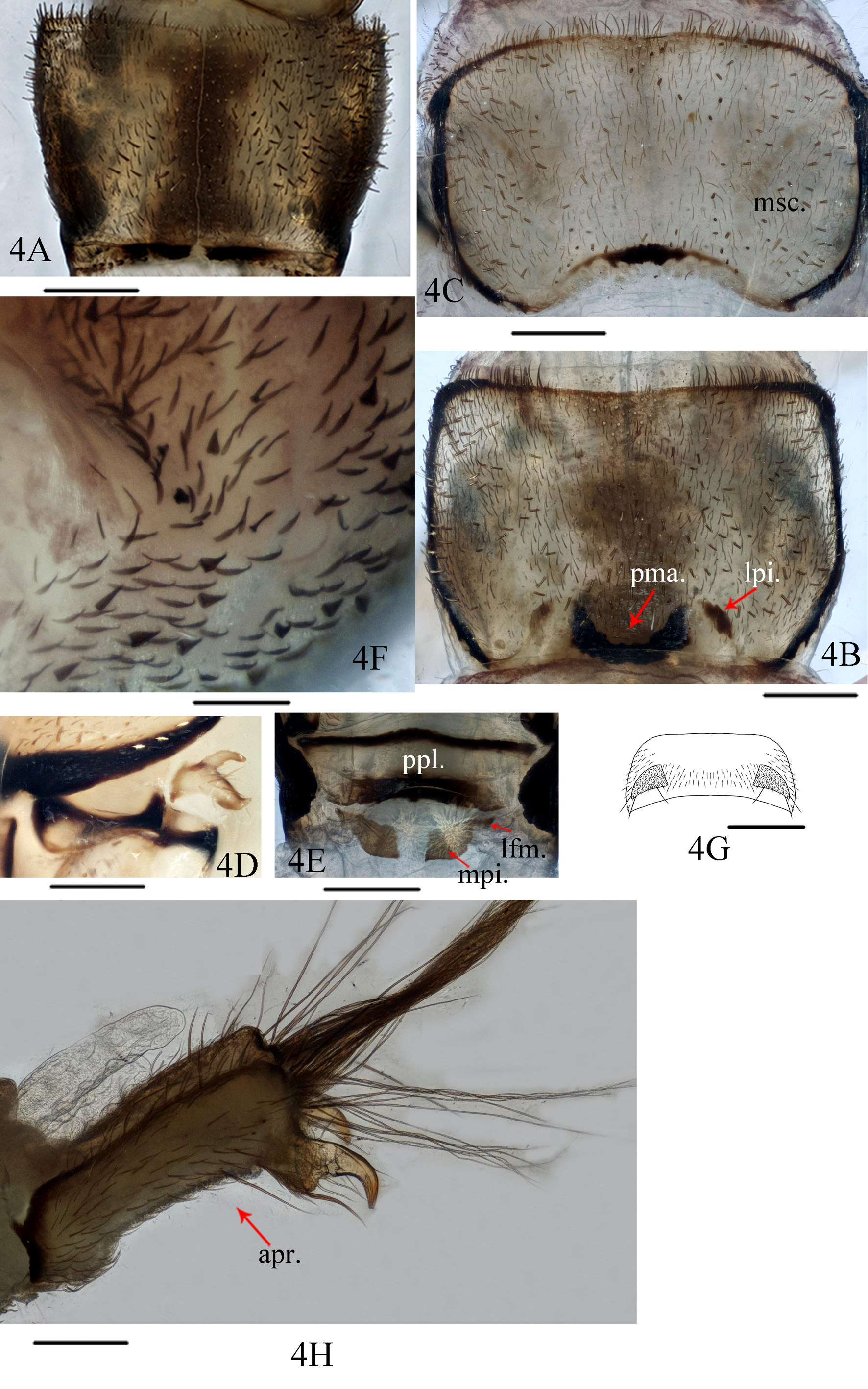
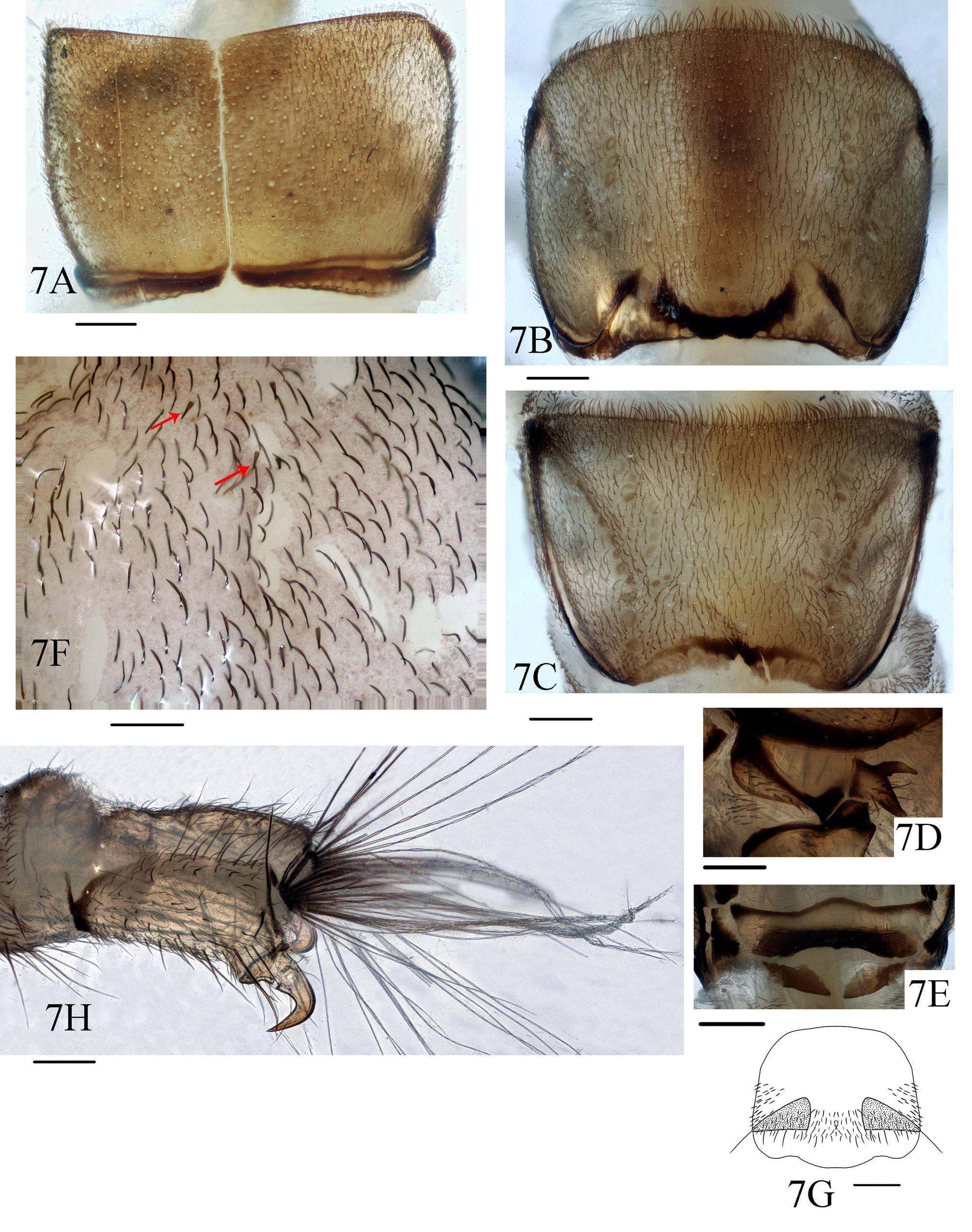
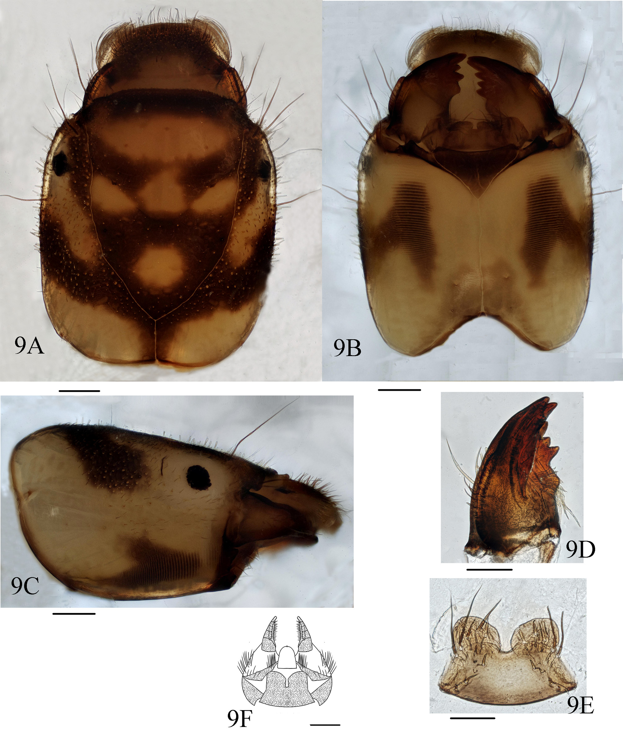
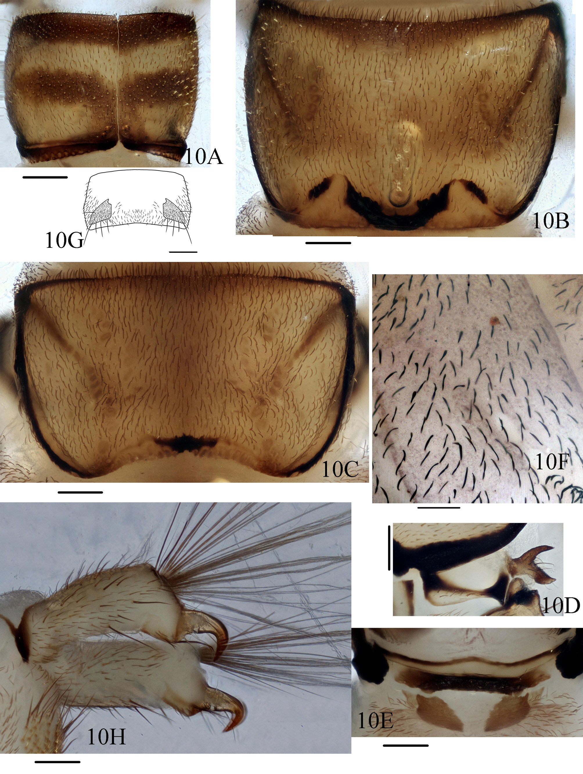
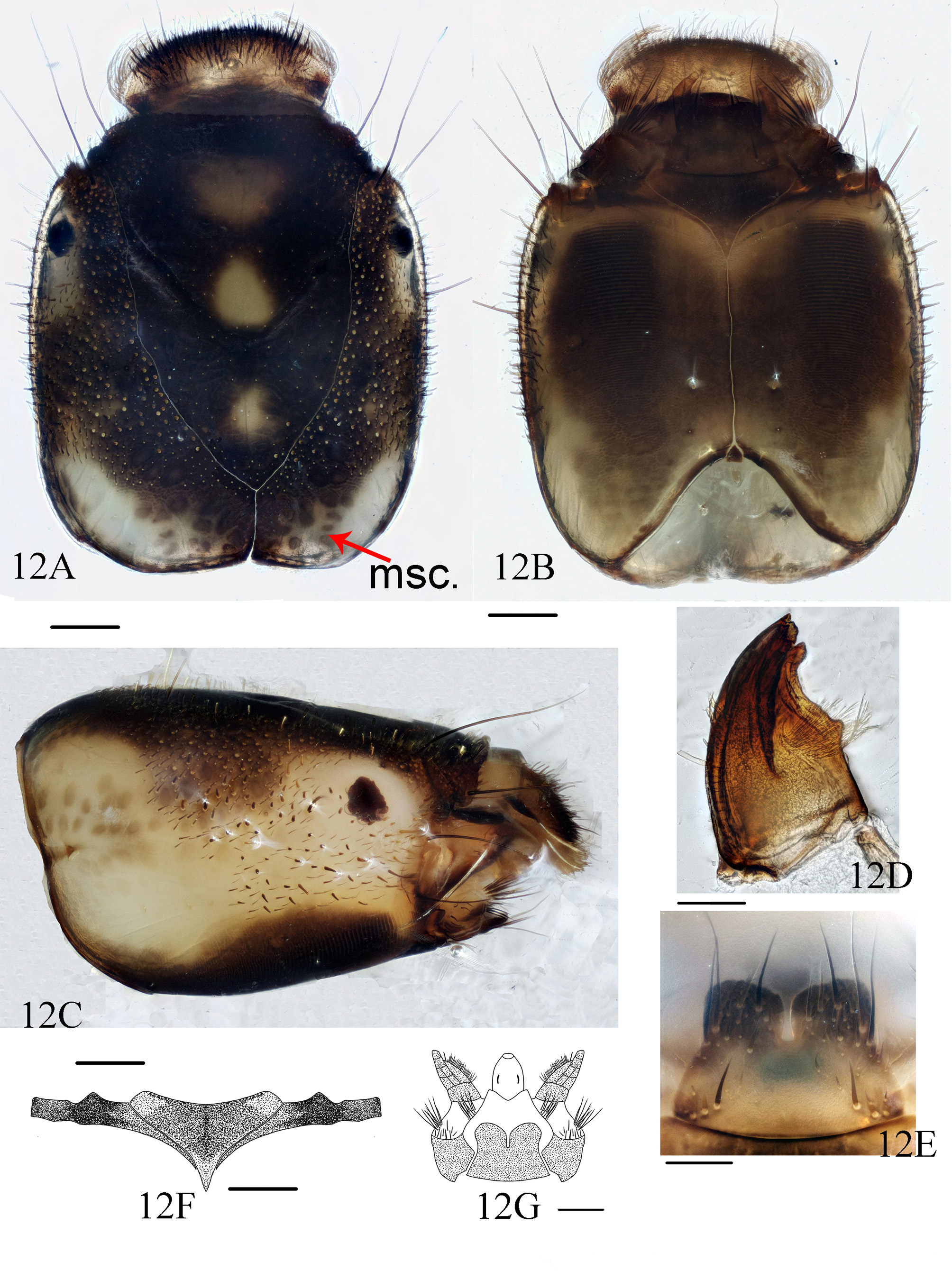
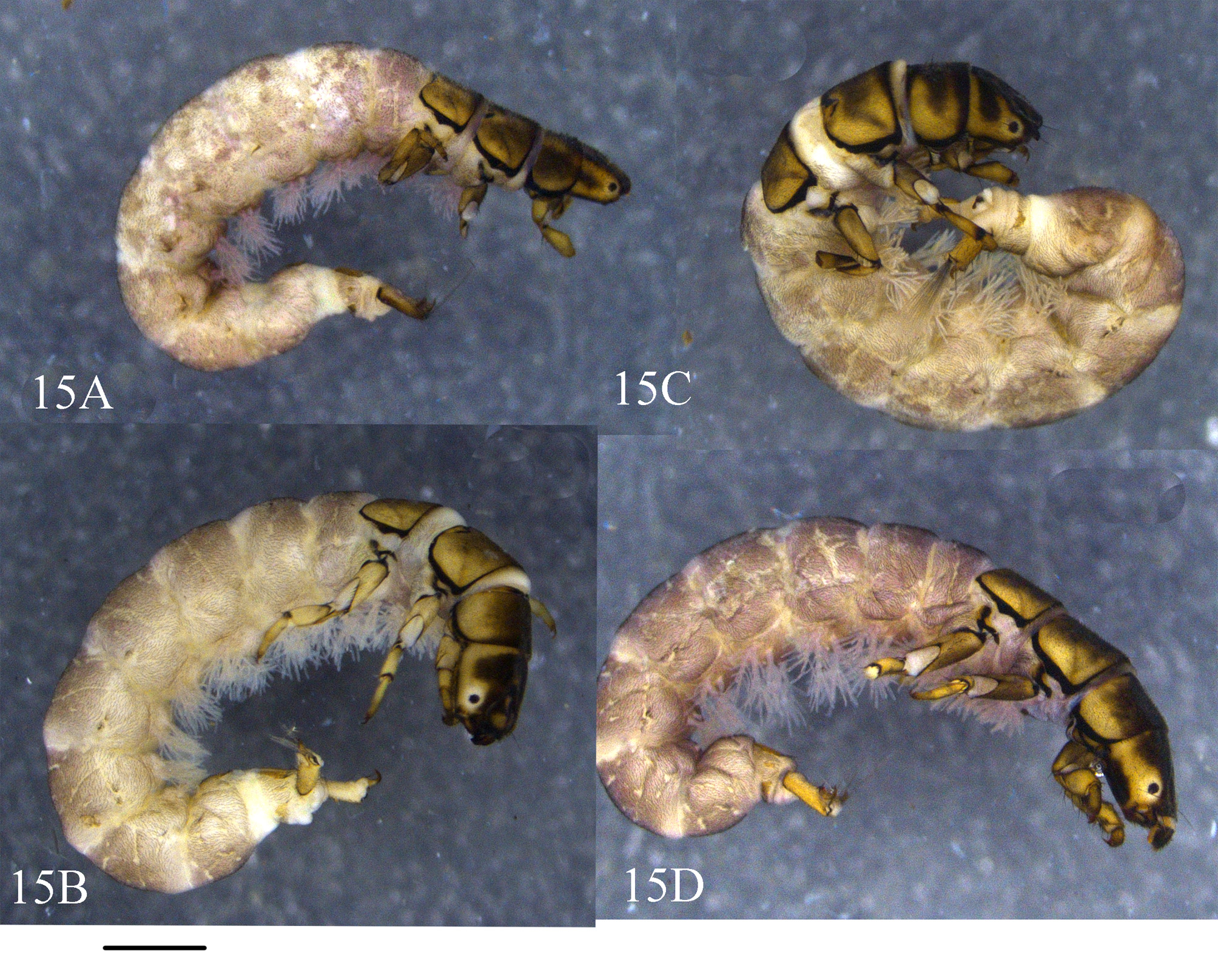
Description. Larvae (5th instar). Body length 10–15 mm (n = 5). Head. Head capsule ( Fig. 12A View FIGURE 12 ) subquadrate in dorsal view, 1.7 mm long, 1.2 mm wide at midlength. Overall coloration mostly dark brown, with 3 yellow oval patches longitudinally arranged on frontoclypeal apotome, of which anterior one largest, middle one second, and posterior one smallest; in dorsal view parietals each with oblique pale brown stripe along posterolateral margin; in lateral view each stripe and yellow area around each eye fused into U-shaped patch, collectively resembling shoulders, neck, and head of bird ( Fig. 12C View FIGURE 12 ); muscle scars on parietals dark brown ( Fig. 12A View FIGURE 12 , msc), and those on postgenae light brown. Eyes oval, black. Frontoclypeal apotome ogival, with frontoclypeal suture arms slightly sinuous. Dorsum of head with hair-like setae, dark brown truncate peg-like setae, and acuminate peg-like setae. Labrum yellow, with its anterior margin round and hair brush at each anterolateral corner. Mandibles ( Fig. 12D View FIGURE 12 ) yellow, triangular in ventral view, each with 5 apical teeth; left mandible with about 20 setae at basal half of lateral margin and with brush of stiff hairs at middle of inner side; right mandible with more than 20 setae at basal half of lateral side. Maxillae each with cardo transverse and sub-rectangular, brown ( Figs 12B, 12F View FIGURE 12 ); stipes oblique and subrectangular, palpifer with about 29 setae, palp 5-segmented, and maxillary lobe adjacent to mesal margin of palp ( Fig. 12G View FIGURE 12 ). Labium ( Fig. 12G View FIGURE 12 ) triangular and short in ventral view. Submentum ( Fig. 12E View FIGURE 12 ) in ventral view with basal 2/3rds somewhat trapezoidal and distal 1/3rd incised mesally, forming two lobes; posterior margin slightly convex posteriorly; each anterolateral corner with 3–5 long, strong setae and many short setae; each posterolateral corner bearing stout, brown setae. Anterior ventral apotome ( Fig. 12B View FIGURE 12 ) nearly triangular, brown, with anterior margin slightly concave and anterolateral angles rounded. Ventral ecdysial line about twice as long as anterior ventral apotome. Posterior ventral apotome tiny, triangular, brown.
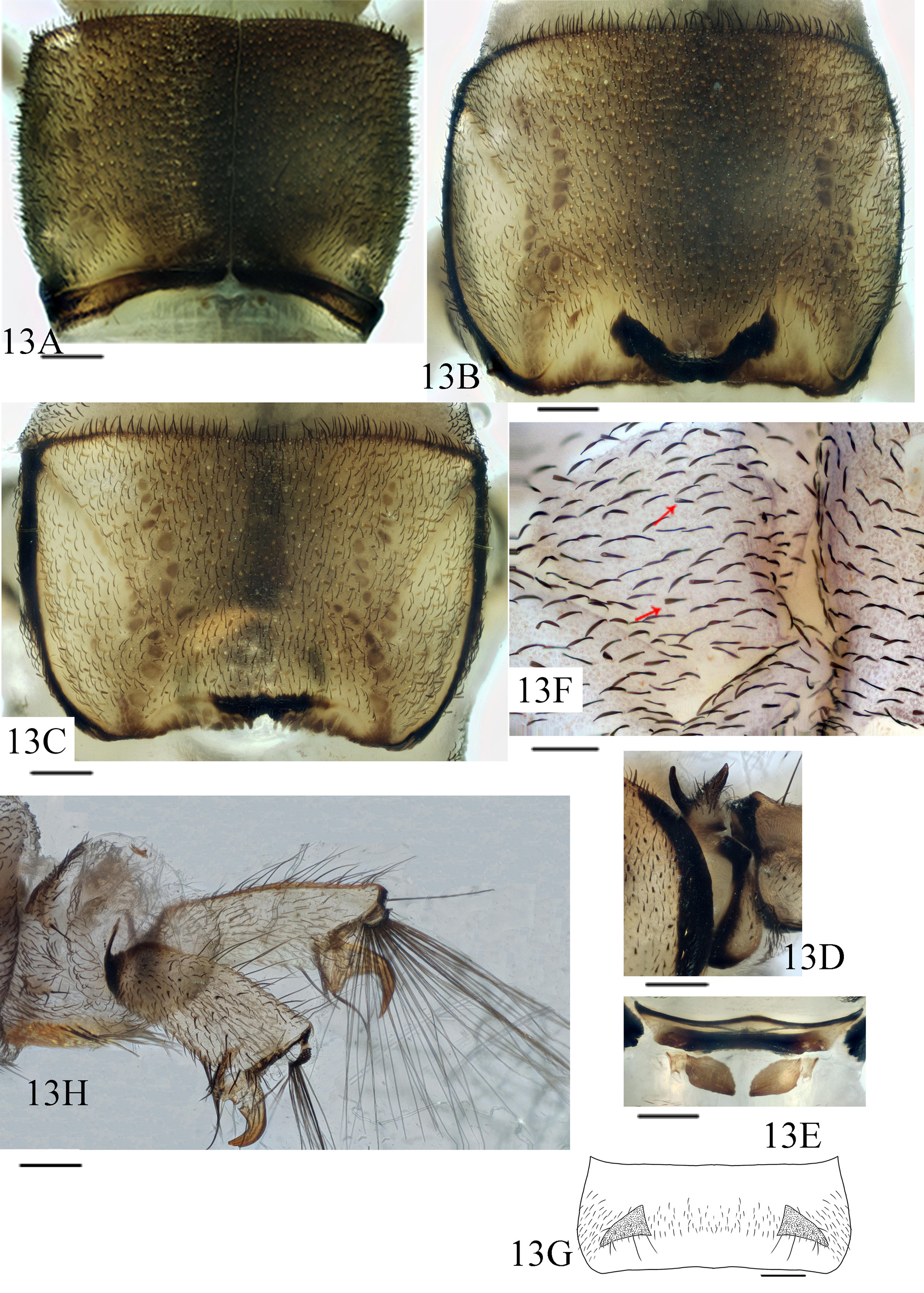
Thorax. Pronotum ( Fig. 13A View FIGURE 13 ) dark brown, sub-rectangular and subdivided longitudinally, covered with hairlike setae, truncate peg-like setae, and acuminate peg-like setae; three types of setae alternatively arranged along anterior margin, with 2 types of peg-like setae as long as hair-like setae; muscle scars distinct; prosternal plate ( Fig. 13E View FIGURE 13 ) dark brown in posterior 2/3rds, pale in anterior 1/3 except with dark anterior border, with its width about 7 times its length, anterior margin sinuous and convex anteromesally, posterior margin slightly sinuous and concave posteromesally; lateral piece and median piece behind each end of posterior prosternal sclerite fused into subrectangular piece on each side, each with anterolateral and posteromesal corners acute. Mesonotum ( Fig. 13B View FIGURE 13 ) dark brown dorsomesally, then gradually paler laterally, but lateral borders black, anterior margin with lateral portion black, middle portion brown, U-shaped posterior marking black and pair of lateral fusiform marks indistinct; muscle scars darker, distinct, longitudinally arranged; hair-like setae and 2 types of peg-like setae scattered over surface of notum. Metanotum ( Fig. 13C View FIGURE 13 ) lighter than pro- and mesonota; color patterns similar to those of mesonotum, but dorsomesal stripe much narrower; posterior marking straight; anterior margin mostly with hair-like setae and dotted with truncate peg-like setae; posteromesal margin with 6–7 teeth.
Legs. Legs yellow to brown. Forelegs shorter and stouter than mid- and hind legs ( Figs. 14A View FIGURE 14 – 15C View FIGURE 15 ). Each foretrochantin bifurcate ( Fig. 13D View FIGURE 13 ), two branches divergent about 70°, with about 15 setae. Forecoxae somewhat conical; those of mid- and hind legs cylindrical. Trochanters triangular and 2-segmented, each with basal segment subtriangular and shorter than subtriangular apical segment, ventral trochanteral brush present on forelegs, with more than 15 spike-like setae. Forefemora in lateral view pentagonal, each with dorsal margin angulate and lower margin possessing dense long-slender setae and spike-like setae, mid- and hind femora cylindrical, with each lower margin bearing spike-like setae. Each tibia and tarsus more slender than its femur. Tarsal claw of each foreleg stout, about half as long as its tarsus, curved slightly downwards, its basal seta slender; that of each midleg about half as long as its tarsus, with its base thick, curved slightly downwards, with apex acute; that of each hind leg about 1/3rd as long as its tarsus, curved about 90°.
Abdomen. Abdominal segments I–IX ( Fig. 13F View FIGURE 13 ) covered with black hair-like setae and sparsely scattered with flattened scale -hair setae (arrows). Dorsolateral areas (sa 2) of segments I–VII with 3 long setae on each side. Tergum IX with distinct pair of triangular tergites covered with hair-like setae and each with 4 long-slender setae at posterior margin ( Fig. 13G View FIGURE 13 ); sterna of segments VIII–IX each with pair of ventral plates, covered with spike-like setae. Anal prolegs slightly sclerotized, heavily setose apically; anal claws yellowish brown, hook-like, angled about 90° ( Fig. 13H View FIGURE 13 ).
Diagnosis. See diagnosis for H. columnata .
Materials examined. CHINA, An-hui Province : 6 larvae, Huang-shan , Tang-kou Town, Fu-xi-cun, Fu-xi, 30.24°N, 118.54°E, alt. 614 m, 13 Jul. 2015, by Bei-xin Wang & Shu-zhao Gao GoogleMaps . Zhe-jiang Province : 3 males, Lin-an , Gao-hong Town, Mt. Da-shan, 30.39°N, 119.62°E, alt. 507 m, 9 May 2015, by Ji-hua Xu, Si-wen He & Shu-zhao Gao. GoogleMaps
Known distribution. China (An-hui, Gui-zhou, He-nan, Jiang-xi, Shaan-xi).
Key to 5th instar larvae of 15 Chinese Hydropsyche View in CoL species
1. Dorsum of head uniformly black or blackish brown, except for small area around eye ( Hur et al. 2000, fig. 1)............ 2
- Dorsum of head with marks or stripes ( Figs. 3A View FIGURE 3 , 6A View FIGURE 6 , 9A View FIGURE 9 , 12A View FIGURE 12 )................................................. 3
2. Posterolateral corners of parietals black to pale brown, with distinct muscle scars near coronal suture, Yunnan ( Zhou 2007, fig. 24a).......................................................................... H. grahami View in CoL (morphotype c1)
- Posterolateral corners of parietals light yellow, without muscle scars ( Hur et al. 2000, fig. 1)... H. orientalis Martynov 1934
3. Anterior margin of frontoclypeal apotome not straight, with teeth or notably convex ( Lepneva 1964, fig. 678; Zhou 2007, fig. 28a)................................................................................................ 4
- Anterior margin of frontoclypeal apotome straight or slightly concave ( Figs. 3A View FIGURE 3 , 6A View FIGURE 6 , 9A View FIGURE 9 , 12A View FIGURE 12 )....................... 5
4. Anterior margin of frontoclypeus with upturned tooth or denticle on each side ( Lepneva 1964, fig. 678)............................................................................................... H. ornatula McLachlan 1878 View in CoL
- Anterior margin of frontoclypeus asymmetrical, more convex on right side than on left side ( Zhou 2007, fig. 28a).................................................................... H. quadrata ( Li & Dudgeon 1990, in Li et al. 1990)
5. Frontoclypeal apotome without marks, stripes or spots; but with paired longitudinal stripes along frontoclypeal sutures from base to anterior ends ( Fig. 3A View FIGURE 3 ; Zhou 2007, fig. 25a).......................................................... 6
- Frontoclypeal apotome with diverse marks, stripes or spots; without longitudinal stripes along frontoclypeal sutures from base to anterior ends ( Figs. 6A View FIGURE 6 , 9A View FIGURE 9 , 12A View FIGURE 12 ; Lepneva 1964, fig. 678).................................................. 7
6. Metanotum with distinct longitudinal stripes (Zhou 2007, figs. 25a, 25c)..................... H. formosana Ulmer 1911 View in CoL
- Metanotum yellow, without stripes ( Fig. 4B View FIGURE 4 ).............................. H. arion Malicky & Chantaramongkol 2000
7. Triangular anterior ventral apotome nearly isosceles; anterior margin of ventral ecdysial line with shallow notch ( Lepneva 1964, fig. 678)................................................................. H. pellucidula (Curtis 1934) View in CoL
- Triangular anterior ventral apotome nearly equilateral; anterior margin of ventral ecdysial line with deep notch ( Figs. 6B View FIGURE 6 , 9B View FIGURE 9 , 12B View FIGURE 12 )............................................................................................... 8
8. Dark marks on frontoclypeal apotome A-shaped from base to mediotransversal folding (Zhou 2007, figs. 21a, 22a)....... 9
- Dark marks on frontoclypeal apotome not A-shaped, but with oval marks ( Figs. 6A View FIGURE 6 , 9A View FIGURE 9 , 12A View FIGURE 12 ), or more than two pairs of stripes ( Zhou 2007, fig. 26a)........................................................................... 10
9. Center of frontoclypeal apotome without peg-like setae ( Zhou 2007, fig. 22a).................. H. furcula Tian & Li 1985 View in CoL
- Center of frontoclypeal apotome with numerous truncate peg-like setae, Guangdong, Guangxi ( Zhou 2007, fig. 21a).......................................................................... H. grahami Banks 1940 View in CoL (morphotype c10)
10. Anterior margin of submentum with shallow notch, much shorter than half of mesal line of basal submentum ( Zhou 2007, fig. 26b)........................................................................ H. polyacantha Li & Tian 1989 View in CoL
- Anterior margin of submentum with deep notch, at least half as long as mesal line of basal submentum ( Figs. 6E View FIGURE 6 , 9E View FIGURE 9 , 12E View FIGURE 12 ) 11
11. Trochantin each with two branches divergent less than 90° ( Fig. 13D View FIGURE 13 )........................................... 12
- Trochantin each with two branches divergent about 90° ( Figs. 7D View FIGURE 7 , 10D View FIGURE 10 )......................................... 13
12. Prosternal plate with its width about 6 times its length ( Fig. 13E View FIGURE 13 )........................................ H. trifora
- Prosternal plate with its width about 4 times its length (Zhou 2007, fig. 19f)................. H. fukienensis Schmid 1965
13. Frontoclypeal apotome with 7–9 light marks ( Lepneva 1964, fig. 703)........................ H. newae Kolenati 1858
- Frontoclypeal apotome with fewer marks ( Figs. 6A View FIGURE 6 , 9A View FIGURE 9 )..................................................... 14
14. Pronotum with two dark brown stripes and two yellow transverse stripes ( Fig. 10A View FIGURE 10 )........................ H. simulata View in CoL
- Pronotum yellow or dark brown, without stripes or marks ( Fig. 7A View FIGURE 7 )............................................ 15
15. Frontoclypeal apotome with three longitudinal oval marks ( Fig. 6A View FIGURE 6 )................................... H. columnata
- Frontoclypeal apotome with two prominent marks ( Zhou 2007, fig. 20a).................... H. serpentina Schmid 1965


No known copyright restrictions apply. See Agosti, D., Egloff, W., 2009. Taxonomic information exchange and copyright: the Plazi approach. BMC Research Notes 2009, 2:53 for further explanation.
|
Kingdom |
|
|
Phylum |
|
|
Class |
|
|
Order |
|
|
Family |
|
|
Genus |