
Nousia (Araucophlebia) latifolia, Kluge, Nikita J., 2014
|
publication ID |
https://doi.org/ 10.11646/zootaxa.3754.4.9 |
|
publication LSID |
lsid:zoobank.org:pub:CECF329A-F13D-4E5F-A314-807211781D08 |
|
DOI |
https://doi.org/10.5281/zenodo.6141119 |
|
persistent identifier |
https://treatment.plazi.org/id/03A58780-FFCC-FFB5-FF41-F88110FCFE60 |
|
treatment provided by |
Plazi |
|
scientific name |
Nousia (Araucophlebia) latifolia |
| status |
sp. nov. |
Nousia (Araucophlebia) latifolia sp. n.
( Figs 1 View FIGURES 1 – 2 –30)
Material. Holotype: L-S-I♂ {specimen [IV] (11), ZIN}, Chile: VII region (Maule), prov. Cauquenes, rio Curanilahue near Reserva Nacional Los Ruiles, 3.I.2012, coll. N. Kluge and L. Sheyko. Paratypes: the same locality, 30.XII.2011 – 3.I.2012: 1 L-S-I♂ (specimen [IV] (13), MNHN), 6 larvae. IX region (Araucania), prov. Malleco, rio Picoiquen, El Manzano (15 km W. Angol), 7–12.II.2012, coll. N. Kluge and L. Sheyko: 3 L-S-I♂, 1 L- S-I ♀, 21 larvae ( ZIN); the same locality, 4 larvae ( MNHN).
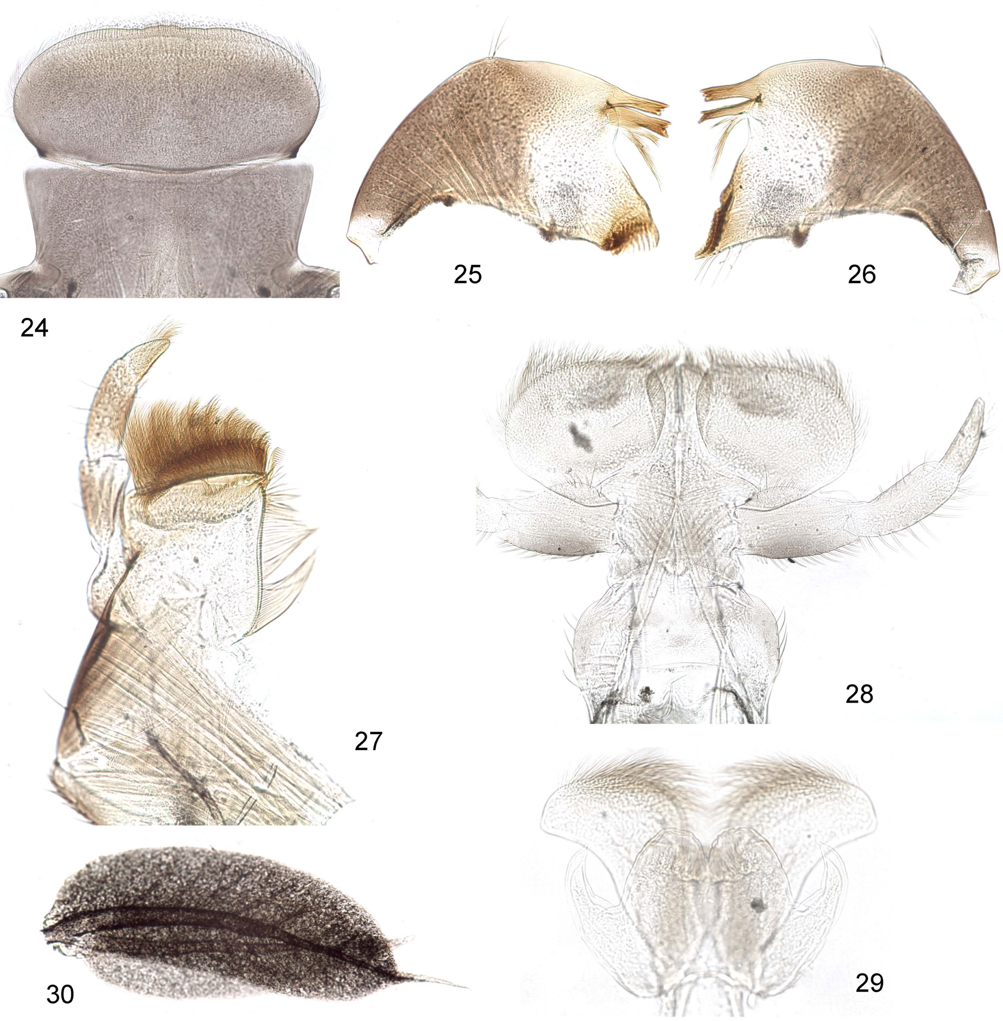
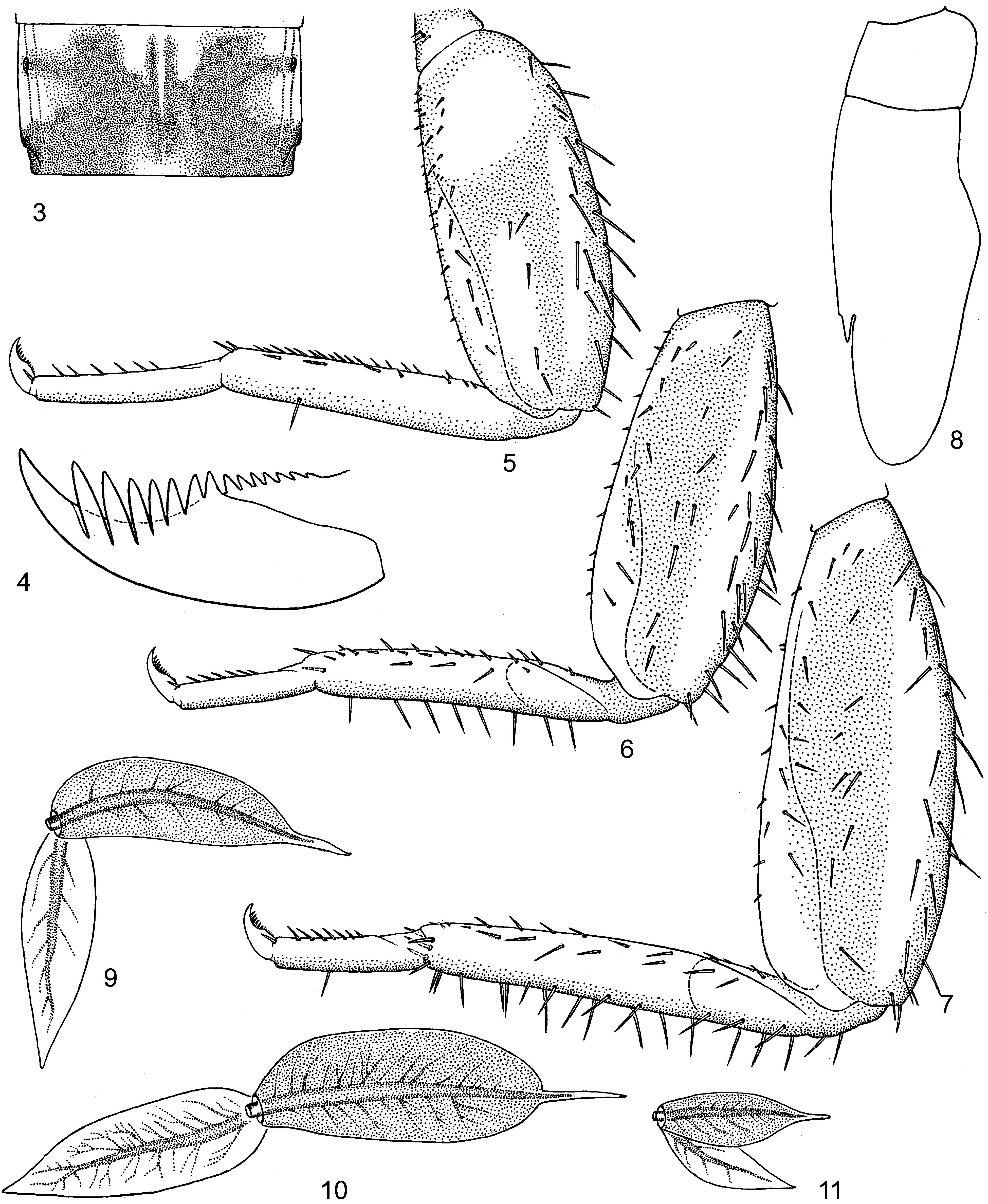
Larva. CUTICULAR COLORATION: Head brown, with brown labrum and lateral areas of mandibles ( Figs 25, 26 View FIGURES 24 – 29 ). Pronotum, mesonotum and fore protoptera brown, nearly unicolor. Femora lighter brownish, with diffusive lighter longitudinal blanks; fore femur also with large diffusive roundish blank near base ( Figs 5–7 View FIGURES 3 – 11 ). Abdominal terga and sterna ocher; terga with more or less developed diffusive darker brownish maculae adjacent to anterior margin, lateral margins and bases of tergalii.
HYPODERMAL COLORATION: Head and thorax with ocher and brown markings. Legs pale, each femur with small brown spot at apex. Abdominal terga dark brown, with ocher blanks as in imago. Abdominal sterna ocher, either unicolor, or with brown maculae. Tergalii dark brown, with dorsal lamella darker and ventral lighter (Fig. 30).
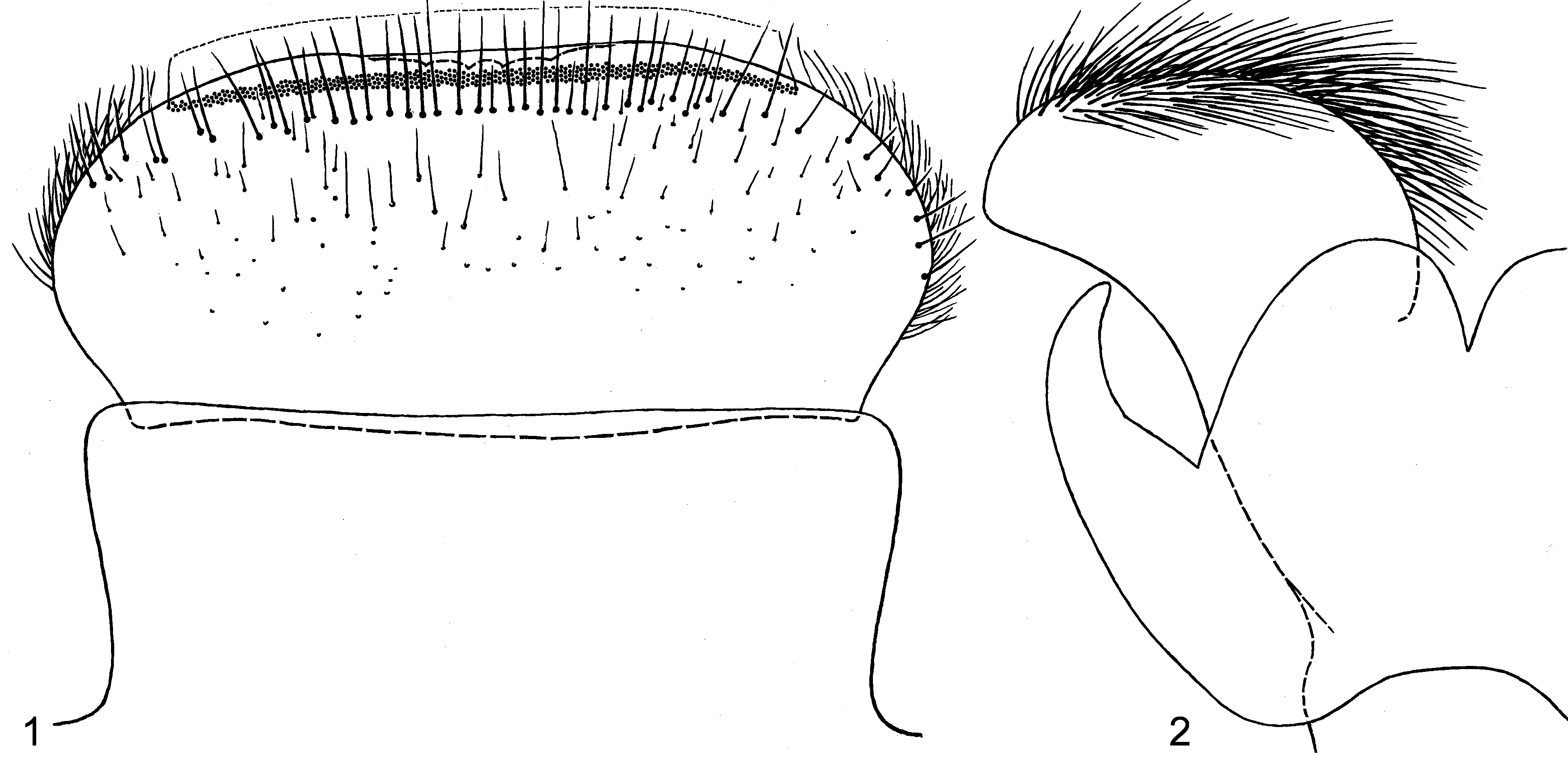
SHAPE AND SETATION: Clypeus with lateral margins nearly parallel or slightly diverging anteriorly ( Figs 1 View FIGURES 1 – 2 and 24 View FIGURES 24 – 29 ). Labrum without median incision; among five median denticles usual for Nousia , two lateral ones smoothed out and non-expressed, and three median ones can be more or less expressed; distal transverse setal row locates close to anterior margin, dense and irregular, its setal bases are situated close one to another and form a stripe 5–6 setal bases width; proximal transverse setal row nearly regular and locates close to distal row ( Fig. 1 View FIGURES 1 – 2 ). Mandibles as in Figs 25, 26 View FIGURES 24 – 29 . Maxilla as in Fig. 27 View FIGURES 24 – 29 , with 8–9 pectinate setae in ventro-apical row. Labium as in Fig. 28 View FIGURES 24 – 29 ). Hypopharynx with deep median cleft ( Figs 2 View FIGURES 1 – 2 , 29 View FIGURES 24 – 29 ).
Mesonotum with a pair of pointed projections on hind margin between protoptera ( Fig. 8 View FIGURES 3 – 11 ). Femora of all legs widened, widest in distal part ( Figs 5–7 View FIGURES 3 – 11 ). Femur of each leg with irregularly situated long spine-like setae on outer margin, smaller setae on dorsal surface and smaller setae on inner margin. Patella-tibial suture absent on fore leg and present on middle and hind legs. Fore tibia with moderately dense, irregularly situated, pectinate spine-like setae on inner side; with or without one or several long spine-like setae on outer margin (1 seta in Fig. 5 View FIGURES 3 – 11 ). Middle tibia with fewer, irregularly situated, spine-like setae on inner side and with a row of long spine-like setae on outer margin ( Fig. 6 View FIGURES 3 – 11 ). Hind tibia with irregularly situated, spine-like setae on inner side, with irregularly situated, long, spine-like setae on anterior side and with two rows of long, spine-like setae on outer margin ( Fig. 7 View FIGURES 3 – 11 ). Tarsus with row of spine-like setae on inner side; tarsus of hind leg with or without one or several long, spine-like setae on outer margin (1 seta in Fig. 7 View FIGURES 3 – 11 ). Claw with 6–7 denticles on main part and 4–5 denticles on proximal portion; denticles progressively larger apically, apical denticle not much larger ( Fig. 4 View FIGURES 3 – 11 ).
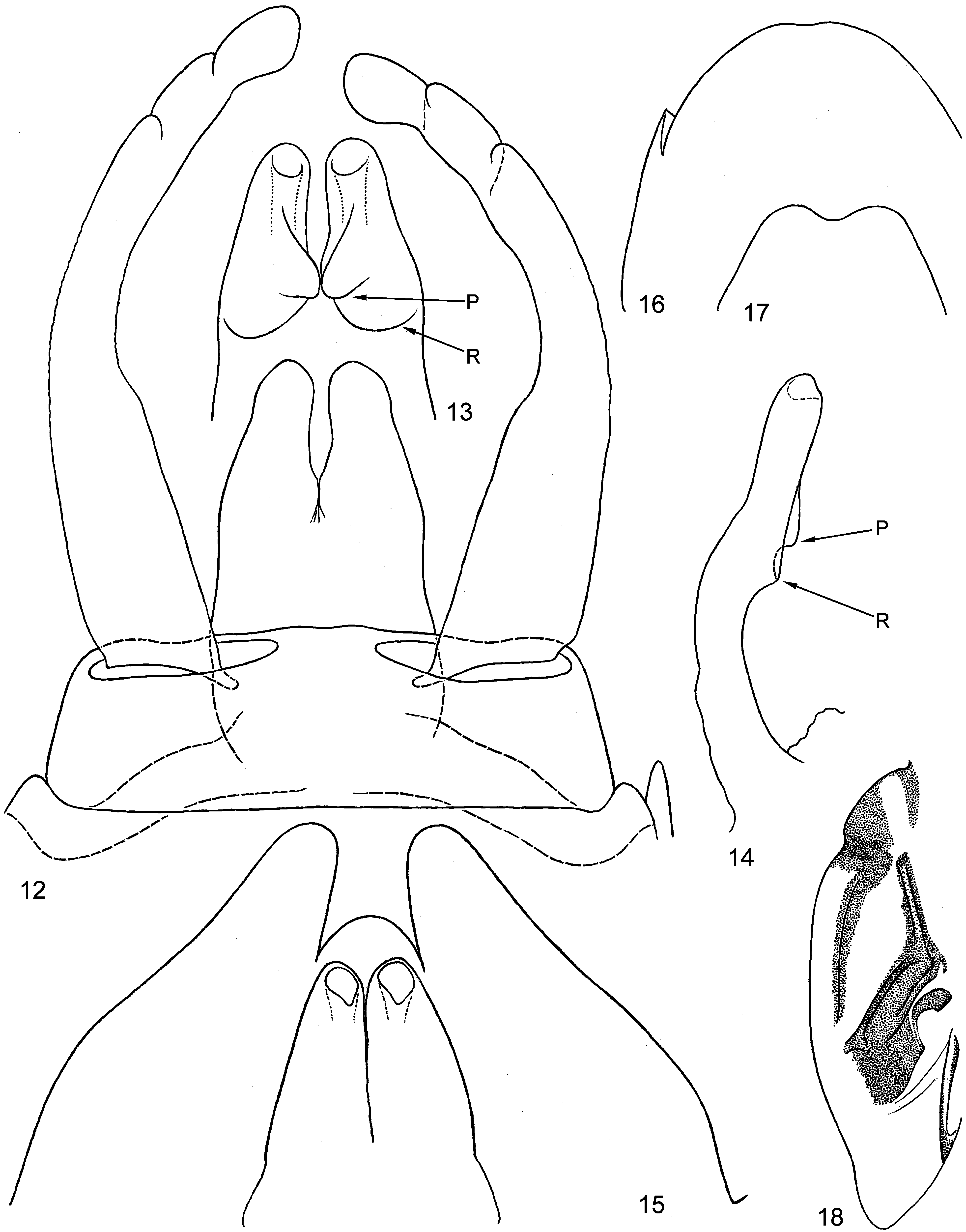
Abdomen with blunt posterolateral projections on segments VI–IX. Only abdominal tergum X with a regular row of pointed denticles on hind margin; hind margins of other terga with minute, widely spaced spine-like vestiges of denticles; sterna without denticles or their vestiges. Tergalius of each pair I–VII has dorsal lamella oval with thin terminal filament; ventral lamella lanceolate; tergalius I smaller than next ones, with dorsal lamella arched; tergalii II–IV subequal; tergalii V–VII gradually smaller ( Figs 9–11 View FIGURES 3 – 11 , 30). Larval protopenes with distal part of gonoducts lined by cuticle ( Fig. 15 View FIGURES 12 – 18 ); on exuviae shed at larval/subimaginal molt, proximal unpaired part of larval protopenis is inverted, so that protopenis looks much shorter. In female larva posterior margin of sternum IX with median incision poorly expressed ( Fig. 16 View FIGURES 12 – 18 ).
Subimago. CUTICULAR COLORATION: Head colorless. Pronotum brownish. Mesonotum with contrasting dark brown sclerites on colorless background; pigmented area anteriad of mesonotal suture represents a narrow stripe along medioparapsidal suture ( Fig. 18 View FIGURES 12 – 18 ). Thoracic sterna and pleura have brownish sclerites and colorless membranes. Fore femur brownish, with apical 1/3 darker brown; middle and hind femora light, with dark brown apex. All tibiae light with contrasting brown base. Tarsi slightly darker than tibiae. Abdominal terga and sterna brownish, with paired submedian blanks corresponding muscle bases. Styliger, gonostyli and penis of the same brownish color.
HYPODERMAL COLORATION: Similar to imago (see below).
TEXTURE: An all legs of male and female first tarsal segment (fused with tibia) covered with microtrichiae, like other parts of legs and body; tarsal segments II–V covered with pointed microlepides.
Imago, male. Head brown. Upper eyes not elevated, contiguous medially, brownish-red. Thorax on all sides (dorsal, lateral and ventral) brown. On fore wing basal sclerites brownish, veins ocher-brownish, membrane colorless; pterostigma whitish, with simple oblique cross veins. Hind wing with basal sclerites brownish. Fore leg: femur reddish brown, with dark brown apex; tibia ocher, with reddish brown base and inner side and brown spot on apex; tarsus light ocher. Middle and hind legs: femur reddish or ocher-brown with dark brown apex; tibia ocher, reddish brown proximally; tarsus light ocher. Middle and hind legs with patella-tibial suture well-developed. Tarsus with apical thorn on each of 4 proximal segments. Both claws pointed. Abdominal terga brown; each tergum II–VII with a pair of small submedian ocher blanks adjacent to anterior margin and with a pair of larger ocher blanks at anterior-lateral angles ( Fig. 3 View FIGURES 3 – 11 ). Abdominal sterna lighter brownish. Styliger, gonostyli and penis ocherbrownish. Penis lobes brought together, with smooth outer sides and with a pair of dorso-median projections directed proximally ( Figs 12–14 View FIGURES 12 – 18 ). Caudalii ocher-brownish with dark brown joints of segments.
Imago, female. Coloration similar to that of male. Posterior margin of sternum IX with shallow median incision ( Fig. 17 View FIGURES 12 – 18 ).
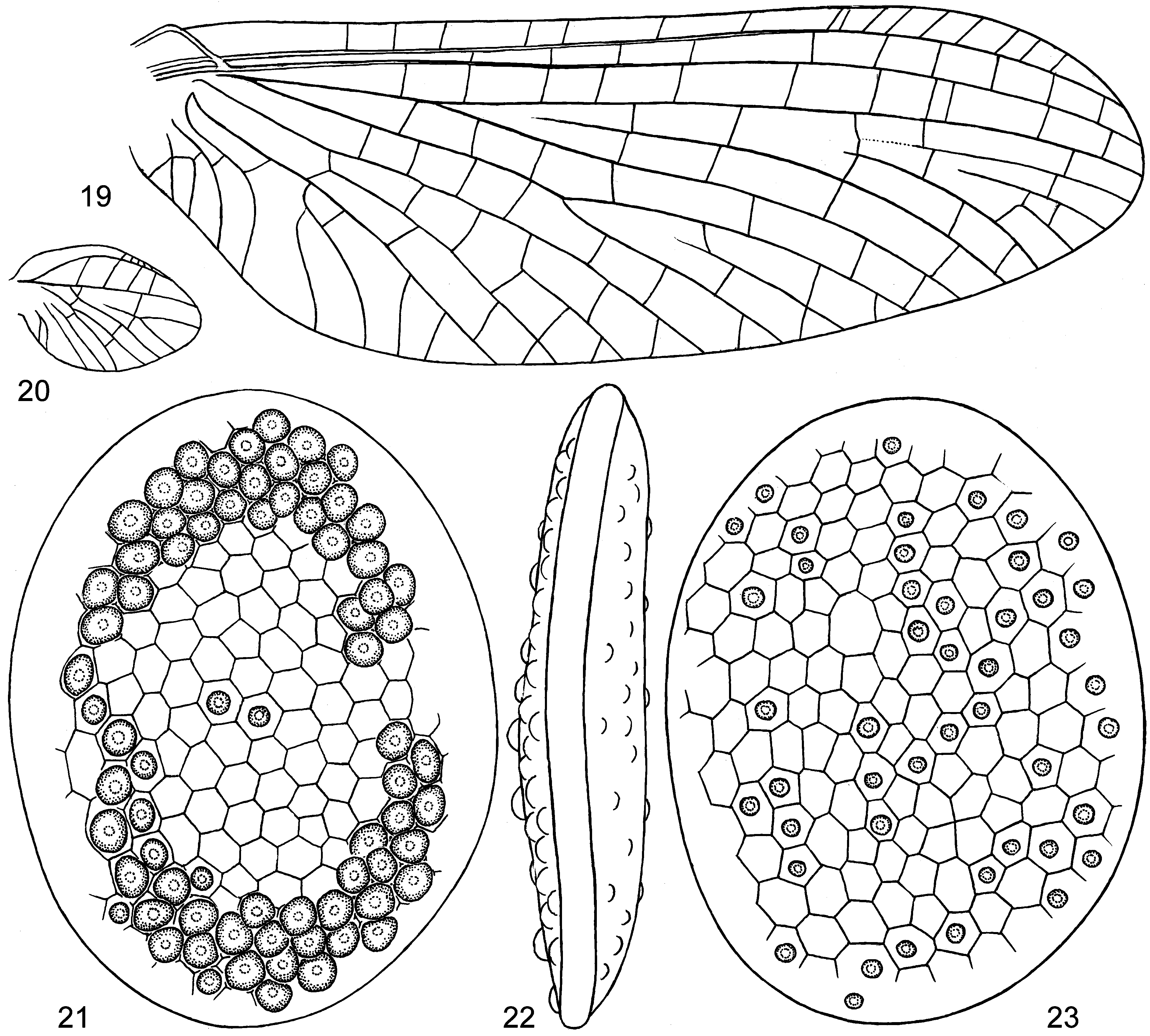
Egg ( Figs 21–23 View FIGURES 19 – 23 ). Length 0.25 mm. Has a form of flat disc with rigid and smooth margin. Each of two flat sides with net-like relief of hexagonal and pentagonal cells not reaching margin; some of these cells bear hemispheric papillae—one papilla per cell. On one side, papillae near margins are dense and large, so that contiguous one with another ( Fig. 21 View FIGURES 19 – 23 ). On another side papillae are smaller and irregularly dispersed ( Fig. 23 View FIGURES 19 – 23 ). Dimensions. Fore wing length (and approximated body length) 6–7 mm.
No known copyright restrictions apply. See Agosti, D., Egloff, W., 2009. Taxonomic information exchange and copyright: the Plazi approach. BMC Research Notes 2009, 2:53 for further explanation.