
Hovops
|
publication ID |
https://doi.org/10.11646/zootaxa.3780.3.6 |
|
publication LSID |
lsid:zoobank.org:pub:B392A186-1712-48AB-B67F-226F0B4BAC8E |
|
DOI |
https://doi.org/10.5281/zenodo.6125947 |
|
persistent identifier |
https://treatment.plazi.org/id/03A58793-9013-F16D-FF26-AB96F7AEFC82 |
|
treatment provided by |
Plazi |
|
scientific name |
Hovops |
| status |
|
Key to species of Hovops View in CoL View at ENA (only females)
This completes the key provided by Corronca and Rodriguez Artigas (2011) and incorporates the new species described here.
1. Spiders with 6 or fewer ventral spines on tibiae I–II and not more than 4 ventral spines on metatarsi I–II................. 2
– Spiders with more than 6 ventral spines on tibiae I–II and up to 6 ventral spines on metatarsi I–II...................... 7
2. Epigyne with MF represented as a cavity.................................................................... 3
– Epigyne with MF represented as a sclerite................................................................. 4
3. Epigyne sub-triangular, CO posterior to midline of epigyne, widely separated, EP well developed, elongated, postero-lateral, long and slender CD going into centre of vulva; multilobulated spermathecae with bilobed head (figs 7a–c, Corronca & Rodri- guez Artigas 2011).................................................................... H. mariensis (Strand) View in CoL
– Epigyne sub-quadrangular, CO anterior to epigyne midline and EP developed, sub-circular and in anterior-lateral position; short and divergent CD in midline, spermathecae multilobulated with largest number of lobes in middle portion (figs 4e–f, Corronca & Rodriguez Artigas 2011).............................................. H. madagascariensis (Vinson) View in CoL
4. MF of small to medium size............................................................................. 5
– MF covering almost entire epigyne; LL of epigyne cordiform, approaching at midline; anterior CO, long and slender CD and
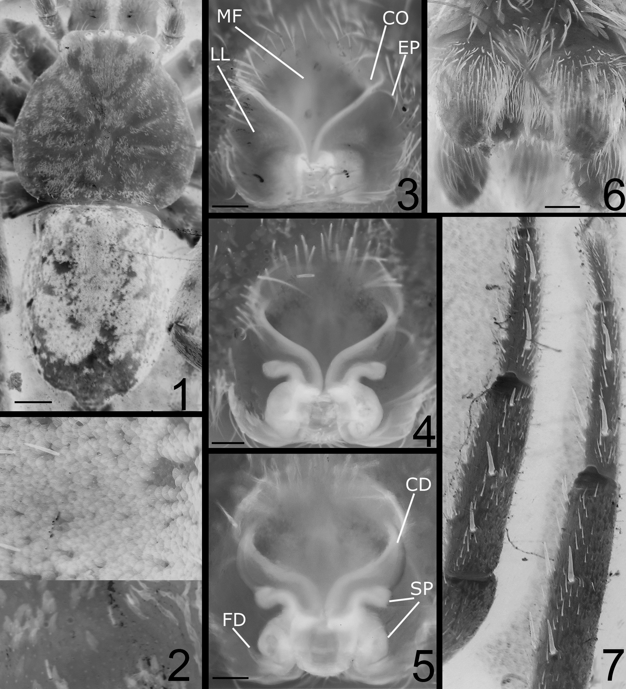
small spermathecae in posterior position (figs 8d-e, Corronca and Rodriguez Artigas 2011)............ H. pusillus (Simon) View in CoL 5. Sub-rhomboidal MF with a sharp and elongated posterior apex, LL of epigyne well developed and near midline; anterior EP situated at the sides of the CO ( Figs 3–5 View FIGURES 1 – 7 ).................................................. H. antakarana View in CoL sp. n.
– MF, LL, CO and EP otherwise.......................................................................... 6
6. Sub-circular MF; EP in midline; CO near in anterior position; CD initially straight, then divergent (figs 5d–e, Corronca and Rodriguez Artigas 2011)................................................ H. merina Corronca & Rodriguez Artigas View in CoL
– Sub-pentagonal MF; EP posterior to midline, with anterior and separated CO; CD diverging from beginning (figs 2e–f, Corronca & Rodriguez Artigas 2011)....................................... H. betsileo Corronca & Rodriguez Artigas View in CoL
7. MF anterior to the midline of the epigyne.................................................................. 8
– Sub-hexagonal and elongated MF posterior to midline of epigyne; EP separated in midline and CO separated in anterior position of epigyne (figs 6d–e, Corronca & Rodriguez Artigas 2011).................................. H. legrasi (Simon) View in CoL
8. EP in the middle portion of the epigyne.................................................................... 9
– EP behind the middle portion of the epigyne.............................................................. 10
9. Sub-pentagonal MF anterior to midline; EP widely separated in midline of epigyne by more than a width of the MF, and CO anterior and close together (figs 3f–g, Corronca & Rodriguez Artigas 2011)........ H. lidiae Corronca & Rodriguez Artigas View in CoL
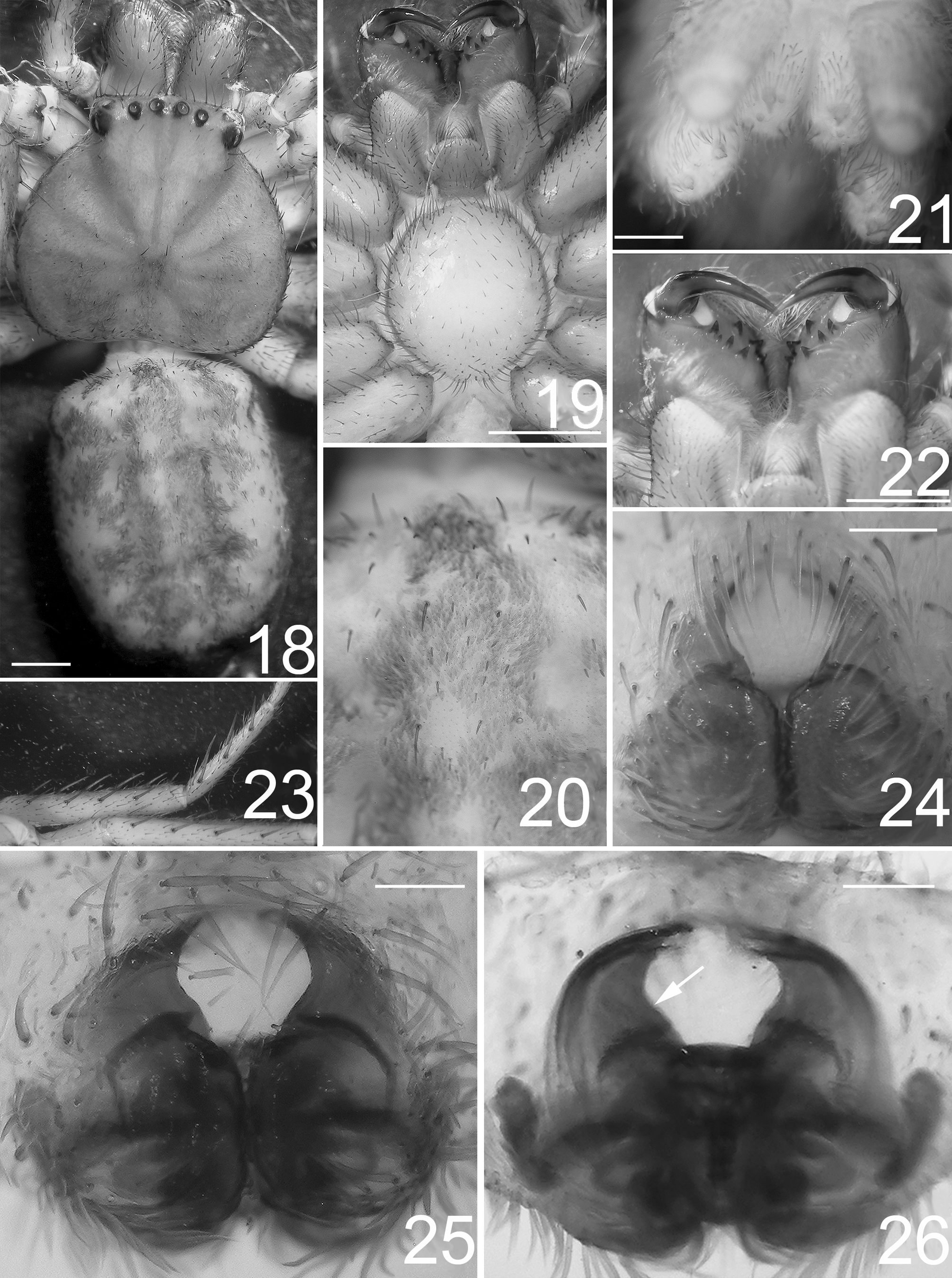
– Sub-hexagonal MF ( Fig. 25 View FIGURES 18 – 26 ); EP separated by less than a width of the MF, and anterior CO scarcely separated ( Figs 25–26 View FIGURES 18 – 26 )......................................................................................... H. menabe View in CoL sp. n.
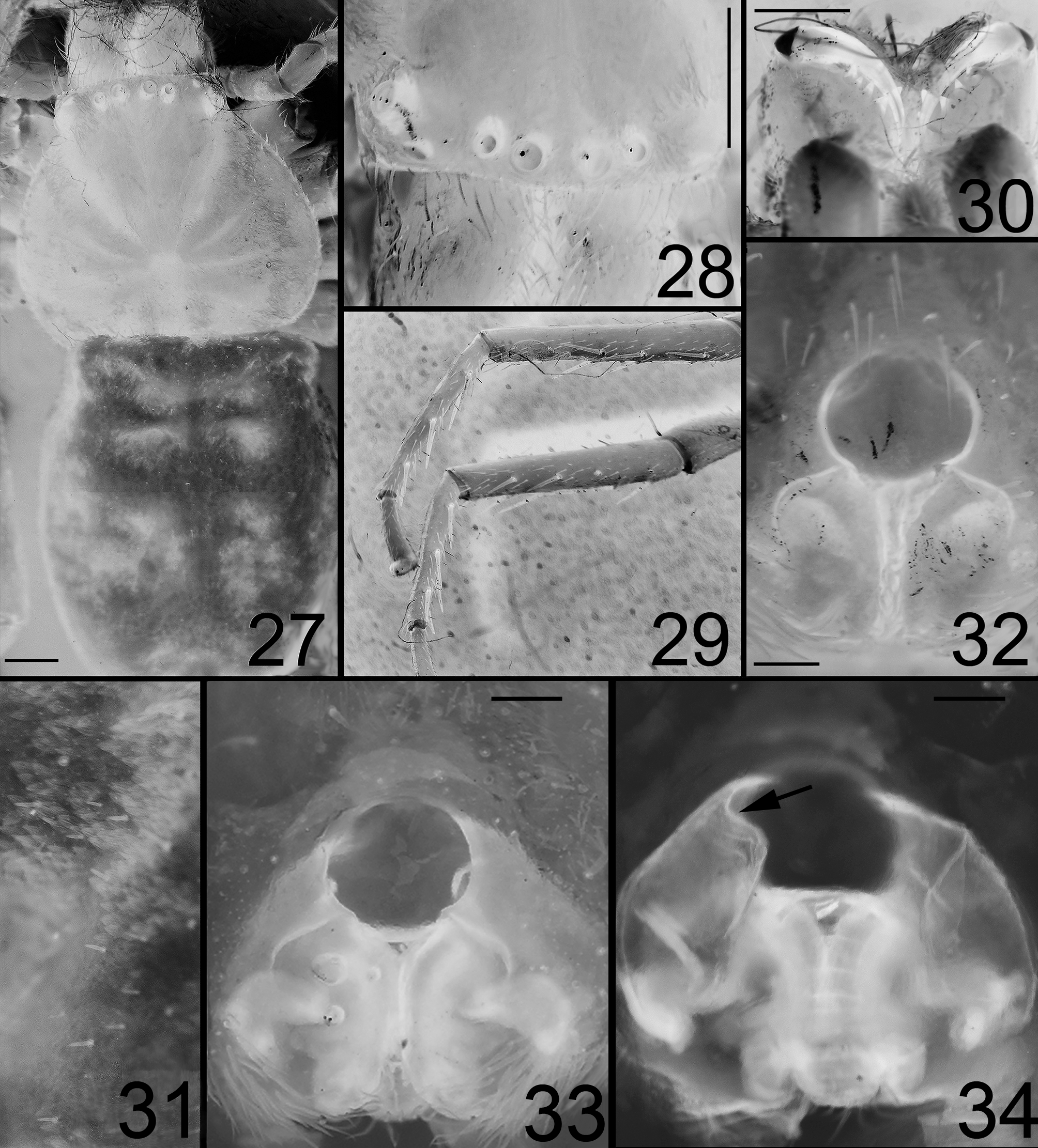
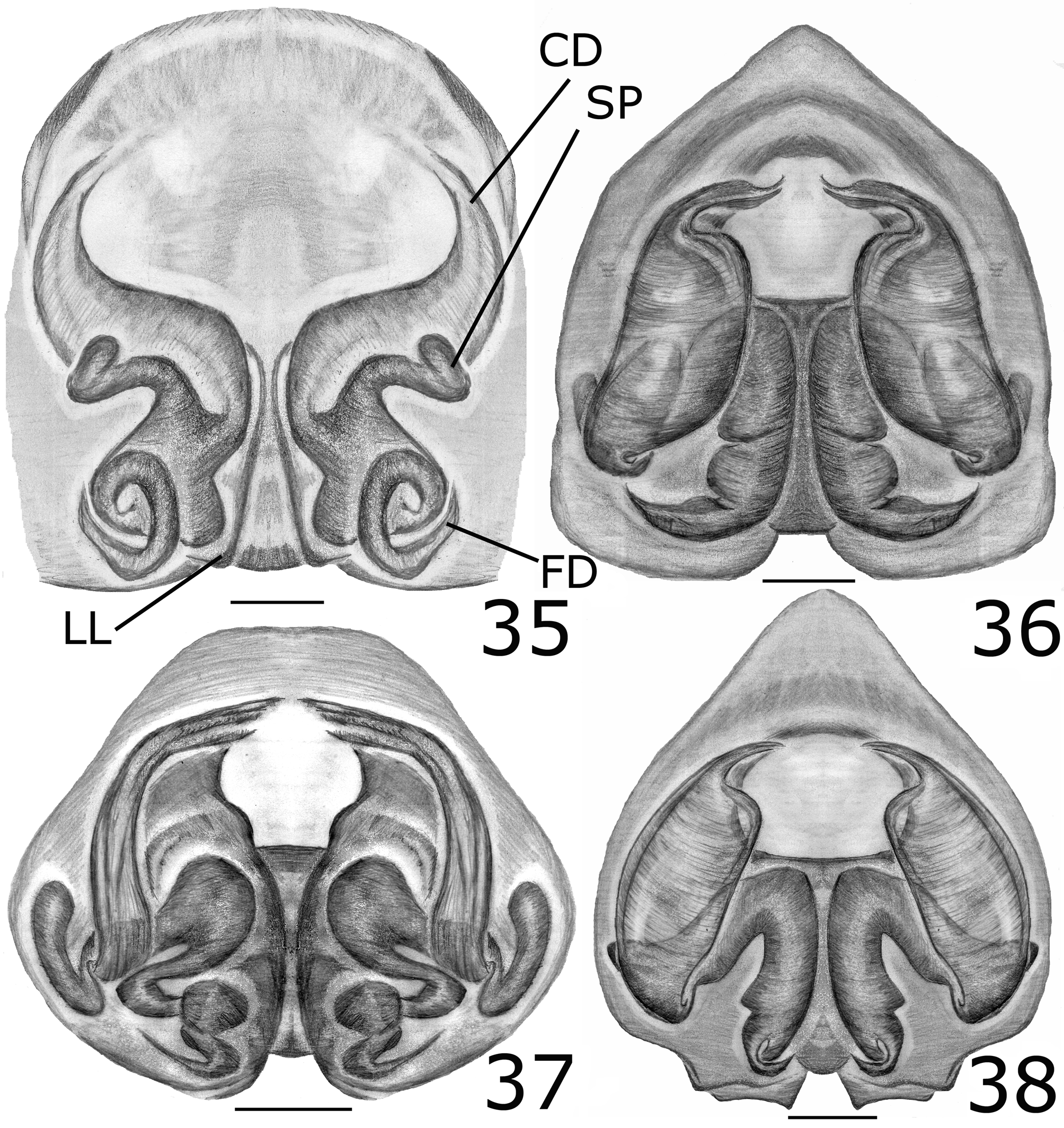
10. Sub-circular MF ( Fig. 32 View FIGURES 27 – 34 ); anterior CO separated by more than the half of the width of the MF ( Fig. 33 View FIGURES 27 – 34 ); oblique EP ( Figs 32– 33 View FIGURES 27 – 34 ); CD ducts thin in the beginning and later enlarged forming a large internal lobe and external walls nearly straight ( Figs 34 View FIGURES 27 – 34 , 38 View FIGURES 35 – 38 )...................................................................................... H. vezo View in CoL sp. n.
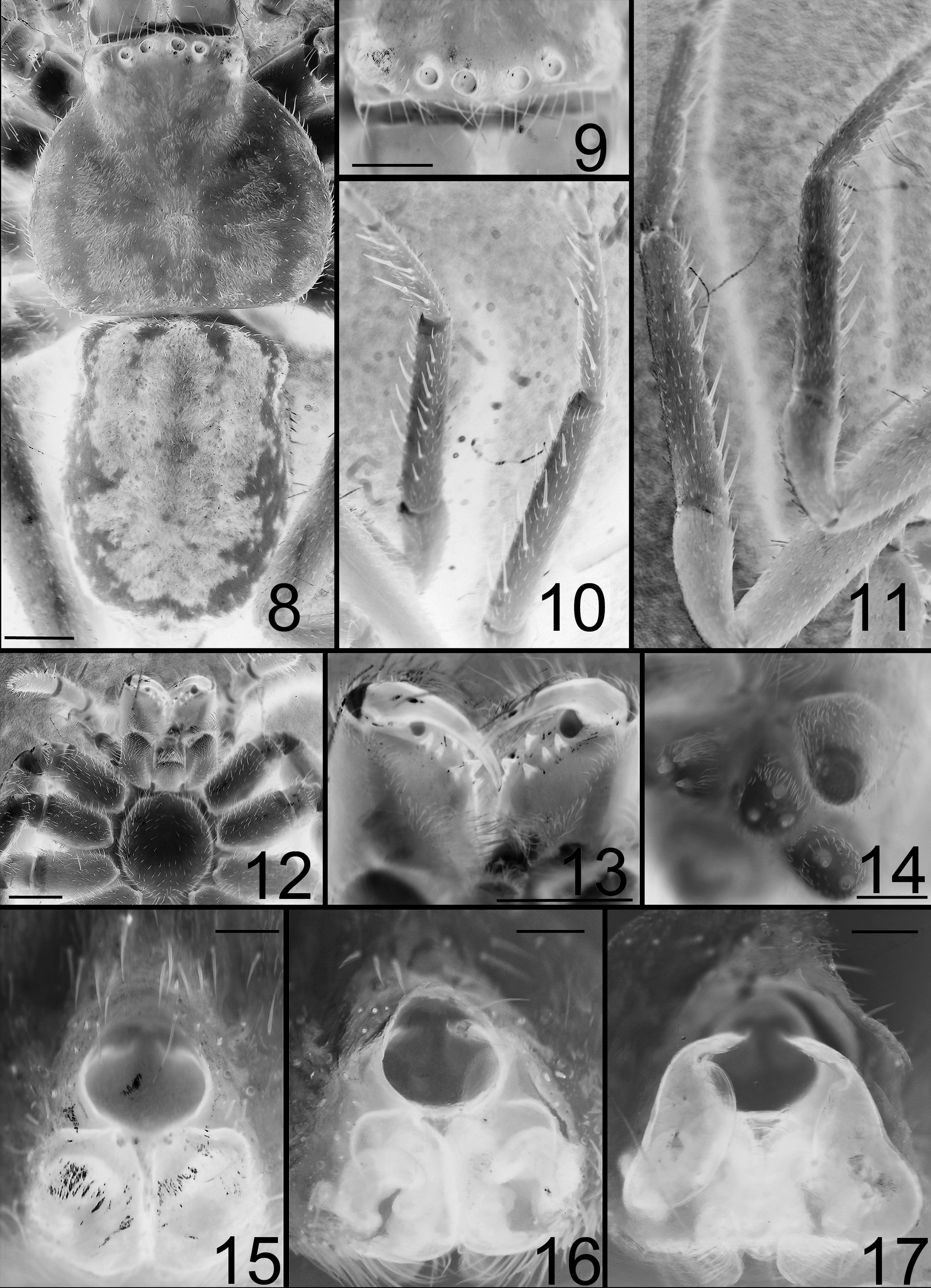
– Ovoid MF with the greater cross axis ( Fig. 15 View FIGURES 8 – 17 ); anterior CO separated by less than half the width of the MF ( Fig. 17 View FIGURES 8 – 17 ); straight EP ( Figs 15–16 View FIGURES 8 – 17 ); CD thin in the beginning and later with a deep notch that widens to form a large internal lobe and external walls curved ( Fig. 17 View FIGURES 8 – 17 )..................................................................... H. ikongo View in CoL sp. n.
No known copyright restrictions apply. See Agosti, D., Egloff, W., 2009. Taxonomic information exchange and copyright: the Plazi approach. BMC Research Notes 2009, 2:53 for further explanation.