
Cosmocephalus podicipis, Mutafchiev, Yasen, Halajian, Ali & Georgiev, Boyko B., 2010
|
publication ID |
https://doi.org/10.5281/zenodo.193420 |
|
DOI |
https://doi.org/10.5281/zenodo.6208267 |
|
persistent identifier |
https://treatment.plazi.org/id/03A587D4-FF84-295C-9187-095C0E76F85B |
|
treatment provided by |
Plazi |
|
scientific name |
Cosmocephalus podicipis |
| status |
sp. nov. |
Cosmocephalus podicipis n. sp.
Type host: Great Crested Grebe, Podiceps cristatus (L.) ( Podicipediformes , Podicipedidae ).
Other host: Black-necked Grebe, Podiceps nigricollis Brehm.
Type locality: vicinity of the town of Ramsar (36°55’N, 50°40’E), Mazandaran Province, Iran.
Other locality: Durankulak Lake (43°40´N, 28°33´E), Dobrich Region, Bulgarian Black Sea coast.
Site: Oesophagus.
Intensity of infection: 1 male and 4 females in P. cristatus collected on 18 February 2008 and 7 males and 3 females in P. cristatus collected on 5 January 2009 from Iran; 1 male in P. nigricollis collected on 9 October 1989 from Bulgaria.
Type-material: From P. cristatus : Holotype: ZMB Vermes Entozoa 7459 (male). Paratypes: ZMB Vermes Entozoa 7460 ( 1 male and 1 female); BMNH 2009.11.6.1 – 2 ( 1 male and 1 female); AHC 45437 ( 1 male and 1 female); CLGE-BAS N000.461 ( 1 male); in the collection of AH ( 3 males and 3 females).
From P. nigricollis : Paratype: ( 1 male) CLGE-BAS N000.661.
Voucher: CLGE-BAS N001.067 ( 1 male and 1 female), SEM stub.
Etymology: The name of the new species reflects the generic name of its definitive host species.
Description ( Figs 1A–G View FIGURE 1 A – G ; 2A–G; 3A–F)
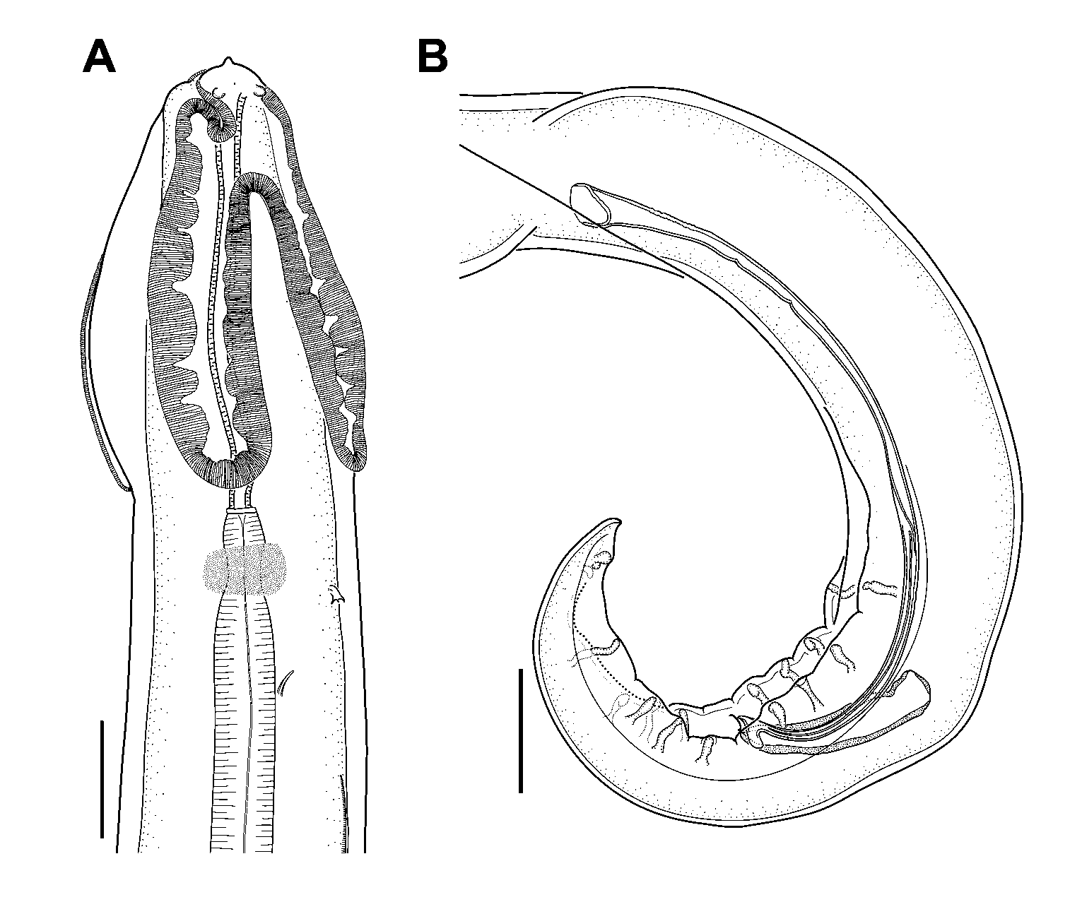
General. Medium-sized acuariids. Anterior end with two triangular pseudolabia, each bearing single amphid and pair of prominent papillae ( Fig. 2B, C View FIGURE 2 A – G ). Pair of swellings situated dorsally and ventrally between bases of pseudolabia ( Fig. 2B, C View FIGURE 2 A – G ). Cordons arise dorsally and ventrally between pseudolabia; each cordon forming loop adjacent to its base and then continuing its course along longitudinal body axis; at level of posterior end of buccal cavity, cordons recurrent in anterior direction and reach level of anterior quarter of buccal cavity where they anastomose laterally ( Figs 1A, B View FIGURE 1 A – G ; 2A). Cordons consisting of a single row of cuticular plates (each plate c. 1.5 µm long) and longitudinal cuticular ridge along outer rims of plates; deep, almost closed longitudinal groove between plates and longitudinal ridge. Plates of descending cordon arm of unequal width, thus forming scalloped appearance ( Figs 1A, B View FIGURE 1 A – G ; 2E). Deirids 18–30 long, bicuspid, situated behind cordons ( Fig. 2A, D, F View FIGURE 2 A – G ). Postdeirids, c. 5 long, with bifurcate tip ( Fig. 2G View FIGURE 2 A – G ). Lateral alae welldeveloped, extending from level just posterior of deirids to level about middle of body ( Fig. 2D View FIGURE 2 A – G ). Buccal cavity elongate ( Fig. 1A, B View FIGURE 1 A – G ). Excretory pore situated posteriorly to deirids ( Figs 1A, B View FIGURE 1 A – G ; 2D). Nerve ring surrounding anterior portion of muscular oesophagus. Phasmids subterminal ( Fig. 3B, D View FIGURE 3 A – F ).
Male (from P. cristatus from Iran, n=8 except otherwise stated). Body length 8.1–13.2 mm ( 10.6 mm). Maximum body width 224–275 (249), about mid-body; width 98–127 (111) at level of cloaca. Tail 228–373 (309) long. Cordons 376–527 (437) long, recurrent in anterior direction to 107–143 (121) from anterior body end, 21–25 wide. Deirids and excretory pore at 416–573 (488) and 483–631 (555), respectively, from anterior end of body. Left postdeirid and right postdeirid at 6.1–8.9 mm ( 7.4 mm, n=6) and 5.6–7.9 mm ( 6.8 mm, n=6), respectively, from anterior body end. Buccal cavity 358–527 (442) long and 14–16 wide. Muscular oesophagus 665–1,032 (881) long and 48–81 (66) wide. Glandular oesophagus 2,682–3,872 (3,305) long and 107–143 (123) wide. Nerve ring at 380–573 (476) from anterior body end. Cuticle 16–19 thick. Distance between cuticular striations 4–5 μm. Lateral alae extending to 5.3–8.8 mm ( 6.8 mm) from anterior body end, maximum width 47–50. Caudal alae 607–921 (745) long ( Fig. 3C View FIGURE 3 A – F ). Single ventral cuticular ridge extending between level at 1,356–2,613 (1,895) from posterior body end and beginning of caudal alae ( Fig. 3E View FIGURE 3 A – F ). Single median sessile precloacal papilla present. Nine pairs of pedunculate caudal papillae, 4 precloacal and 5 postcloacal ( Figs 1D View FIGURE 1 A – G ; 3D, E). Anterior four pairs of postcloacal pedunculate papillae with almost equal distance between them; fifth pair situated near posterior extremity of tail. Sixth pair of postcloacal papillae sessile, smaller, situated between bases of last pedunculate papillae. Left spicule 400–446 (423) long, 10–12 wide, with small projection on its distal end ( Fig. 3F View FIGURE 3 A – F ). Right spicule 125–150 (143) long, 23–28 (25) wide ( Fig. 1G View FIGURE 1 A – G ). I CL/BL 0.038–0.046 (0.042); I mOE/gOE 0.237–0.304 (0.268); I OE/BL 0.358–0.432 (0.397); I CA/BL 0.061– 0.078 (0.068); I LSP/RSP 2.770–3.360 (2.977).
Female (from P. cristatus from Iran, n=7 except otherwise stated). Body length 10.9–17.8 mm ( 15.5 mm). Maximum body width 254–435 (358), width 86–143 (116) at anus and 237–435 (349) at vulva. Tail 250–335 (307) long. Cordons extending to 514–814 (635) from anterior end, recurrent in anterior direction to 104–157 (133) from anterior body end, 34–54 (43) wide. Deirids and excretory pore at 545–814 (676) and 612–917 (769), respectively, from anterior body end. Left postdeirid and right postdeirid at 6.4–9.5 mm ( 8.3 mm, n=4) and 6.1–8.7 mm ( 7.5 mm, n=4), respectively, from anterior body end. Buccal cavity 478–699 (574) long and 20–25 (23) wide. Muscular oesophagus 745–1,220 (957) long and 57–90 (73) wide. Glandular oesophagus 3,084–4,200 (3,575) long and 131–175 (154) wide. Nerve ring at 507–734 (606) from anterior body end. Cuticle 15–22 thick. Distance between striations 5–7 μm. Lateral alae extending to level of vulva, 54–65 wide. Vulva at 5.6–8.9 mm ( 7.7 mm) from anterior body end ( Fig. 3A View FIGURE 3 A – F ). Reproductive system didelphic. Vagina vera short, posteriorly directed, separated from vagina uterina by well-developed circular musculature ( Fig. 1C View FIGURE 1 A – G ). Vagina uterin а longer, with muscular walls. Posterior extremity of tail with nipple-like projection ( Figs 1E View FIGURE 1 A – G ; 3B). Eggs elongate-oval, 41–45 × 19–20 (43 × 20, n=20), containing first stage larva ( Fig. 1F View FIGURE 1 A – G ). I CL/BL 0.036–0.047 (0.040); I mOE/gOE 0.191–0.301 (0.239); I OE/BL 0.271–369 (0.309); I V/BL 0.480–0.515 (0.496).
Remarks. Several studies have reported species of the genus Cosmocephalus as parasites of grebes ( Podiceps spp.): C. obvelatus from Podiceps sp. from the Sea of Azov and Azerbaijan ( Gushanskaya 1950) and from Aechmophorus occidentalis (Lawrence) in Canada (Gallimore 1964, cited after Storer 2000); Cosmocephalus sp. from Podiceps grisegena (Boddaert) from West Siberia ( Spasskaya 1949); and C. obvelatus magnus Vasil’kova, 1926 from Podiceps cristatus in Turkmenistan ( Kibakin 1965). None of these records was accompanied by descriptions or illustrations.
The specimens from P. cristatus from Iran and those from P. nigricollis from Bulgaria exhibit very similar morphology, including their metrical data (Table 1). They obviously belong to the same species, which we name C. podicipis n. sp. They can be distinguished from their congeners (see below for a survey of Cosmocephalus spp.) on the basis of the following comparisons:
C. podicipis can be differentiated from C. obvelatus by its longer cordons in both males and females (Table 1). The relative length of the cordons (as a proportion of the body length) of the new species is also greater than that in C. obvelatus , i.e. I CL/BL is 0.038–0.046 vs 0.029–0.036 in males and 0.036–0.047 vs 0.026– 0.035 in females. The cordons of the new species are narrower than those in C. obvelatus ( Fig. 2A View FIGURE 2 A – G and Fig. 6A View FIGURE 6 A – I ). C. podicipis has shorter spicules (Table 1). The females of C. podicipis are characterised by a longer tail and smaller body width at the level of the anus ( Fig. 1E View FIGURE 1 A – G and Fig. 5E View FIGURE 5 A – H ). The eggs of C. podicipis are markedly more elongate ( Fig. 1F View FIGURE 1 A – G and Fig. 5G View FIGURE 5 A – H ).
The new species differs from C. imperialis [as described by Morishita (1930), see Table 1] by the longer body of males and the shorter spicules. The females of C. podicipis are narrower and their eggs are larger than those of C. imperialis .
The males of C. podicipis can be distinguished from those of C. faridi [as described by Khalil (1931), see Table 1] by their longer body and shorter spicules. Furthermore, the cordons of C. faridi are characterised by their elongate loops reaching to one third of the total length of the cordons ( Khalil 1931).
The new species is differentiated from C. capellae [as described by Yamaguti (1935)] by its longer body length in males (Table 1). In addition, C. capellae has tricuspid deirids while those of C. podicipis are bicuspid.
The male specimens of C. podicipis differ from those of C. jaenschi (compared with data obtained in the present study, Table 1) by their shorter left spicules. C. podicipis has bicuspid deirids, in contrast with the tricuspid deirids of C. jaenschi . In addition, C. jaenschi has a prominent appendage at the distal end of the left spicule ( Fig. 7B View FIGURE 7 A, B ), absent in C. podicipis .


No known copyright restrictions apply. See Agosti, D., Egloff, W., 2009. Taxonomic information exchange and copyright: the Plazi approach. BMC Research Notes 2009, 2:53 for further explanation.
|
Kingdom |
|
|
Phylum |
|
|
Class |
|
|
Order |
|
|
Family |
|
|
Genus |