
Dermestocyphon Pic, 1918
|
publication ID |
https://doi.org/ 10.5281/zenodo.5740395 |
|
DOI |
https://doi.org/10.5281/zenodo.5740817 |
|
persistent identifier |
https://treatment.plazi.org/id/03A587E0-611F-8853-FE60-FAFEC9365544 |
|
treatment provided by |
Marcus |
|
scientific name |
Dermestocyphon Pic, 1918 |
| status |
|
Dermestocyphon Pic, 1918: 18 (subgenus of Cyphon Paykull, 1799 View in CoL , type species: Cyphon drianti Pic,1918 (subsequent designation by SASAGAWA (1985)).
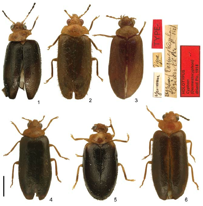
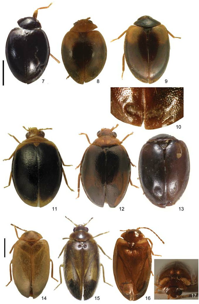
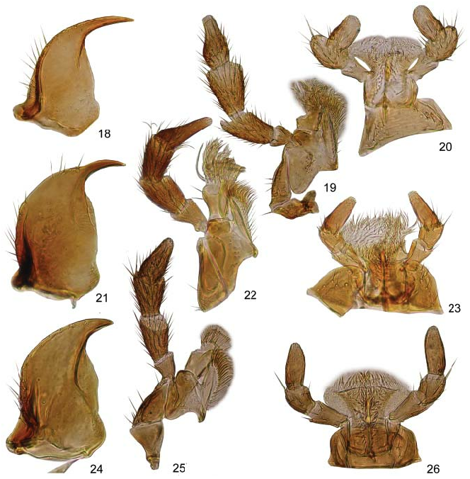
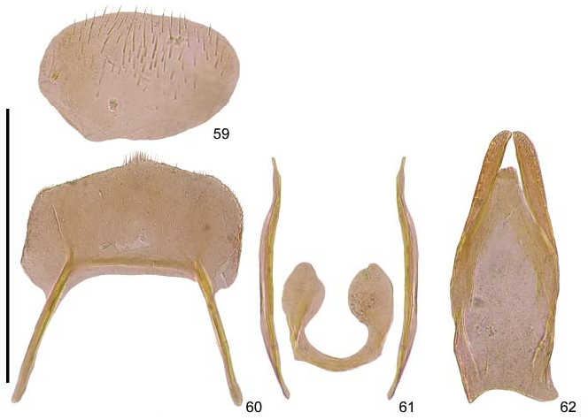
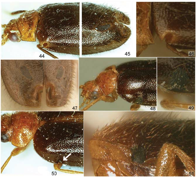
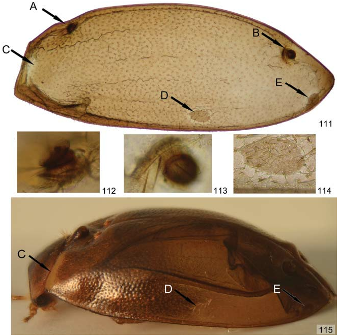
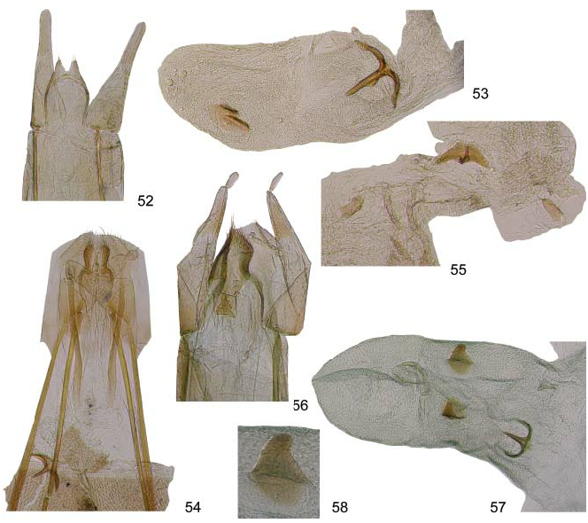
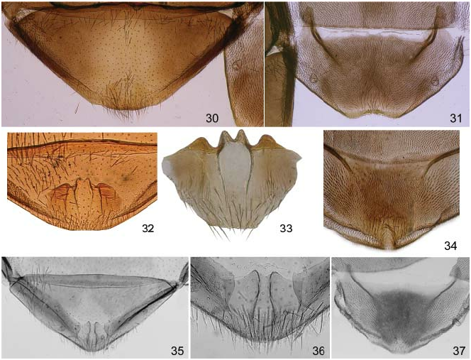
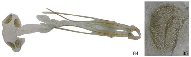
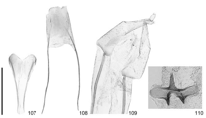
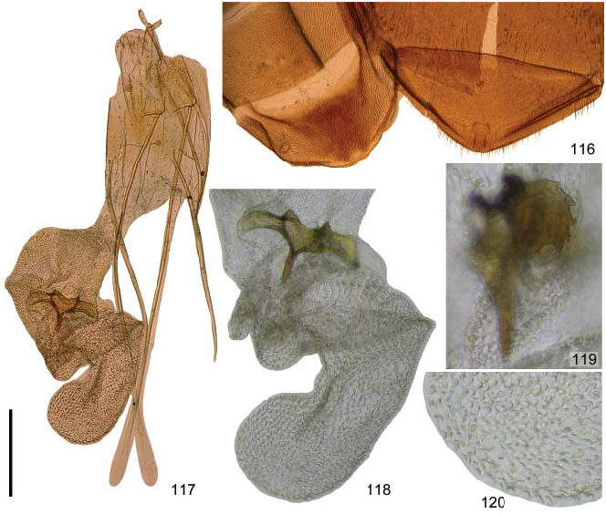
Diagnosis. Body oval to oblong ( Figs 1–17 View Figs 1–6 View Figs 7–17 ); coloration yellowish-brown to black, dorsum often with colour pattern, very variable in some species. Head and pronotum covered with subtle, granulate punctures; elytra with normal punctation. Antennomere I cylindrical, antennomere II globular to cylindrical, remaining antennomeres more or less conical; segment III of labial palpi arising from the lateral ( Fig. 20 View Figs 18–26 ) or terminal ( Figs 23, 26 View Figs 18–26 ) part of segment II; mandibles ( Figs 18, 21, 24 View Figs 18–26 ) symmetrical, simply pointed at apices. Hind wing ( Figs 27–29 View Figs 27–29 ) with long MP 4 connected with CuA + AA 1+ 2 in median portion (form 2 sensu YOSHITOMI 2005). Mesoventral process long and narrow, subparallel-sided, bilobed at apex in Himacyphon subgen. nov. and Oreocyphon . Apodemes of male tergite VII small, not protruding; apodemes of male tergite VIII connected with a transverse rod-like structure in basal portion of plate (e.g., Fig. 60 View Figs 59–62 ); tegmen small, U-shaped, with long lateral rod-like projections (e.g., Fig. 61 View Figs 59–62 ); penis elongate, with well developed trigonium and elongate, sometimes apically widening parameroids (e.g., Fig. 62 View Figs 59–62 ). Female: excitators on the elytra ( Figs 10, 13, 15–17 View Figs 7–17 , 44–51 View Figs , 111–115 View Figs 111–115 ) either only in apical portion or both in apical and humeral portions of elytra (excitators at outer margin of humeral region of elytra are found among Scirtidae only in Dermestocyphon ); ovipositor in some species with a relatively well sclerotized proctiger, which is composed of paired thumb-like projections; baculus simple, long, without branchlet ( Figs 52, 54, 56 View Figs 52–58 ); in Himacyphon subgen. nov. and Oreocyphon sternite VII with a membranous flap ( Figs 32–33, 35–36 View Figs 30–37 ) having species-specific morphology; bursella with proximal ( Himacyphon subgen. nov.), distal ( Oreocyphon ) or both proximal and distal sclerites ( Dermestocyphon s. str.).
Remarks. The synapomorphies of Dermestocyphon are as follows: 1) hind wing morphology (MP 4 connected with median portion of CuA + AA 1+2 at right angle; radial cell subtriangular, proximal portion of RA3+4 joining RA1+2 at right angle); 2) bauplan of male genitalia (small tegmen with elongate lateral rods, penis with elongate trigonium and well developed parameroids).
Dermestocyphon has a relatively uniform morphology of the male genitalia, but several other features vary remarkably, allowing a distinction of three subgenera:
1. Segment III of labial palpi arising from the side of segment II ( Fig. 20 View Figs 18–26 ); body elongate (TL/EW 1.9–2.2), sides subtly curved to subparallel ( Figs 1–6 View Figs 1–6 ); mesoventral process narrow, tempered at apex; elytra of females with humeral excitators, rarely also with apical excitator ( Figs 44–51 View Figs ); bursella with tricornate proximal sclerite and two small oval distal sclerites ( Figs 53, 55, 57 View Figs 52–58 ). .......................................................... Dermestocyphon s. str.
– Segment III of labial palpi arising from the apex of segment II ( Figs 23, 26 View Figs 18–26 ); body oval to oblong oval; elytra of females with adscutellar or apical excitators (present in all species with known females); bursella either with tricornate proximal sclerite or with 4 distal sclerites. .......................................................................................................................... 2
2. Body oval (TL/EW 1.3–1.5, Figs 7–9, 11–13 View Figs 7–17 ); mesoventral process moderately narrow, distinctly bilobed at apex; elytra of females with apical excitators ( Figs 10, 13 View Figs 7–17 ; uncertain if in all species); bursella with 4 sclerites in distal portion ( Fig. 85 View Figs 84–85 ). ............................... .......................................................................................... Oreocyphon Klausnitzer, 2009
– Body oblong oval (TL/EW 1.6–1.8, Figs 14–16 View Figs 7–17 ); mesoventral process narrow, subtly bilobed at apex; elytra of females with fungiform adscutellar excitators ( Figs 15, 16 View Figs 7–17 ) and microreticulated areas in basal portion of elytra ( Figs 17 View Figs 7–17 , 111, 115 View Figs 111–115 ; in some species also in lateral and apical portions of elytra); bursella with tricornate proximal sclerite ( Figs 110 View Figs 107–110 , 118 View Figs 116–120 ). .............................................................................. Himacyphon subgen. nov.

No known copyright restrictions apply. See Agosti, D., Egloff, W., 2009. Taxonomic information exchange and copyright: the Plazi approach. BMC Research Notes 2009, 2:53 for further explanation.
|
Kingdom |
|
|
Phylum |
|
|
Class |
|
|
Order |
|
|
Family |
Dermestocyphon Pic, 1918
| Ruta, Rafał, Yoshitomi, Hiroyuki & Klausnitzer, Bernhard 2013 |
Dermestocyphon
| PIC M. 1918: 18 |