
Arisubathynella cheongmiensis, Park & Eun, 2012
|
publication ID |
https://doi.org/10.11646/zootaxa.3368.1.15 |
|
DOI |
https://doi.org/10.5281/zenodo.5254044 |
|
persistent identifier |
https://treatment.plazi.org/id/03A587F8-FFCF-7676-AFEF-7970FE04E36E |
|
treatment provided by |
Felipe |
|
scientific name |
Arisubathynella cheongmiensis |
| status |
sp. nov. |
Arisubathynella cheongmiensis n. sp.
( Figs. 1 – 4 View FIGURE 1 View FIGURE 2 View FIGURE 3 View FIGURE 4 )
Type material. Holotype: male, dissected and mounted on six slides. South Korea, Kyeonggi-Do, Yeoju-Gun , Jumdong-Myon, Dori ( 37°13’54.6” N, 127°43’4.1” E), 12. March 2010, ( J.-L. Cho) ( NIBR IV0000242682 View Materials ) GoogleMaps . Allotype: female, dissected and mounted on six slides, same data as for holotype ( NIBR IV0000242683 View Materials ) GoogleMaps . Paratypes: 3 males and 2 females each mounted as a whole specimen on a slide ( NIBR IV0000242684 View Materials – IV0000242688 View Materials ) , one male and one female each dissected and mounted on six slides ( NIBR IV0002242689 View Materials , IV0000242690 View Materials ), same data as for holotype GoogleMaps .
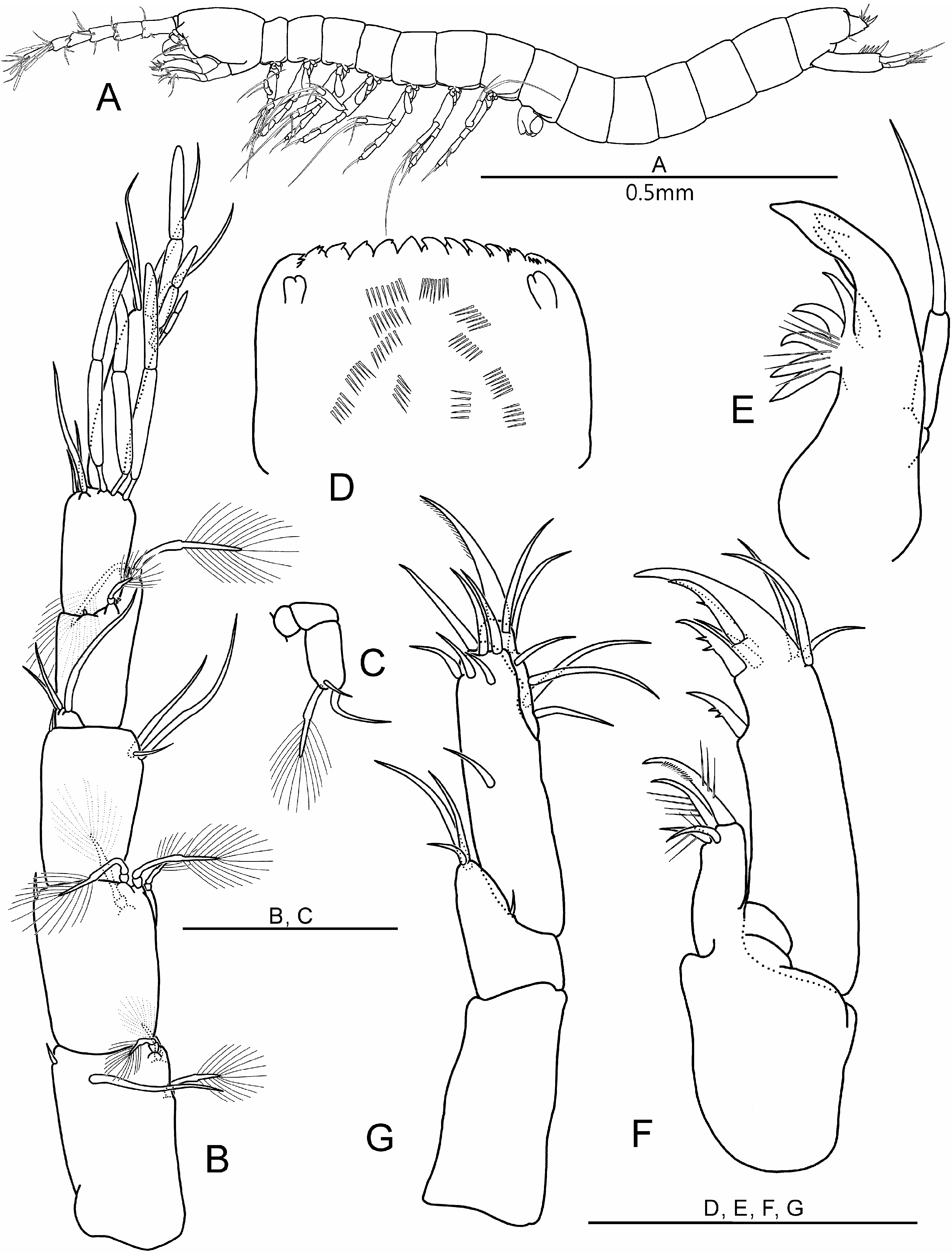
Description of adult male ( Holotype). Body length 1.03 mm (other males: 0.94–1.01 mm). Approximately 16 times wide. Head slightly longer than anterior two thoracic segments together ( Fig. 1A View FIGURE 1 ).
Antennule ( Fig. 1B View FIGURE 1 ) six-segmented. First segment with one seta on inner distal corner, with one simple dorsal seta and with one dorso-lateral, one medio-lateral and one ventro-lateral plumose seta. Second segment with one group of four plumose setae and with one simple seta on inner distal corner. Third segment with one seta on inner distal corner, two lateral simple setae of very unequal size and one simple ventro-lateral seta. Inner flagellum of third segment with three simple setae. Fourth segment with one stub seta and one plumose seta on dorsal margin, and with two stub setae and two plumose setae on outer distal apophysis. Fifth segment with three simple setae on inner distal margin and three dorsal aesthetascs. Sixth segment with three subterminal aesthetascs of different size and four simple setae.
Antenna ( Fig. 1C View FIGURE 1 ) three-segmented, as long as first antennular segment. Two proximal segments without setae, distal segment with two simple setae of different size and one plumose seta terminally. Plumose seta as long as long simple seta.
Labrum ( Fig. 1D View FIGURE 1 ) flat, with eight median teeth of more or less similar size flanked by two smaller distally dentate teeth on both sides. Inner surface with one nipple-like protrusion and six rows of ctenidia on both sides.
Mandible ( Fig. 1E View FIGURE 1 ) with incisor process of three teeth. Tooth of the ventral edge absent. Lobe with row consisting of five spines. Palp of one segment, four times as long as wide, with one apical seta.
Maxillule ( Fig. 1F View FIGURE 1 ) two-segmented. Proximal segment with four setae on inner distal margin. Distal segment with two terminal smooth spines, three dentate spines on inner edge and with three simple setae of different size on outer distal margin.
Maxilla ( Fig. 1G View FIGURE 1 ) four-segmented, setal formula 0-4-7-7.
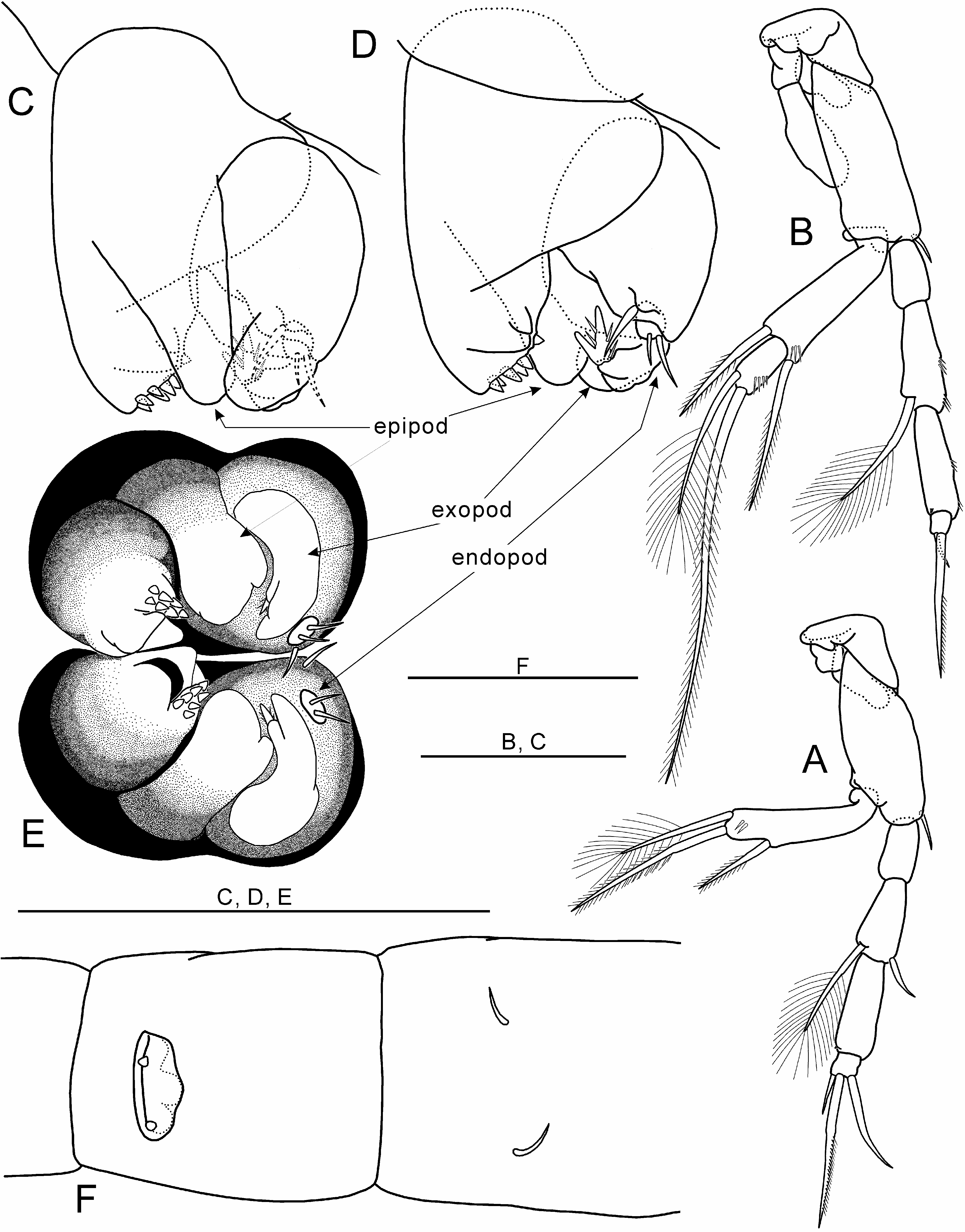
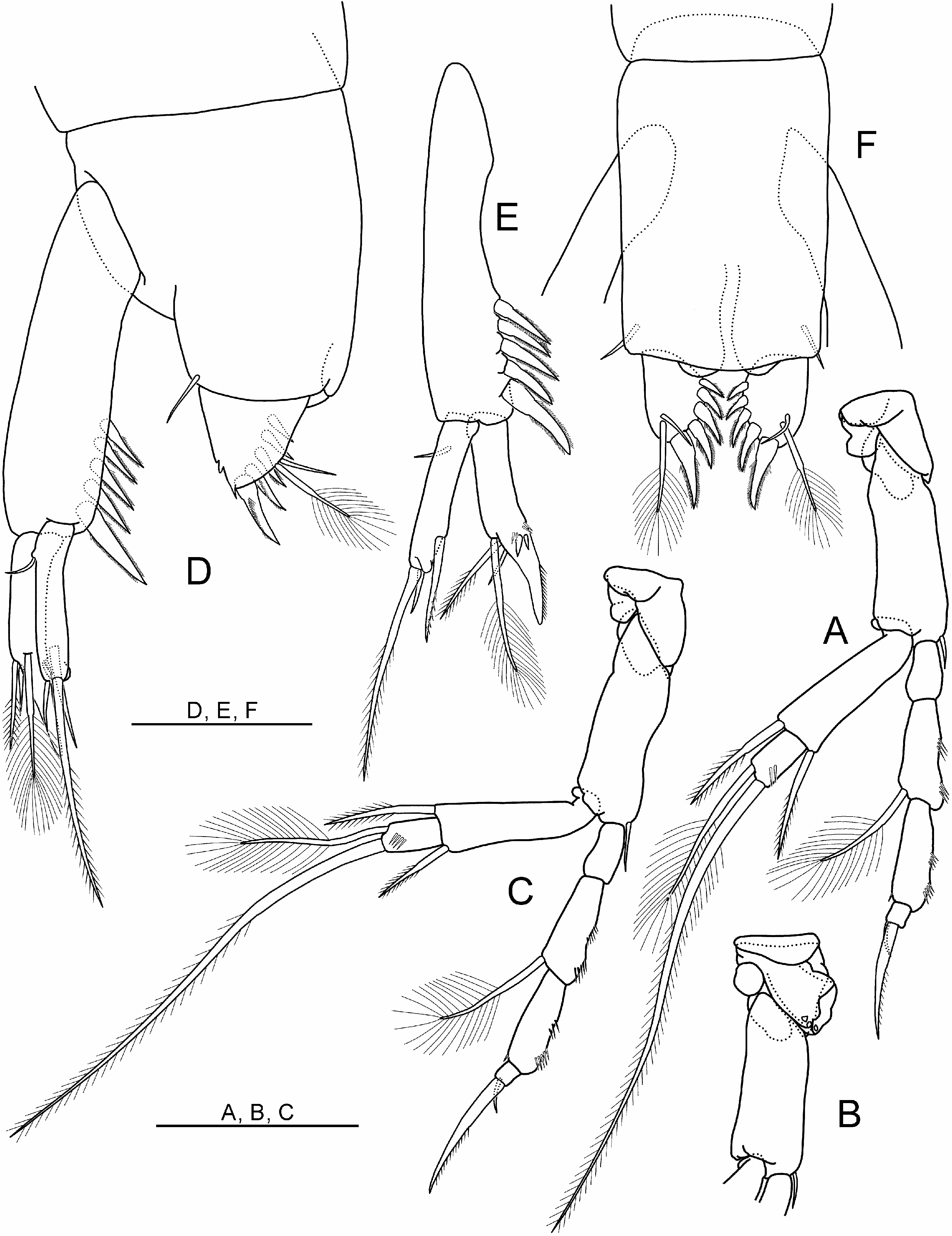
Thoracopods I – VII ( Figs. 2A, B View FIGURE 2 , 3A, B, C View FIGURE 3 , 4A, B, C View FIGURE 4 ) similar in size except thoracopod I. Thoracopods II – V each with one epipod. Thoracopods I, VI-VII without epipod. Basipod of thoracopods I – VII with one seta at inner distal corner. Exopod of thoracopod I one-segmented, with one medial seta on ventral edge and terminally with one long seta and one shorter plumose seta. Exopods of thoracopods II – VII two-segmented. Proximal segment being longer than distal one and with one dorsal and one ventral seta. Distal segment with one long seta and one plumose seta terminally. Endopods of thoracopods I – VII four-segmented, setal formulae:
Thoracopod I 0+0/1+1/0+1/3(1)
Thoracopod II – VII 0+0/0+1/0+1/2(1)
Thoracopod VIII ( Figs. 2C, D, E View FIGURE 2 ) bell-shaped in lateral view, 1.2 times longer than wide. Main axis tilting backwards. Protopod massive with prominent penial region displaying distal opening encircled by frontal, dentate and with inner lobe. Frontal and inner lobe smooth. Dentate lobe with seven tiny denticles. External lobe (epipod) conical in lateral view, balloon-shaped in ventral view, exceeding penial region in lateral view. Basipod as large as half protopod, with one basipodal seta near base of endopod. Exopod one third the size of basipod, two-lobed distally; upper lobe smooth, lower lobe with bifurcate tooth. Endopod small with two termintal setae.
First pleopod in form of seta (cf. Fig. 2F View FIGURE 2 ).
Uropod ( Fig. 4D, E View FIGURE 4 ) with sympod bearing five spines. Most distal spine thicker and longer than the others which are of similar size. Endopod 40 % as long as sympod, with one large, fused, distal spur and two much smaller spines at its base, two simple setae and one plumose seta on outer distal margin. Exopod longer than endopod, with one basi-ventral seta and distally with one subterminal latero-internal seta, one projection and two terminal setae, of which the outer seta is six times longer than the inner one.
Pleotelson ( Figs. 4D, F View FIGURE 4 ) with one seta near base of furcal rami. Anal operculum slightly convex. Furcal rami slightly longer than wide, with five spines and two setae, of which the longer one is plumose.
Description of adult female ( Allotype). The female differs from the male in the protopod of thoracopod VI and in thoracopod VIII. Body length1.05 (other females: 0.95 – 1.05mm). Protopod of thoracopod VI ( Fig. 4B View FIGURE 4 ) with tubular protrusion directed inwards. Distal margin of protrusion undulate. Thoracopod ( Fig. 2F View FIGURE 2 ) form of two tiny teeth on hemispherical plate.
Etymology. The specific epithet is derived from a tributary of the Han-River (Cheongmi Stream), where the new species was discovered.

| NIBR |
National Institute of Biological Resources |
No known copyright restrictions apply. See Agosti, D., Egloff, W., 2009. Taxonomic information exchange and copyright: the Plazi approach. BMC Research Notes 2009, 2:53 for further explanation.
|
Kingdom |
|
|
Phylum |
|
|
Class |
|
|
Order |
|
|
Family |
|
|
Genus |