
Philactinoposthia ischiae, Nilsson, Karin Sara, Wallberg, Andreas & Jondelius, Ulf, 2011
|
publication ID |
https://doi.org/ 10.5281/zenodo.277458 |
|
DOI |
https://doi.org/10.5281/zenodo.5683933 |
|
persistent identifier |
https://treatment.plazi.org/id/03A6456D-FFB3-AE44-FF4B-95D2FED9FE1A |
|
treatment provided by |
Plazi |
|
scientific name |
Philactinoposthia ischiae |
| status |
sp. nov. |
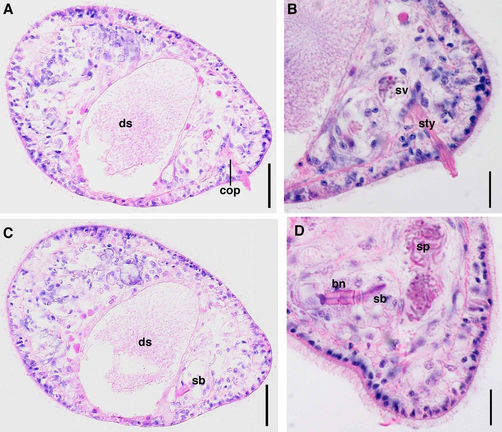
Philactinoposthia ischiae sp.nov.
( Figs. 3 View FIGURE 3 , 4, 5, 6)
Type Material: Holotype: Type SMNH-8052. Paratype 1: SMNH Type-8053, Paratype 2: SMNH Type-8054.
Type Locality. Ischia, Bay of Naples, Italy, at the volcanic carbon dioxide vent (station N3) with mean surface pH: 7.09 ( Hall-Spencer et al. 2008), from 2-3 m water depth in fine sand. (40° 43' 55" N, 13° 57' 50" E).
Other Material examined. Living specimens in squeeze preparations; 3 sets of 4-5 μm serial sagittal sections of paraffin-embedded specimen.
Etymology. Species epithet refers to name of type locality.
Description. Active swimmers. Living specimens ~400-600 μm long and ~180 μm wide. Anterior and posterior end rounded. Body cylindrical. Epidermis completely ciliated. Uncolored rhabdoid glands uniformly scattered across body, very small, appearing white in incident light, not visible in sections (Fig. 4AB). Body colorless in transmitted light, except some light brown coloration of digestive syncytium in some specimens (Fig. 4A). Statocyst located 50 μm behind anterior end, at U12. Frontal organ present, cell bodies of frontal glands extend from U20 to U30. Mouth opening ventral, middle of body, at U50. Digestive central syncytium extends from U40 to U70 ( Fig. 5 View FIGURE 5 AC). One specimen with large needle-like structures, 40–50 μm long, located around middle of body which is the same location as digestive syncytium thus possibly food material (Fig. 4C).
Ovaries paired, ventral; each lateral band with up to 7 oocytes, extending from middle of body to bursal nozzle, U50 to U80 (Fig. 4A). No female gonopore detected. Well-developed seminal bursa, at U85, with well-defined ~18 μm long bursal nozzle directed ventrally to the anterior (Fig. 4AB, 5CD). Testes paired, dorsal; separate from ovary. Testes extend anterior from middle of body to copulatory organ, from U40 to U80. Male gonopore positioned sub-terminally slightly lateral in dorsal view, opens directly to male copulatory organ composed of a long, ~100 μm, curved, conical sclerotized stylet-like structure (Fig. 4AB, 5AB). Stylet needles strongly stained by eosin ( Fig. 5 View FIGURE 5 B).

| SMNH |
Saskatchewan Museum of Natural History |
No known copyright restrictions apply. See Agosti, D., Egloff, W., 2009. Taxonomic information exchange and copyright: the Plazi approach. BMC Research Notes 2009, 2:53 for further explanation.