
Philactinoposthia multipunctata, Nilsson, Karin Sara, Wallberg, Andreas & Jondelius, Ulf, 2011
|
publication ID |
https://doi.org/ 10.5281/zenodo.277458 |
|
DOI |
https://doi.org/10.5281/zenodo.5683935 |
|
persistent identifier |
https://treatment.plazi.org/id/03A6456D-FFBD-AE49-FF4B-95D2FB7FFCC1 |
|
treatment provided by |
Plazi |
|
scientific name |
Philactinoposthia multipunctata |
| status |
sp. nov. |
Philactinoposthia multipunctata sp.nov.
( Figs. 3 View FIGURE 3 , 7 View FIGURE 7 , 8 View FIGURE 8 , 9 View FIGURE 9 )
Type material: Holotype: SMNH Type-8046. Paratype 1: SMNH Type-8047, Paratype 2: SMNH Type-8048.
Type locality. Punta Ala Torre Balbo in Italy (42° 48' 25" N, 10° 44' 34" E), at 3 m water depth in fine sand.
Other material examined. Living specimens in squeeze preparations; 1 set of 4–5 μm serial sagittal sections of paraffin-embedded specimen.
Etymology. Name refers to the numerous rhabdoid glands. Multi = many, punctum = dot, in latin.
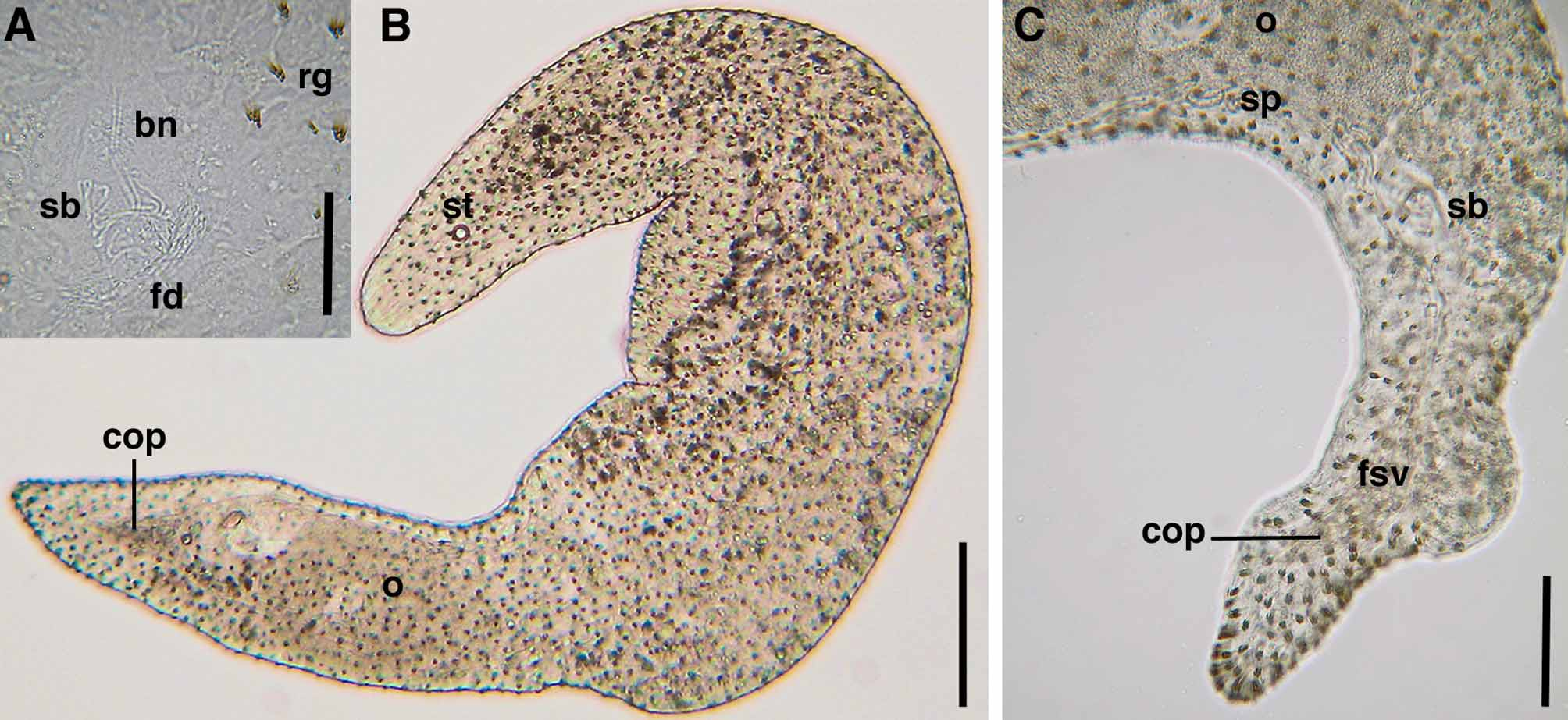
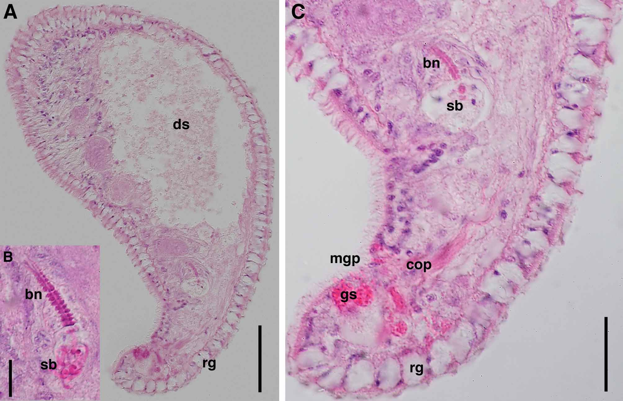
Description. Active and fast swimmer. Living specimens ~ 3 mm long and ~340–500 μm wide. Body shape cylindric, both ends tapering. Epidermis completely covered with 5 μm long cilia. Numerous red-colored rhabdoid glands in body wall, uniformly scattered across body ( Fig. 7 View FIGURE 7 BC). Each rhabdoid gland with up to 9 strokes and 10 μm long ( Fig. 7 View FIGURE 7 A). Epidermis uncolored in transmitted light. Statocyst 8 μm in diameter, located anteriorly at level U7,5. Frontal organ well developed, cell bodies of frontal glands located posterior to statocyst, extending from U10 to U20. Mouth ventral, located in middle of body. Digestive central syncytium extends from U25 to U70.
Ovary unpaired, ventral, with up to 5 oocytes, extends posterior from level of testes, to bursal nozzle, U55–60 to U80. Female gonopore ventral, at level U85, opens directly to seminal bursa with wall and a fibrous disc in distal end; visible in pressed specimens ( Fig. 7 View FIGURE 7 A). Seminal bursa connects anteriorly to well defined and clearly visible bursal nozzle with appearance like a stack of hats, with ventro-anterior direction ( Fig. 8 View FIGURE 8 ABC). Bursal nozzle is long and slender, 60 μm long and consists of at least 13 “hats”.
Testes paired, lateral to ovary, extends anteriorly from level of mouth, to male copulatory organ posteriorly, U45 to U90. Male gonopore positioned in posterior end, ventral, at level U95, opens to male copulatory organ with v-shaped sclerotized penis-like structure built of 100 μm long stylet needles. Penis-like structure filled with spermatozoa, easily detected with moderate pressure. Needles weakly stained by eosin. Eosinophilic glands surround male antrum ( Fig. 8 View FIGURE 8 C). False seminal vesicle located at proximal end of stylet-like structure.


| SMNH |
Saskatchewan Museum of Natural History |
No known copyright restrictions apply. See Agosti, D., Egloff, W., 2009. Taxonomic information exchange and copyright: the Plazi approach. BMC Research Notes 2009, 2:53 for further explanation.