
Planaeschna tsuchi, Kompier & Karube & Futahashi & Phan, 2021
|
publication ID |
https://doi.org/10.11646/zootaxa.5027.1.1 |
|
publication LSID |
lsid:zoobank.org:pub:CCF10500-01A3-476C-A4BE-57161E164EBD |
|
DOI |
https://doi.org/10.5281/zenodo.5492686 |
|
persistent identifier |
https://treatment.plazi.org/id/03A687CB-FFC7-FFF5-E0E7-7577FCC4FC4B |
|
treatment provided by |
Plazi |
|
scientific name |
Planaeschna tsuchi |
| status |
sp. nov. |
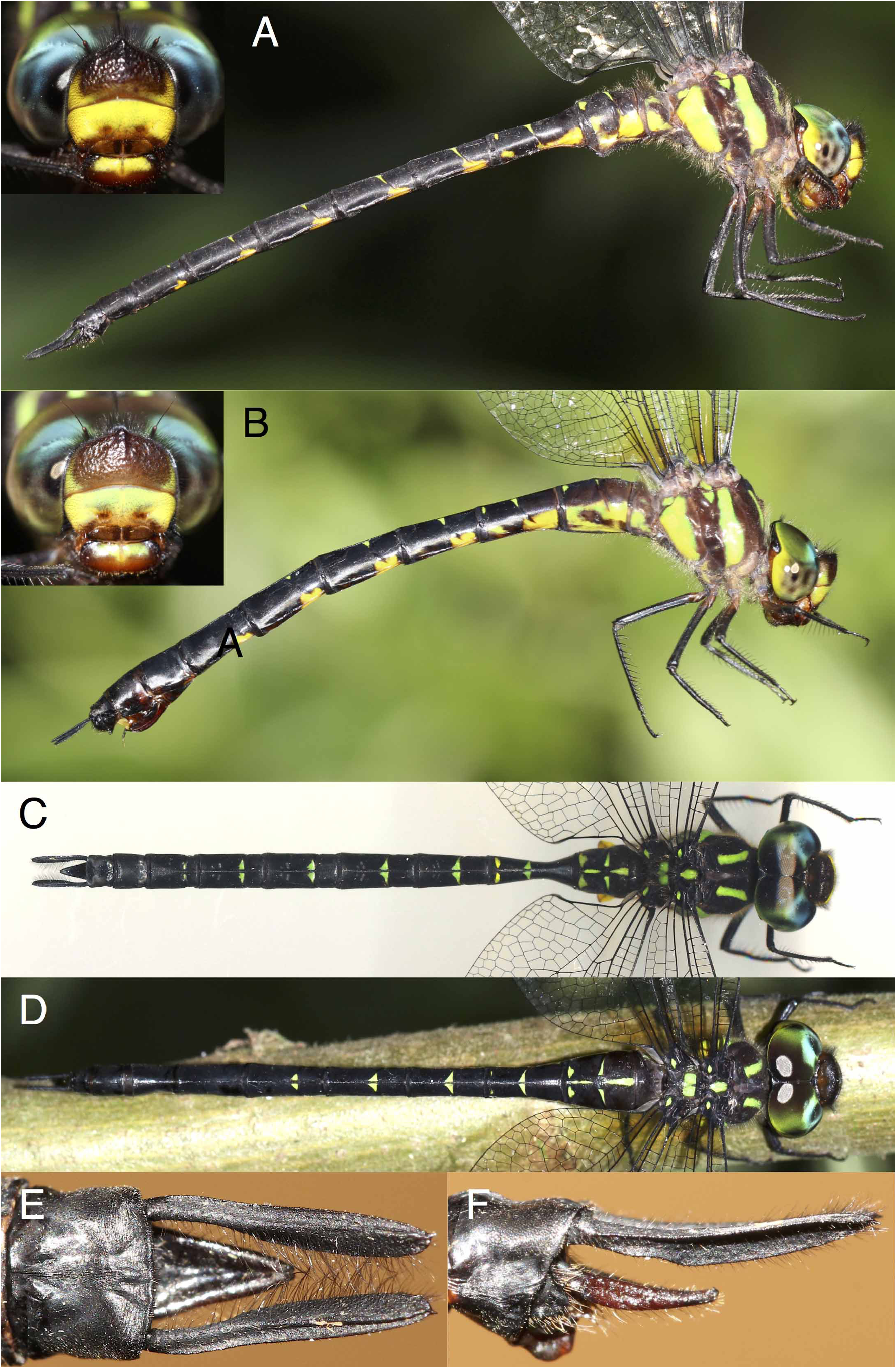
3. Planaeschna tsuchi sp. nov.
( Fig. 5 View FIGURE 5 )
Holotype. ♂, Xuan Son NP, Phu Tho Prov. ( N 21.139, E 104.938), 22-X-2016, TK leg. GoogleMaps
Paratypes. 1 ♂, same location and collector as holotype, 26-X-2014 ; 1 ♂, same location, 13-X-2015, H. Karube leg. ; 2 ♀♀, same location and collector as holotype, 15-X-2015 ; 1 ♀ (specimen used for description), same location and collector, 24-X-2015 .
Etymology. The species takes its name from the pattern on the dorsum of S2, which is reminiscent of the Japanese character for ground “±”, pronounced “tsuchi”. A noun in apposition.
Description of holotype ( Fig. 5C, E–F View FIGURE 5 ).
Head. Labium and base of mandibles orange. Upper half of labrum yellow, lower half orange, anteclypeus orange-brown, darker medially, postclypeus yellow with two orange dimples, and covered in sparse dark hairs. Antefrons yellow along lower margin and at sides, with large brown mark on central part, covered in dark hairs. Postfrons blackish brown, covered in denser dark hairs. Antennae blackish brown. Vertex and occiput black. Compound eyes green with some bluish and grey tones, yellow at lower posterior margin.
Thorax. Prothorax dark brown, somewhat paler at the sides. Pterothorax brown with greenish yellow markings as follows: two green dorsal stripes on the mesepisternum (but lacking markings on the antealar sinus), mesepimeron dark brown along humeral suture, but largely covered by greenish yellow broad lateral stripe. Mesinfraepisternum pale brown. Metepisternum with small greenish yellow triangle near base of wing. Metepimeron greenish yellow, except for brown adjacent to metapleural suture. Metinfraepisternum with posterior fourth yellow. Venter brownish. Trochanters brown, but hind pair with some yellow posteriorly. Legs including spikes and claws blackish brown, but base of fore and middle femora orange-brown on the outer surface and femora of fore legs yellow for proximal half on the inside.
Wings hyaline, veins black. Nodal index: 23–26:29–22/24–20:21–24; Pt blackish brown covering 3–4 underlying cells in FW and HW. Triangle 6-celled in FW, 4–5 celled in HW. Anal triangle 3-celled. Anal loop 12–14 celled.
Abdomen ( Fig. 5C View FIGURE 5 ) blackish brown with markings as follows: S1 laterally with lower two-thirds yellow, invaded from the anterior margin by a large brown mark. S2 laterally lower half yellow, with yellow auricle and blackish brownish blotch on posterior half. Dorsally with clear green wedge-shaped AD mark, the point facing posteriorly, a green stripe over posterior part of mid-dorsal line, leaving centre and posterior margin free, two MD spots in the form of wedges with apices pointing outward, and two triangular PD markings along the posterior margin, expanding laterally a little more than the MD wedges. S3 with yellow AL mark, two triangular green MD and two oval yellow PD spots, and a small greenish yellow PL spot. S4–8 with green MD spots progressively reduced until tiny on S8, and laterally with a yellow AL spot. S9–10 black.
Secondary genitalia apical segment of vesica spermalis of usual type with two somewhat triangular spheres, but not much separated by apical cleft, and with ears at base.
Anal appendages ( Fig. 5E–F View FIGURE 5 ) black. Cerci in dorsal view with apical two-thirds clearly paddle-shaped and with prominent dorsal carina, in lateral view slightly upcurving, apically pointed and with a slight basal swelling. Epiproct extending just beyond midpoint of cerci, in lateral view slender at base, quickly thickening towards middle and tapering again, apex dorsally with two prominences.
Measurements (in mm). HW 46.0; abdomen incl. appendages 54.0.
Variation in male paratype ( Fig. 5A View FIGURE 5 ). Virtually identical to holotype. Dorsal stripes on pterothorax slightly wider. Yellow PD spots on S2 smaller, but MD spots on S8 slightly more prominent. Ax in FW 26–30 and in HW 19–23, Px in FW 22–23 and in HW 23–26. Anal loop 10–13 celled. Triangles 6–7 celled in FW, 5–6 celled in HW. Pt covering 3–3.5 cells .
Measurements (in mm). HW 47.3–48.1; abdomen incl. appendages 55.4–57.8.
Description of female paratype ( Fig. 5D View FIGURE 5 ). As holotype male, unless specified.
Head. Yellow areas of labrum, postclypeus and antefrons with greenish wash. Brown central mark on antefrons extending to lower margin.
Wings amber at base, extending to the level of the third Ax. Wings with somewhat smoky-brown wash. Nodal index 21–30:28–20/23–19:18–23, triangle in FW 6-celled and in HW 4–5 celled, anal loop 11–12 celled, Pt covering 3–4 cells.
Abdomen. S1 with only a triangular yellow PL spot. S2 laterally with lower half yellowish green area invaded by a broad dark brown stripe extending from the anteroventral corner obliquely upward. Dorsal pattern of S2 with somewhat longer MD wedges and shorter PD triangles. S3 with hardly discernible PD spots. S4 with greenishyellow PL spot narrowly connected to yellow kidney-shaped AL mark, S5–6 with kidney-shaped AL mark and small greenish-yellow PL spot. S7–8 lacking MD spots.
Anal appendages. Cerci black and as long as S8 (shorter than S9–10), ovipositor blackish brown.
Variation in female paratypes ( Fig. 5B View FIGURE 5 ). Little variation, dorsal pattern on S2 with MD wedges reduced in both individuals. Amber in wing extending as far as arculus in one specimen, brownish wash absent from wings in the other. Ax in FW 28–30 and in HW 19–23, Px in FW 19–23 and in HW 21–23. Anal loop 10–12 celled. Triangles 5–7 celled in FW and 4–6 celled in HW. Pt covering 2.5–3.5 cells .
Measurements (in mm). HW 47.0; abdomen incl. appendages 53.0–56.5.
Differential Diagnosis. Both the combination of the pattern on S2 and lack of a longitudinal line over S3 and the robust paddle-shaped cerci are unique among the other known Vietnamese Planaeschna species. Together with the long epiproct of the male, extending beyond the middle of the cerci, and the cerci of the female being shorter than S9+S10, this is enough the conclusively identify the species.
Closest is P. nankunshanensis Zhang, Yeh & Tong, 2010 of Guangdong Province in China, which has similar maculation and of which the male likewise has robust paddle-shaped cerci. The male of P. nankunshanensis can be distinguished by the curved outer edge of the cerci, which have a less prominent dorsal carina, and the much shorter epiproct, and the female by details of maculation of S2 and particularly the very long cerci, as long as S9 and S10 combined ( Zhang 2019: p. 197, pp. 211–212).
Ecology. All records are from the same well-vegetated shallow side stream under heavy forest cover springing from karst rocks at Xuan Son NP of several hundred meters at appr. 380 m asl. It shares this habitat with at least five other Planaeschna species. Males were caught in the afternoon hovering over small open areas of water amongst the rocks, whereas females were observed either over the stream or hanging up in nearby bushes. Although the area was visited from spring to late autumn in consecutive years, it was only encountered in October.
Distribution. Endemic to Vietnam ( Phu Tho Prov.)
| PD |
Dutch Plant Protection Service, Culture Collection of Plant Pathogenic Bacteria |
| MD |
Museum Donaueschingen |
No known copyright restrictions apply. See Agosti, D., Egloff, W., 2009. Taxonomic information exchange and copyright: the Plazi approach. BMC Research Notes 2009, 2:53 for further explanation.
|
Kingdom |
|
|
Phylum |
|
|
Class |
|
|
Order |
|
|
Family |
|
|
Genus |