
Diameza ( Miniseraphs ) isabella (Bernay, 1889)
|
publication ID |
https://doi.org/10.5252/g2010n3a4 |
|
persistent identifier |
https://treatment.plazi.org/id/03A69848-FFB6-1E7A-FF36-FCACFDA2FCB8 |
|
treatment provided by |
Marcus |
|
scientific name |
Diameza ( Miniseraphs ) isabella |
| status |
|
Diameza ( Miniseraphs) isabella (Bernay in Deshayes, 1865) ( Figs 20 View FIG K-O; 23)
Terebellum isabella Bernay View in CoL in Deshayes, 1865: 470, 471, pl. 92, figs 14-16.
Terebellum isabella View in CoL – Bosatta et al. 1973: 189.
Terebellum ( Seraphs) isabellae – Cossmann 1889: 98. — Oppenheim 1896: 195 partim. — Cossmann 1904: 46. — Cossmann & Pissarro 1911: pl. 32, fig. 158-6.
Diameza ( Miniseraphs) isabella – Jung 1974: 30, 31, pl. 9, figs 13-18, text-figs 20, 21. — Le Renard 1992: 6. — Le Renard & Pacaud 1995: 112. — Pacaud & Le Renard 1995: 162.
TYPE LOCALITY. — Chaussy, Les Garennes (Val d’Oise, France), Lutetian (Middle Eocene).
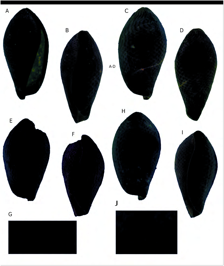
TYPE MATERIAL. — Lectotype designated by Jung (1974) from the Bernay collection, and now deposited at UCBL, not found; paralectotype ( UCBL, Deshayes coll.), not found. Accordingly, a neotype designation is needed to clarify the taxonomic status of this species. A neotype from the type locality ( MNHN A28935, leg. Pacaud, Fig. 23 View FIG A-D) is chosen here. Th is initiative conforms to the rules specified by the ICZN (1999: art. 75), as regards the designation of a neotype.
OTHER MATERIAL EXAMINED. — See Appendix 1.
DESCRIPTION
Shell
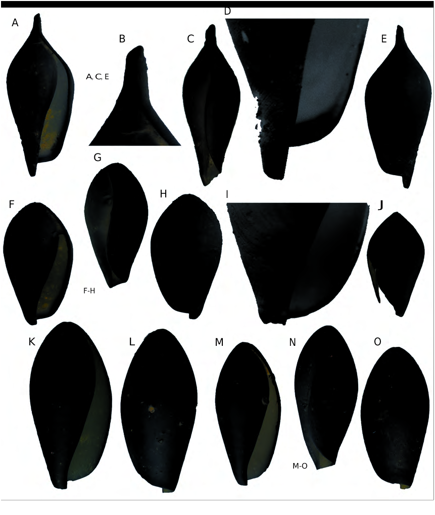
Small shell up to 16 mm in height and up to 8 mm in diameter, inflated with wide apical angle. Aperture long and narrow. Callus of the inner lip clearly delineated, but weakly developed. Columella slightly bent backwards near the base. Outer lip thickened and extending to the apex ( Fig. 20K View FIG ). Outer lip prosocyrt on the abapical part and opisthocyrt on the adapical part in labral view ( Fig. 20N View FIG ). Siphonal notch moderately deep. Oblique grooves near the base of the shell.
Colour pattern
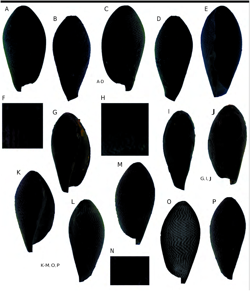
The colour pattern of the species is composed of 2 levels of pigmentation: dark axial wavy lines on a lighter background. Th e axial lines form some zigzags ( Fig. 23 View FIG ). The zigzags cover mainly the median part of the shell. On the base and the apex, the lines remain straight or slightly sinuous, showing very weak curves ( Fig. 23 View FIG I-M, O, P). Th e width of the lines is almost homogenous. Th e space between them is equivalent to their width.
VARIABILITY
The specimens bear zigzags of greater ( Fig. 23D View FIG ) or lesser amplitude ( Fig. 23I View FIG ). Some specimens bear a gradual transition from wavy lines to a zigzag pattern ( Fig. 23H, I View FIG ), while others show an abrupt transition, probably due to a growth interruption ( Fig. 23F, G View FIG ). Wavy axial lines crossing and forming a localized grid, called meshwork ( Fig. 23M, N View FIG ) as defined by Meinhardt (1998: fig. 5.3c, d), have been observed too.
COMPARISONS OF THE THREE SPECIES OF DIAMEZA Shell
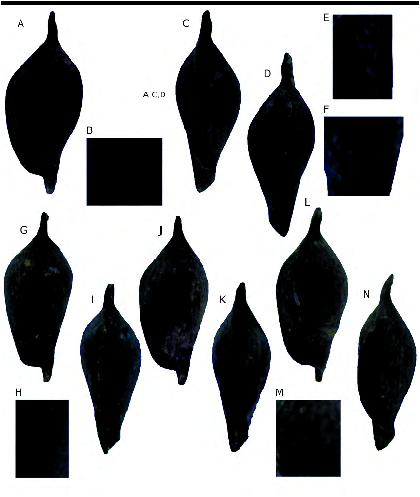
Diameza View in CoL ( s.s.) fragilis displays a distinctive feature, the acute apical structure ( Fig. 20B View FIG ). According to Jung (1974), this feature is used to distinguish Diameza View in CoL from Miniseraphs View in CoL . Diameza View in CoL ( s.s.) fragilis is however closely related to D. ( Miniseraphs View in CoL ) in sharing a small and inflated shell, a wide apical angle and a sinuous outer lip ( Fig. 20 View FIG ). Diameza ( Miniseraphs) isabella is very similar to D. ( M.) eratoides . Diameza ( M.) isabella is more slender ( Fig. 20F, K View FIG ) and the adult specimens are twice as large as those of D. ( M.) eratoides ( Jung 1974) . Furthermore, D. ( M.) eratoides shows a small protuberance ( Fig. 20J View FIG , rarely preserved) on the apex, missing in D. ( M.) isabella .
Residual colour pattern
All species share a pattern composed of two levels of pigmentation. Th e colour pattern of Diameza ( s.s.) fragilis ( Fig. 21 View FIG ) is closely related to that of D. ( Miniseraphs) eratoides ( Fig. 22 View FIG ). It shares with D. ( M.) eratoides a combination of two morphologies:1) dark wavy lines visible on the extremities of the shell and near the outer lip; and 2) a chessboard pattern on the median part of the shell. Nevertheless, some shells of Diameza ( s.s.) fragilis bear a residual pattern only composed of axial lines ( Fig. 21 View FIG J-K), lacking in D. ( Miniseraphs) eratoides . Diameza ( M.) eratoides differs from D. ( M.) isabella by a chessboard pattern covering a large part of the shell. Moreover, the amplitude of the wave of each line is lower than that observed on Diameza ( Miniseraphs) isabella ( Fig. 23 View FIG ).
No known copyright restrictions apply. See Agosti, D., Egloff, W., 2009. Taxonomic information exchange and copyright: the Plazi approach. BMC Research Notes 2009, 2:53 for further explanation.
|
Kingdom |
|
|
Phylum |
|
|
Class |
|
|
Order |
|
|
Family |
|
|
Genus |
Diameza ( Miniseraphs ) isabella
| Caze, Bruno, Merle, Didier, Pacaud, Jean-Michel & Saint Martin, Jean-Paul 2010 |
Diameza ( Miniseraphs ) isabella
| LE RENARD J. & PACAUD J. - M. 1995: 112 |
| LE RENARD J. & PACAUD J. - M. 1995: 162 |
| LE RENARD J. 1992: 6 |
| JUNG P. 1974: 30 |
Terebellum isabella
| BOSATTA G. & FERRERO M. & PICCOLI G. 1973: 189 |
Terebellum ( Seraphs ) isabellae
| COSSMANN M. 1904: 46 |
| OPPENHEIM P. 1896: 195 |
| COSSMANN M. 1889: 98 |
Terebellum isabella
| DESHAYES G. - P. 1865: 470 |