
Culeolus recumbens Herdman 1882
|
publication ID |
https://doi.org/ 10.11646/zootaxa.4996.3.2 |
|
publication LSID |
lsid:zoobank.org:pub:A1982CE0-AD2F-496B-80AB-FB3C4FA69F7A |
|
DOI |
https://doi.org/10.5281/zenodo.5074707 |
|
persistent identifier |
https://treatment.plazi.org/id/03A6FD6C-FF8F-E82D-55E3-379BFE7AFE61 |
|
treatment provided by |
Plazi |
|
scientific name |
Culeolus recumbens Herdman 1882 |
| status |
|
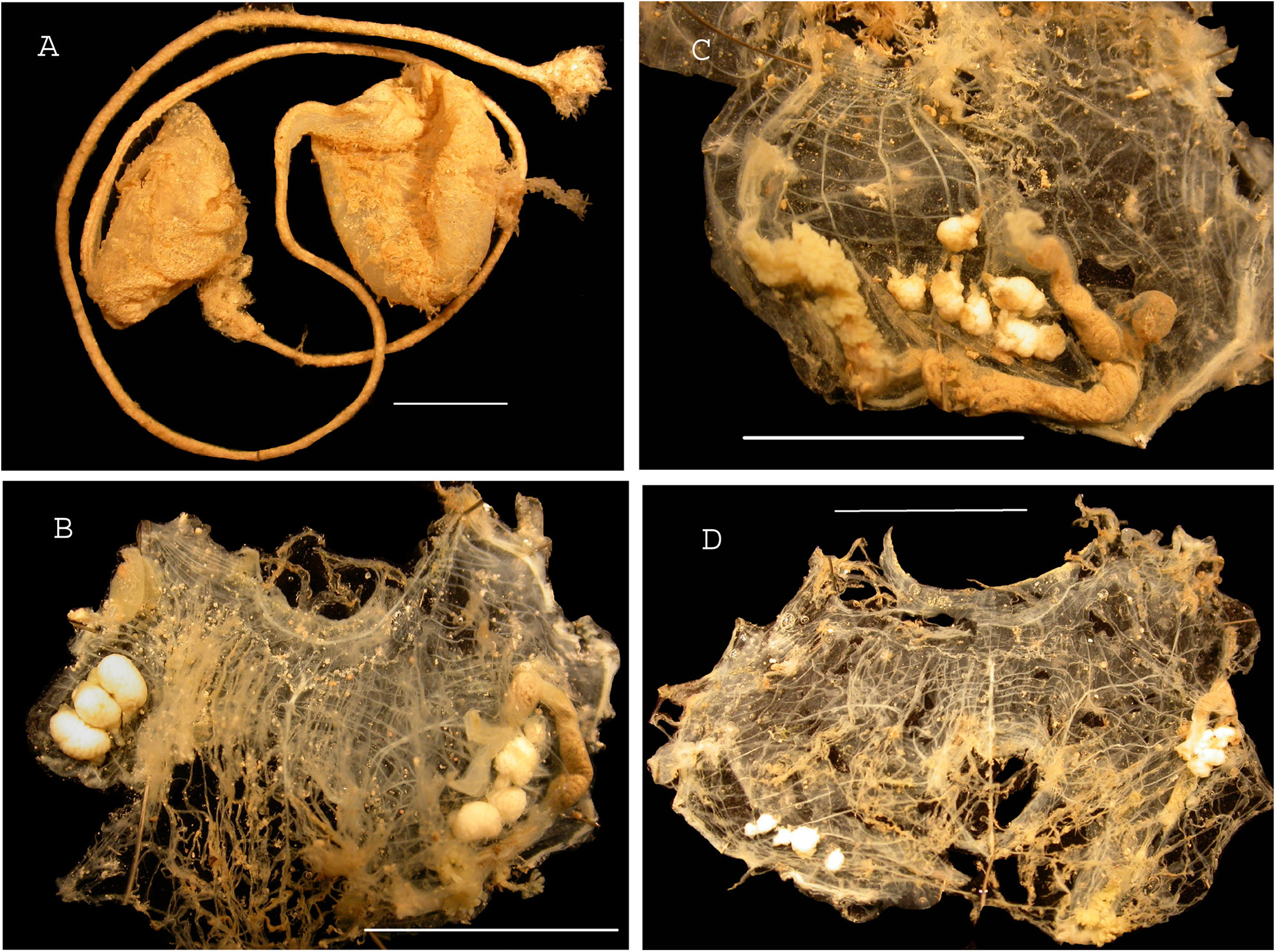
Culeolus recumbens Herdman 1882
Figure 20 View FIGURE 20
Stations: Kanadeep CP 5062; cp 5087; cp 5092; cp 5093. Eight specimens.
Culeolus recumbens Herdman 1882 : South West Indian Ocean; Monniot & Monniot 1982: Antarctic; Monniot & Monniot 199: New Caledonia; Monniot & López-Legentil 2017: Papua New Guinea; Sanamyan & Sanamyan 1999: North New Zealand.
All specimens have the same shape with a triangular body held on a long stiff peduncle ending in a tuft of filaments ( Fig. 20A View FIGURE 20 ). They are different sizes; the largest has a 3cm head and a peduncle 15cm long. The tunic is thin and soft on the head, spotted with button-like papillae with some adhering sand. A half ring of larger papillae encircles ventrally the anterior part of the body at some distance from the atrial aperture. The peduncle is flexible but harder, totally encrusted inserted at the side of the oral aperture. The body wall is thin and transparent with well spaced muscular bundles extending from the siphons and crossing the body sides ( Fig. 20B–D View FIGURE 20 ). All specimens are damaged with the branchial sac and often the gonads and gut ejected through the atrial siphon. The oral tentacles are particularly long overlapping within the oral aperture. The dorsal tubercle opens in a slit above an elongated neural ganglion. The prepharyngeal band curves in a deep dorsal V. The dorsal lamina has large flat languets.
It is difficult to determine if the branchial longitudinal vessels form three or four low folds on each side; in a specimen a tentative formula on the left side is:
DL – 1 (4) 2 (4) 3 (3) 6 E
The digestive loop is long and opened ( Fig. 20B, C View FIGURE 20 ). The narrow stomach is covered by a digestive gland divided into several papillated lobes. The anus has numerous lobes. The gonads ( Fig. 20B–D View FIGURE 20 ). are variable in number on each side according to the specimens, they can be from three to nine on the right side and from four to six on the left inside the gut loop. Their shape is uncommon for the genus they are similar to polycarps of the genus Polycarpa : testis and ovary are joined in a round gland with short joined ducts ( Fig. 20B, C View FIGURE 20 ). One large endocarp is located on the body wall anterior to the gonads and another endocarp lies at the top of the intestine loop. No other endocarps were found. No spicules where seen in the tissues.
Culeolus recumbens differs from other species of Culeolus present in New Caledonia by having several gonads on each side in the shape of polycarps. The external body shape may lead to confusion with other sandy pedunculate species of the same region.
No known copyright restrictions apply. See Agosti, D., Egloff, W., 2009. Taxonomic information exchange and copyright: the Plazi approach. BMC Research Notes 2009, 2:53 for further explanation.
|
Kingdom |
|
|
Phylum |
|
|
Class |
|
|
Order |
|
|
Family |
|
|
Genus |
Culeolus recumbens Herdman 1882
| Monniot, Francoise 2021 |
Culeolus recumbens
| Herdman 1882 |
Culeolus recumbens
| Herdman 1882 |