
Afroeudesis Franz
|
publication ID |
https://doi.org/ 10.11646/zootaxa.4048.4.2 |
|
publication LSID |
lsid:zoobank.org:pub:7D3B40E7-1AF7-4AA9-8416-6035840E8FEC |
|
DOI |
https://doi.org/10.5281/zenodo.6095559 |
|
persistent identifier |
https://treatment.plazi.org/id/03A72D51-FB3A-CB42-FF79-1BA7FCB1FDF2 |
|
treatment provided by |
Plazi |
|
scientific name |
Afroeudesis Franz |
| status |
|
Afroeudesis Franz View in CoL , status n.
Afroeudesis Franz, 1963: 16 View in CoL (as subgenus of Pseudoeudesis View in CoL ). Type species: Pseudoeudesis basilewskyi Franz, 1963 (des. orig.).
Revised diagnosis. Head in dorsal view subtriangular, with eyes located behind middle; median dorsal area on head elevated and demarcated laterally by ridges, forming elongate subtriangular field with median longitudinal impression; occipital constriction only slightly narrower than vertex; submentum lacking lateral sutures; pronotum broadest in front of middle, with transverse antebasal groove connecting one pair of lateral pits; basisternal part of prosternum distinct, not rudimentary; notosternal sutures complete; procoxal sockets broadly closed and distant from lateral margins of prosternum; prosternal process indistinctly carinate; hypomeral ridges absent; mesoventral intercoxal process subtriangular, broadest at base and narrowing caudad where it meets distinct anterior metaventral process; metaventrite with lateral longitudinal carinae; metaventral intercoxal process narrowly separating metacoxae, with two long spines; each elytron with two rudimentary asetose basal foveae; aedeagus with slender parameres.
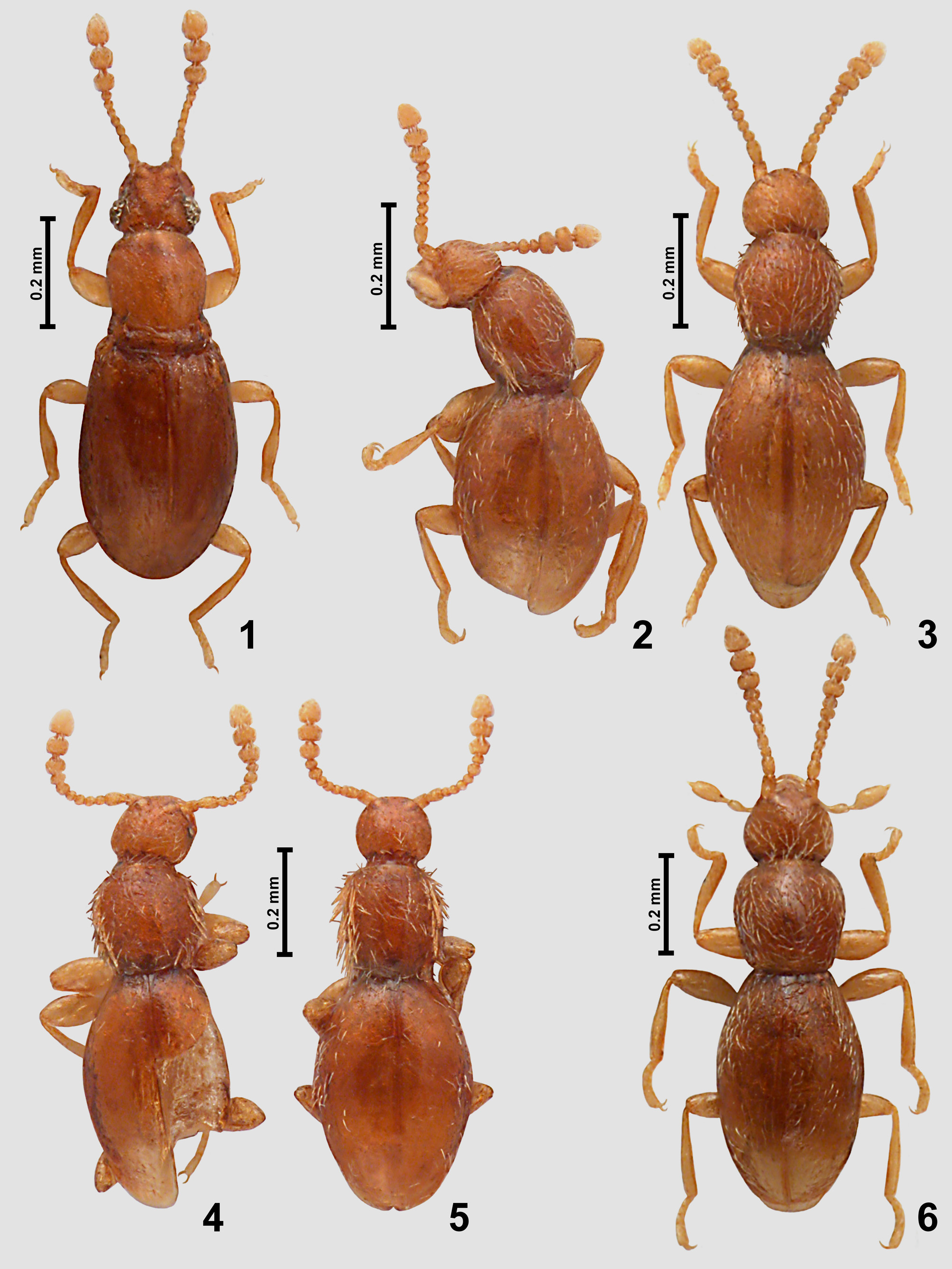
Redescription. Body ( Fig. 1 View FIGURES 1 – 6 ) strongly convex, elongate and moderately slender, strongly constricted between head and pronotum but weakly so between pronotum and elytra; appendages moderately long, pigmentation light brown, cuticle sparsely setose.
Head ( Figs 1 View FIGURES 1 – 6 , 7–8 View FIGURES 7 – 10 ) with occipital constriction ( Fig. 7 View FIGURES 7 – 10 ; occ) only slightly narrower than vertex and dividing head capsule into exposed subtriagular anterior part and posterior 'neck' region ( Fig. 7 View FIGURES 7 – 10 ; nr) retracted into prothorax; eyes large in male ( Fig. 1 View FIGURES 1 – 6 ) and rudimentary in female ( Figs 7–8 View FIGURES 7 – 10 ) located behind middle; tempora ( Fig. 7 View FIGURES 7 – 10 ; tm) short but distinct in male and long in female, lacking bristles; vertex broader than long, with convex sides, not bulging dorsocaudad; frons subtrapezoidal; median area on frons and vertex forming subtriangular elevated field demarcated laterally by ridges and bearing median longitudinal impression broadest between weakly raised supraantennal tubercles and narrowing posteriorly; frontoclypeal groove absent; antennal insertions narrowly separated; sides of clypeus at base with lateral angulate expansion.
Labrum ( Fig. 7 View FIGURES 7 – 10 ; lb) transverse. Mandibles ( Figs 7–8 View FIGURES 7 – 10 ; md) symmetrical, subtriangular, each lacking subapical tooth ( Afroeudesis s. str.) or with one robust tooth (Nanoscydmus). Each maxilla ( Fig. 8 View FIGURES 7 – 10 ) with subtriangular basistipes ( Fig. 8 View FIGURES 7 – 10 ; bst) and mediostipes ( Fig. 8 View FIGURES 7 – 10 ; mst), long palpifer ( Fig. 8 View FIGURES 7 – 10 ; ppf), elongate galea ( Fig. 8 View FIGURES 7 – 10 ; gal) and lacinia ( Fig. 8 View FIGURES 7 – 10 ; lac) and strongly elongate maxillary palp ( Fig. 8 View FIGURES 7 – 10 ; mxp) composed of elongate palpomere I, strongly elongate, pedunculate and slender palpomere II, large and strongly elongate palpomere III broadest distally to middle, and small, elongate, subconical and pointed palpomere IV. Labium with broad submentum ( Fig. 8 View FIGURES 7 – 10 ; smn) lacking lateral sutures; subrectangular mentum ( Fig. 8 View FIGURES 7 – 10 ; mn); and short prementum bearing long labial palps. Hypostomal ridges ( Fig. 8 View FIGURES 7 – 10 ; hr) long, recurved and extending up to posterior tentorial pits ( Afroeudesis s. str.) or curved and incomplete (Nanoscydmus).
Gular plate ( Fig. 8 View FIGURES 7 – 10 ; gp) large and subtrapezoidal; gular sutures ( Fig. 8 View FIGURES 7 – 10 ; gs) superficial; posterior tentorial pits ( Fig. 8 View FIGURES 7 – 10 ; ptp) narrow and straight.
Antennae ( Fig. 1 View FIGURES 1 – 6 ) slender, with three antennomeres forming distinct club.
Prothorax ( Figs 1 View FIGURES 1 – 6 , 9 View FIGURES 7 – 10 ) in dorsal view bell-shaped, broadest anterior to middle, with arcuate anterior margin; anterior corners weakly marked, obtuse-angled; sides rounded but with distinct posterior constriction; posterior corners obtuse-angled and rounded; posterior margin arcuate. Pronotum with distinct and long transverse antebasal groove connecting one pair of large lateral pits located close to lateral pronotal margins. Sides of pronotum lacking thick bristles.
Prosternum ( Fig. 9 View FIGURES 7 – 10 ) with basisternal part ( Fig. 9 View FIGURES 7 – 10 ; bst) long and indistinctly demarcated from procoxal cavities ( Fig. 9 View FIGURES 7 – 10 ; pcc); prosternal process ( Fig. 9 View FIGURES 7 – 10 ; psp) indistinctly carinate, narrow; procoxal sockets ( Fig. 9 View FIGURES 7 – 10 ; pcs) closed and distant from lateral sternal margins; hypomera ( Fig. 9 View FIGURES 7 – 10 ; hy) elongate and laterally confluent with pronotum, lacking hypomeral ridges; notosternal sutures ( Fig. 9 View FIGURES 7 – 10 ; nss) complete.
Mesoscutellum (not shown) visible between elytral bases in intact specimens but very small, with oval posterior part, anterior constriction and straight anterior margin; scutoscutellar suture present.
Mesoventrite ( Fig. 10 View FIGURES 7 – 10 ) lacking demarcated anterior ridge, in anterior region with pair of asetose impressions separated at middle; mesoventral intercoxal process ( Fig. 10 View FIGURES 7 – 10 ; msvp) subtriangular, broadest anteriorly and narrowing posteriorly, flattened but with median longitudinal elevation visible at least in its anterior portion; mesanepisternum with short prepectus ( Fig. 10 View FIGURES 7 – 10 ; pre); mesoventrite with very deep ventrolateral fovea at each side ( Fig. 10 View FIGURES 7 – 10 ; vlf); mesocoxal sockets concealed in ventral view ( Afroeudesis s. str.) or exposed (Nanoscydmus).
Metaventrite ( Fig. 10 View FIGURES 7 – 10 ; v3) subtrapezoidal, anteriorly fused with mesoventrite, posteriorly bisinuate and with narrow subtrapezoidal ( Afroeudesis ) or broadly subtriangular (Nanoscydmus) anterior metaventral process ( Fig. 10 View FIGURES 7 – 10 ; amvp); metaventral intercoxal process ( Fig. 10 View FIGURES 7 – 10 ; mtvp) narrowly separating metacoxae, with two robust spines. Metanepisterna and metepimera narrow.
Metafurca ( Fig. 10 View FIGURES 7 – 10 ) with elongate stalk and divergent lateral furcal arms ( Fig. 10 View FIGURES 7 – 10 ; lmfa).
Elytra ( Fig. 1 View FIGURES 1 – 6 ) oval, each with two rudimentary asetose basal foveae, in Afroeudesis s. str. hardly noticeable, especially subhumeral fovea, in Nanoscydmus very small but both distinct; humeral calli distinct and developed as longitudinal protuberances; subhumeral lines indistinct, basal impressions absent.
Hind wings in males of Afroeudesis s. str. and Nanoscydmus well-developed, in females of Afroeudesis s. str. absent, in females of Nanoscydmus not described.
Legs ( Figs 1 View FIGURES 1 – 6 , 9, 10 View FIGURES 7 – 10 ) moderately long and slender; trochanters broad, femora gradually clavate, tibiae broadening distally; tarsi short and robust.
Aedeagus ( Figs 15−16 View FIGURES 15 – 22 ) thin-walled, elongate, with symmetrical median lobe but asymmetrical endophallus; parameres free and slender, each with one apical seta.
Remarks. The discovery that Afroeudesis from Tanzania and Nanoscydmus from the Himalayas and Yunnan were congeneric raises a question of possible mislabeling, i.e., specimens collected in Nepal might have been erroneously labeled as being of the African origin. However, Afroeudesis was based on specimens collected in 1957, and published in 1963. First Himalayan species described by Franz were published in two papers, both appeared in 1970. The first one ( Franz 1970a) was based on a single specimen collected in 1962 and sent to Franz by H. Freude; in the other paper ( Franz 1970b) a few new species collected by H. Janetschek in 1961 were dealt with. Further Franz's papers focused on the Himalayan Scydmaeninae were based on specimens collected in 1970 and later. It seems that before 1970 Franz had not been working on any large Himalayan material, and therefore a mislabeling can be ruled out as unlikely.
As mentioned previously, Afroeudesis s. str. and Afroeudesis (Nanoscydmus) differ in the following key characters:
- Afroeudesis s. str.: hypostomal ridges complete, recurved and extending posteromesally up to posterior tentorial pits, their posterior portions nearly parallel; the anterior metaventral process narrow, its lateral margins only slightly divergent posteriorly; the mesocoxal sockets concealed in ventral view; the mesoscutellum has a straight anterior margin, sub-basal constriction and broadened oval posterior portion with lateral and posterior margins broadly rounded;
- Afroeudesis (Nanoscydmus) : hypostomal ridges short and incomplete, lacking posterior subparallel portions and not reaching posterior tentorial pits; the anterior metaventral process much broader than long, subtriangular, with lateral margins strongly divergent and extending obliquely toward lateral metaventral margins; the mesocoxal sockets exposed in ventral view; the mesoscutellum subtriangular.


No known copyright restrictions apply. See Agosti, D., Egloff, W., 2009. Taxonomic information exchange and copyright: the Plazi approach. BMC Research Notes 2009, 2:53 for further explanation.
|
Kingdom |
|
|
Phylum |
|
|
Class |
|
|
Order |
|
|
Family |
Afroeudesis Franz
| Jałoszyński, Paweł 2015 |
Afroeudesis
| Franz 1963: 16 |