
Prionacalus atys White, 1850
|
publication ID |
https://doi.org/10.1649/0010-065x-67.3.201 |
|
persistent identifier |
https://treatment.plazi.org/id/03A787D5-FFBC-DB06-0835-AF6FF0B9FA0E |
|
treatment provided by |
Carolina |
|
scientific name |
Prionacalus atys White, 1850 |
| status |
|
Prionacalus atys White, 1850 View in CoL
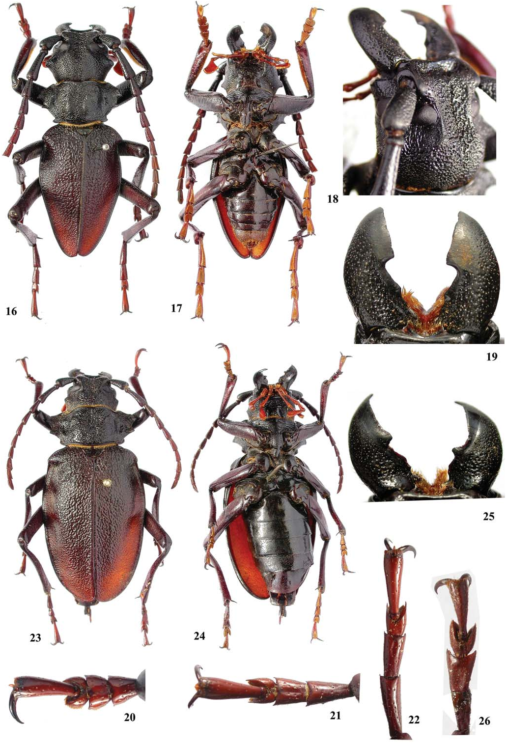
( Figs. 8 View Figs , 16–26 View Figs , 103–105 View Figs , 121–123 View Figs )
Prionacalus Atys White 1850: 11 View in CoL , pl. 13, fig. 4; 1851: 70, 71, 1853: 8; Gemminger and Harold 1872: 2754 (catalogue).
Prionacalus atys View in CoL ; Waterhouse 1880: 485; Monné and Giesbert 1994: 15 (checklist); Monné 1995: 60 (catalogue), 2006: 82 (catalogue), 2012: 7 (checklist); Di Iorio 2003: 3 (distribution); Monné and Hovore 2005: 19 (checklist), 2006: 18 (checklist); Jeniš 2010: 19 ( syntypes male and female), 118 (male), 119 (female).
Prionocalus Atys; Lacordaire 1868: 42 (note); Waterhouse 1872: 261 (key); Thomson 1877: 260 (key).
Psalidognathus ( Prionocalus) Atys ; Lameere 1910: 377, 1913: 66 (catalogue), 1919: 121.
Psalidognathus ( Prionocalus) atys View in CoL ; Blackwelder 1946: 555 (checklist); Duffy 1960: 15, 69 (larva).
Prionocalus atys View in CoL ; Quentin and Villiers 1983: 225 (key); Hüdepohl 1985: 121; Komiya 2001: 32 (key).
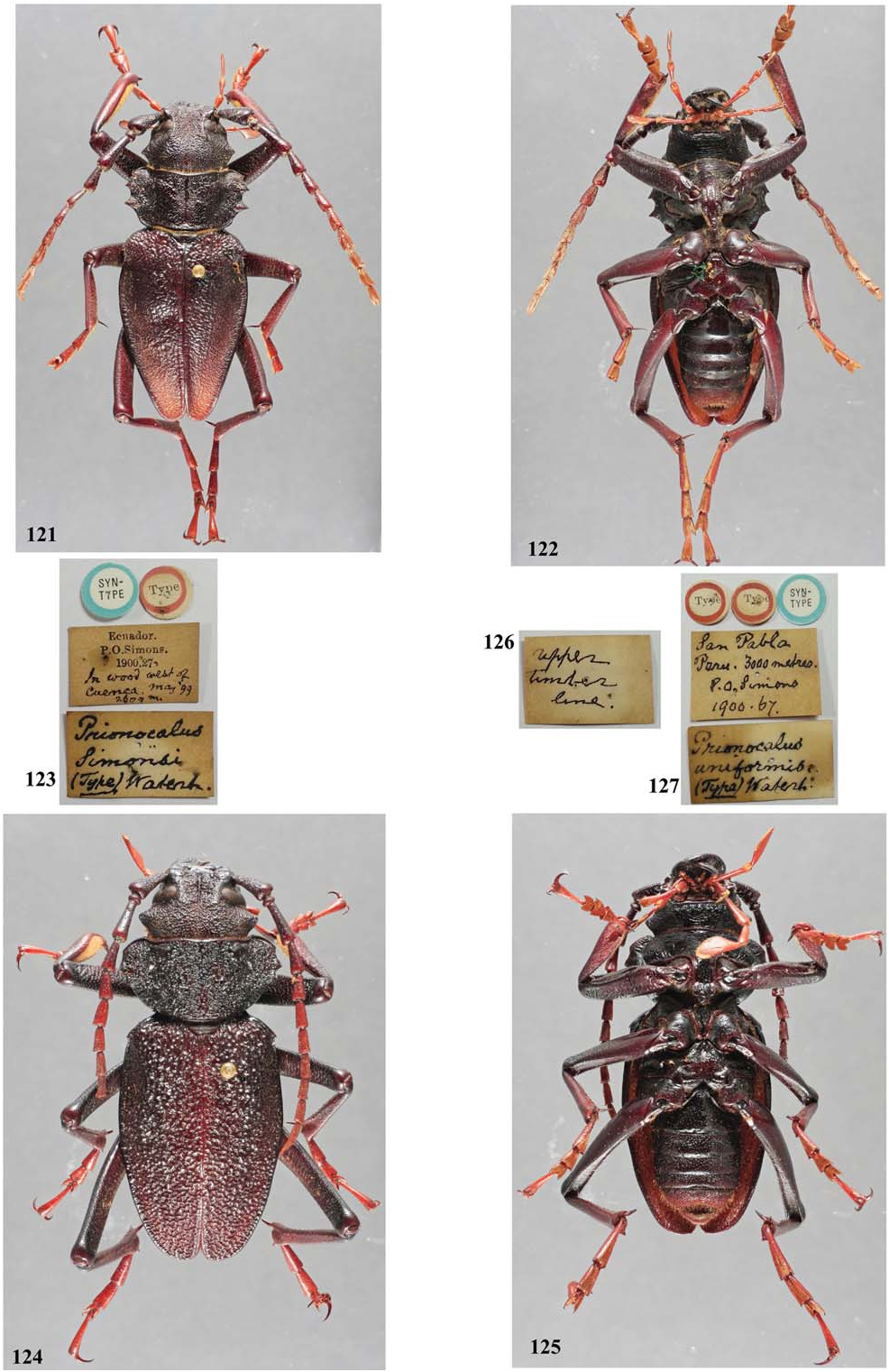
Prionocalus Simonsi Waterhouse 1900: 504 . New synonymy.
Psalidognathus ( Prionocalus) Simonsi ; Lameere 1910: 378, 383 (key), 1913: 66 (cat.), 1919: 121.
Psalidognathus ( Prionocalus) simonsi View in CoL ; Blackwelder 1946: 555 (checklist).
Prionocalus simonsi View in CoL ; Quentin and Villiers 1983: 225 (key); Hüdepohl 1985: 121 (key).
Prionacalus simonsi View in CoL ; Monné and Giesbert 1994: 15 (checklist); Monné 1995: 61 (catalogue), 2006: 83 (catalogue); Di Iorio 2003: 3 (distribution); Monné and Hovore 2005: 19 (checklist), 2006: 18 (checklist); Jeniš 2008: 95 (male), 2010: 18 (males and females), 120 (male), 121 (female), 145 ( syntype male).
Redescription. Male ( Fig. 16 View Figs ). Head and mandibles blackish (occasionally, head dark brown); maxillary and labial palpi reddish; antennae gradually lighter towards apex, usually scape, pedicel, and basal 2/3 of antennomere III blackish, distal 1/3 of antennomere III, and basal 2/3 of IV and V dark brown, distal 1/3 of antennomeres IV and V reddish, antennomeres VI-XI reddish; pronotum blackish (sometimes dark brown); ventral surface of thorax dark brown (usually, prosternum almost blackish); elytra dark brown on basal 1/3, gradually reddish towards apex; ventrites I-IV dark brown, usually distal margin blackish, ventrite V darkbrown on base, reddish towards apex (occasionally entirely reddish); profemora dark brown; meso- and metafemora reddish brown, with some parts dark brown; protibiae dark brown on basal 1/3, gradually reddish towards apex; meso- and metatibiae dark brown on base (sometimes on basal 1/3) reddish on remaining surface; tarsi reddish, except apex of tarsomere V and claws. Head: Coarsely punctate-vermiculate between cephalic carinae, coarser on 1/2 near clypeus; area between apex of cephalic carinae and pronotum coarsely, abundantly, confluently punctate; area on 1/2 near clypeus with longitudinal, well-marked furrow (occasionally present on area between antennal tubercles); distal margin of vertex (close to abrupt declivity) centrally distinctly emarginate. Antennal tubercle moderately coarsely, sparsely punctate on base (sometimes with confluent punctures), gradually becoming smoother towards apex; distinctly far from each other (distance between them equal to its width, or slightly larger). Cephalic carinae ( Fig. 18 View Figs ) very distinct, elevated from base to apex; convergent from antennal tubercle to little after eyes, then divergent towards apex; apex not or slightly elevated, usually not forming triangular tubercle. Area behind eyes variable: not finely striate, with abundant small asperities, mainly between eyes and dorsal surface of lateral tubercle; finely striate, with abundant asperities only behind lower ocular lobes; finely striate, with sparse asperities only behind lower ocular lobes. Lateral tubercle of head moderately large, rounded ( Fig. 16 View Figs ) to distinctly acute ( Fig. 121 View Figs ) at apex. Subhorizontal area of clypeus finely, moderately sparsely punctate, more distinctly basally. Distal area of clypeus distinctly concave. Labrum distinctly concave. Distance between upper ocular lobes 2.0–2.3 times longest width of each lobe. Gena rounded at apex ( Fig. 18 View Figs ), without downward projection. Submentum laterally shallowly, coarsely, abundantly punctate, centrally finely, abundantly punctate or moderately finely, transversely striate (occasionally entire surface of submentum transversely striate, stria can be moderately coarse). Mandible ( Fig. 19 View Figs ) 0.6–0.7 times head length; inner margin laminar; surface coarsely, abundantly punctate, gradually finer towards apex (smooth near inner margin); left mandible with 2 distinct teeth near base (sometimes small and together protracted); right mandible with 2 small, protracted teeth near base and another large tooth close to base of distal plate; distal plate of left mandible wider than that of right mandible (in one specimen examined, right mandible identical to left). Longest width of last maxillary ( Fig. 8 View Figs ) and labial palpomeres equal to about 0.65 times its length. Antennae not reaching distal 1/3 or 1/4 of elytra; scape distinctly enlarged from base to apex; antennomeres III-IV usually rounded at apex on both sides (sometimes slightly projected at outer distal angle); antennomeres V-X usually distinctly dentate at outer distal angle. Thorax: Lateroanterior angles of prothorax slightly projected forward, rounded (occasionally acute); lateroposterior angles of prothorax from acute and distinctly projected sideward (tooth-like) to slightly or not projected (in both cases, rounded); anterior 2 teeth of lateral margin protracted, with anterior tooth smallest (sometimes absent or almost so); latero-basal tooth large, usually slightly curved upwards. Pronotum with distinct median depression and another smaller depression laterally on each side; coarsely, confluently punctate on disc, laterally with coarser and sparser punctures, but with smooth, shiny areas between punctate areas (general appearance of lateral area much more shiny than central area). Elytra moderately coarsely vermiculate on central subtriangular area (larger area on elytral base) from base to middle (sometimes up to base of distal 1/3); remaining surface distinctly finer sculptured; 3.1–3.4 times longer than prothorax; apex individually rounded. Apical tooth of humeral projection absent or distinct. Abdomen: Not surpassing elytral apex. Ventrites I-III finely, sparsely punctate ( Fig. 17 View Figs ); ventrite IV finely, abundantly punctate; ventrite V coarser, distinctly, more abundantly punctate. Legs: Apex of metafemora surpasses elytral apex by about basal 1/3–2/5. Ventral sulcus of protibiae distinct from near base to distal constriction. Protarsomeres I and II ( Fig. 20 View Figs ) short, moderately wide; protarsomeres I-III with very small denticle at apex; mesotarsi shorter than metatarsi ( Fig. 21 View Figs ); metatarsi ( Fig. 22 View Figs ) not notably slender.
Female ( Fig. 23 View Figs ). Color as in males, with same variations, except abdomen entirely dark brown with apex blackish (in one female the body is almost entirely reddish – probably a teneral specimen). Inner margin of mandibles ( Fig. 25 View Figs ) laminar; inner margin as in males, but teeth smaller. Antennae reaching about apex of basal 1/3 of elytra (sometimes almost mid-elytra). Longest width of last maxillary and labial palpomere about 0.4 times length. Cephalic carinae as in males, but less elevated or almost absent, and often less divergent distally. Distance between upper ocular lobes 2.2–3.1 times longest width of 1 lobe. Lateral tubercle of head small, acute at apex. Prothorax as in male. Elytra 3.4–3.9 times longer than prothorax, elytral sculpture as in male or, rarely, uniform throughout. Abdomen distinctly surpassing elytral apex. All ventrites ( Fig. 24 View Figs ) finely, sparsely punctate. Meso- and metatarsi ( Fig. 26 View Figs ) shorter than in male.
Dimensions. Male/female. Total length (including mandibles) = 42.5–56.3/37.4–71.0 mm; prothoracic length = 7.4–9.4/ 6.5–11.1 mm; prothoracic width between apices of anterior angles = 9.7–12.4/ 8.8–14.4 mm; prothoracic width between apices of posterior angles = 9.5–12.0/8.8–15.0 mm; humeral width = 14.3–19.2/13.0– 25.8 mm; elytral length = 23.7–28.9/22.9–44.0 mm.
Geographical Distribution. E c u a d o r (Waterhouse 1900), Peru (White 1850). ( Mexico is not considered a country where this species occurs).
Material Examined. ECUADOR: (no other data), 1 male, 2 females ( ZMHB) ; male (no other data) ( MPCO). Loja: female (no other data) ( IRSN) . Manabí: Portoviejo , female, (ex Nonfried Collection) ( IRSN) . Azuay: Cuenca , female, I.1971 [no collector indicated] ( MPCO) . PERU: 1 male, 1 female, (ex Desbrochers Collection; no other data) ( IRSN) .
Additionally, we examined specimens with the following label: MEXICO, male, (ex Caulle Collection; no other data) ( IRSN); female, (ex E. A. Klages Collection; no other data) ( EMEC); male (no other data) ( EMEC).
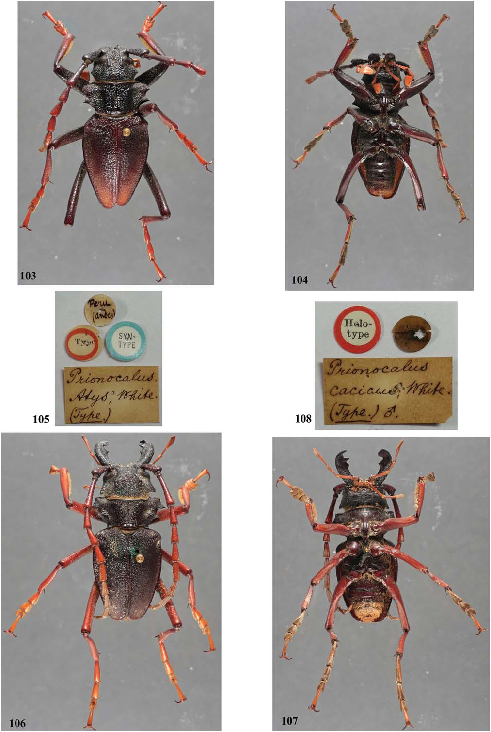
Types and Type Localities. Of Prionacalus atys ( Figs. 103–105 View Figs ): Described from Andes of Peru, apparently, based on a single male, deposited in the BMNH. The descriptions in White (1850, 1851) do not suggest that he had a female of the species. The BMNH website (2013) records one syntype of Prinocalus [ sic] atys . However, Bezark (2013) figured two syntypes in this collection: one male and one female (both photographed by Henry Hespenheide) .
Of Prionocalus [ sic] simonsi ( Figs. 121–123 View Figs ): Described based on males (unknown number) from Ecuador (“in wood west of Cuença [ sic], 2600 m ”). Cuenca is a city in Azuay Province. According to Chubb (1919): “The following notes are based, chiefly, on a collection made by the late Perry O. Simons in the Andean regions of Ecuador, Peru, Bolivia, and Argentina from the latter part of 1898 to November 1901, at varying altitudes up to 5000 metres…The expedition was a private undertaking initiated and financed by Mr. Oldfield Thomas, F.R.S., of the Department of Zoology, British Museum (Natural History), whose enthusiasm is so well known among mammalogists and who has done so much to advance that branch of Zoological Science. His object was to obtain a collection, as complete as possible, of the mammals of the northern portion of the South American Andes, but, with his usual generosity, he allowed Simons to collect birds also during his journey. The specimens thus collected by Simons were acquired by the British Museum and form a particularly welcome addition to the Bird-Room…” We do not know if all specimens of Coleoptera collected by Perry O. Simons are deposited in the BMNH. The website of the BMNH (2013) records only one syntype of Prinocalus [ sic] simonsi . EHN recorded that there is only one male type deposited at BMNH. It is possible that the indication of more than one measure in the original description was a mistake: “Long. 38–47 mm.”
Remarks. According to Waterhouse (1900), “This species [ P. simonsi ] closely resembles P. atys, White , in form, colour, and sculpture, but is at once distinguished by the posterior angles of the thorax being slightly rounded instead of acute and slightly projecting, as they are in P. atys . The head is coarsely and closely rugose, with a short, not very acute, conical tubercle behind the eye. The mandibles are shorter than the head. The thorax is less rugose than the head, and the scape on each side of the disk, although rather closely punctured, is smoother. The elytra are one-third longer than broad, much narrowed towards the apex, vermiculate-rugose at the base, the sides and apex (which are impressed) with much finer rugose sculpture; the apex of each elytron is rounded. The femora are more or less pitchy, the tibiae almost entirely so, the tarsi rather paler.”
Lameere (1910) wrote of P. simonsi (translation): “It does not differ from P. Atys except by the posterior angles of prothorax completely rounded.” And in his key to the species, he separated those species by (translation): “Posterior angles of prothorax projecting. – Peru … P. Atys . / Posterior angles of prothorax rounded. – Ecuador … P. Simonsi .”
Quentin and Villiers (1983) followed the same difference in their key (translation): “Posterior angles of pronotum projecting…. 8 [following to P. atys and P. cacicus ] / Posterior angles of pronotum rounded… P. simonsi Waterh. ” Finally, according to the key in Hüdepohl (1985) (translation): “Lateral margin of pronotum with a small tooth at base ( Peru, Ecuador)… atys WHITE, 1845 / Lateral margin of pronotum rounded at base ( Ecuador)… simonsi WATERHOUSE, 1900 .”
In summary, the authors who treated P. simonsi after the original description only repeated what was said by Waterhouse (1900). The only difference pointed out by Waterhouse (1900), the presence or absence of a tooth at the latero-posterior angles of the prothorax, is a variable character. We have examined males with teeth from very distinct (corresponding to P. atys ) to almost absent (corresponding to P. simonsi ). As there are no other differences between both species, we conclude that P. simonsi is a variation of P. atys , and thus propose its synonymy. This can be confirmed by comparing photographs of syntypes of those species ( Figs. 103 View Figs ( P. atys ), 12 ( P. simonsi )) that show very similar heads, antennae, pronota, elytra, and legs.
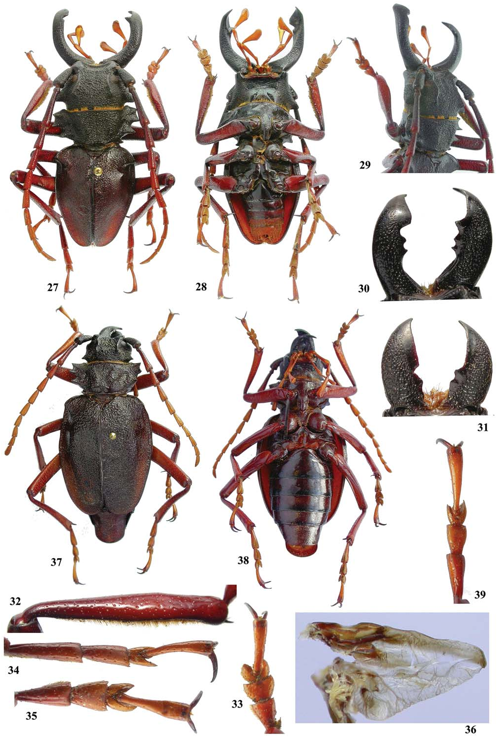
Prionacalus cacicus (White, 1845) ( Figs. 1, 2, 11, 12 View Figs , 27–39 View Figs , 106–108 View Figs , 109–111 View Figs )
Prionus ( Prionacalus) Cacicus White 1845: 110 , pl. 8, figs. 1, 2.
Prionus ( Prionacalus) cacicus ; Monné 2012: 132.
Prionacalus Cacicus ; White 1850: 10, 1851: 70, 1853: 7.
Prionocalus Cacicus ; Thomson 1864: 281, 1877: 260 ( type locality, key).
Psalidognathus ( Prionocalus) cacicus View in CoL ; Reiche 1850b: 265; Lameere 1910: 378, 1913: 66 (catalogue), 1919: 121; Blackwelder 1946: 555 (checklist); Gilmour 1954: 43, pl. 8, fig. 3, pl. 9.
Prionocalus cacicus View in CoL ; Lacordaire 1868: 42 (note); Waterhouse 1872: 261 (key), 262, 1900: 503; Lucas 1886: clxxvii; Quentin and Villiers 1983: 225 (key); Hüdepohl 1985: 121 (key); Pardo- Locarno 2006: 241 (fig. 3).
Prionacalus cacicus View in CoL ; Gemminger and Harold 1872: 2755 (catalogue); Waterhouse 1880: 485; Monné and Giesbert 1994: 15 (checklist); Monné 1995: 60 (catalogue), 2006: 83 (catalogue); Verdcourt 2000: 18; Di Iorio 2003: 3 (distribution); Arai 2005: 34, fig. 1; Monné and Hovore 2005: 19 (checklist), 2006: 18 (checklist); Jeniš 2010: 18 (8 figures, males and females), 19 (5 figures, males and females), 35, 112 (male), 113 (female); Monné et al. 2012: 7 (checklist).
Prionacalus casicus View in CoL [ sic]; Komiya 2001: 29, 30 (fig. 1), 32 (key).
Psalidognathus cacicus View in CoL ; Reiche 1850a: 249.
Psalidognathus Wallisi Taschenberg 1870: 191 View in CoL (female); Lameere 1910: 378 (synonymy).
Prionacalus atys View in CoL (not White, 1850); Lacordaire 1876: pl. 81, figs. 1, 1a; Lameere 1910: 378 (synonymy).
Prionacalus buckleyi View in CoL ; Lameere 1919: pl. 6, fig. 2.
Prionocalus Gunteri Gahan 1894: 221 View in CoL ; Lameere 1910: 376 (synonymy). New synonymy.
Prionacalus gunteri View in CoL ; Jeniš 2010: 16 ( syntype male).
Psalidognathus ( Prionocalus) Buckleyi var. Gunteri; Lameere 1913: 65 (revalidation).
Redescription. Male ( Fig. 27 View Figs ). Head and mandibles blackish (occasionally with anterior margin of submentum reddish); maxillary and labial palpi reddish; antennae gradually lighter towards apex, usually scape and pedicel dark brown (sometimes slightly reddish); antennomere III brown (from dark to lighter); apically reddish from antennomere V or VI; pronotum blackish; ventral surface of thorax dark brown; elytra dark brown (usually blackish at base and, commonly, reddish brown towards apex); ventrite I dark brown (sometimes lighter), ventrite V reddish (frequently almost orange; occasionally dark brown), and intermediate ventrites gradually lighter (often distal margin of ventrites II-IV darker; occasionally entirely dark brown); legs reddish, partly dark brown (sometimes femora entirely blackish), except claws which are reddish on base and blackish on the remainder (occasionally, all tibiae bicolored: dark brown, at least on basal 2/3, and reddish on remainder). Head: Coarsely punctatevermiculate between cephalic carinae, especially apically; area between apex of cephalic carinae and pronotum coarsely, abundantly, confluently punctate (occasionally somewhat vermiculate); area on 1/2 near clypeus with longitudinal, well-marked furrow; distal margin of vertex (close to abrupt declivity) centrally, distinctly emarginate (sometimes emargination only is indicated). Antennal tubercles coarsely punctate near base, gradually smoother towards apex; distinctly separated (distance between them about twice their width). Cephalic carinae ( Fig. 29 View Figs ) very distinct, elevated from base to apex but rarely less prominent; slightly convergent from antennal tubercle to a little after eyes, then divergent towards apex; apex distinctly elevated, forming a triangular tubercle; usually dorsally impunctate (sometimes, distinctly punctate). Area behind eyes with abundant, small asperities, usually closer towards dorsal surface of lateral tubercle, and less distinct towards prothorax. Lateral tubercle of head small and distinctly acute at apex (usually in small specimens) to large ( Fig. 27 View Figs ) and rounded at apex (usually in large specimens). Subhorizontal area of clypeus usually moderately coarsely punctate, mainly basally (occasionally punctures fine and sparse, or coarse and shallow). Distal area of clypeus distinctly concave. Labrum not strongly concave. Distance between upper ocular lobes 2.8–4.6 times longest width of 1 lobe. Gena rounded at apex ( Fig. 29 View Figs ), rarely with weak downward projection. Submentum laterally, shallowly, abundantly punctate, centrally transversely sulcate (sometimes also sparsely punctate centrally; in large specimens, grooves are deep). Mandible in small specimens ( Fig. 31 View Figs ) usually, at most, as long as head; inner margin laminar; surface coarsely, abundantly punctate, gradually finer and sparser towards apex (smooth near inner margin); left mandible with distinct, subtriangluar, large tooth between base and distal plate; right mandible with subtriangular tooth between base and distal plate (usually less distinct), and another close to base of distal plate; distal plate of left mandible wider than that of right mandible. Mandibles in medium sized specimens ( Fig. 30 View Figs ) very variable: as long as head to longer, distinctly laminar to distinctly subcylindrical at basal 1/3; area between base and base of distal plate with 1–2 teeth (on one or both mandibles), and the tooth/teeth placed at middle or closer to base of distal plate. Mandibles in large specimens longer than head; basal 1/2 subcylindrical, gradually more laminar towards base of distal plate; left mandible with one tooth close to the base of distal plate; right mandible with distinct, large, triangular tooth close to base of distal plate. Longest width of last maxillary and labial palpomeres 0.35 ( Fig. 2 View Figs ) to 0.45 ( Fig. 1 View Figs ) times length. Antennae always surpassing elytral apex, usually by last 2 antennomeres, but occasionally only by 1 antennomere (depending, mainly, on elytral length); scape distinctly enlarged from base to apex; antennomeres III-X usually distinctly dentate at apex on both sides (sometimes only at outer distal angle on basal antennomeres), with variable width: narrow ( Fig. 12 View Figs ) or thick ( Fig. 11 View Figs ). Thorax: Latero-anterior angles of prothorax usually acute (sometimes rounded), projected forwards; latero-posterior angles of prothorax distinctly projected sideward, tooth-like (rarely slightly projected and rounded at apex); anterior 2 teeth of lateral margin strongly protracted, anterior tooth smallest; latero-basal tooth large, usually slightly curved upwards. Pronotum longitudinally depressed centrally; coarsely, confluently punctate (sometimes somewhat vermiculate), sometimes almost smooth on depressed area and basal parts; laterally coarsely, confluently punctate (occasionally sparsely punctate), but smooth and shiny between punctate areas. Elytra moderately coarsely vermiculate on basal 1/3 (sometimes only on basal 1/4), gradually distinctly finer towards apex; 2.5–3.5 times longer than prothorax; apices usually individually rounded (occasionally rounded together or with sutural angle distinctly toothlike). Apical tooth humeral projection absent to very distinct. Abdomen: Usually not surpassing elytra. Ventrites I-III ( Fig. 28 View Figs ) finely, sparsely punctate (in very large specimens, punctures more abundant, and somewhat coarser); ventrites IV-V slightly coarser, distinctly more abundantly punctate. Legs: Apex of metafemora usually surpassing elytral apex by 1/2, sometimes by about 1/3 (depending on elytral length). Ventral sulcus of protibiae ( Fig. 32 View Figs ) distinct from near base to distal constriction. Protarsomeres I and II ( Fig. 33 View Figs ) short, moderately wide; protarsomeres I-III with very small denticle at apex; mesotarsi shorter than metatarsi ( Fig. 35 View Figs ); metatarsi normal or slender and long ( Fig. 34 View Figs ).
Female ( Fig. 37 View Figs ). Color as in males, with the same variations, except the abdomen which is entirely dark brown with the apex blackish. Inner margin of mandibles laminar; inner margin as in small males. Antennae reaching distal 1/3 or 1/4 of elytra. Longest width of last maxillary and labial palpomeres 0.25–0.30 times length. Cephalic carinae as in males, usually with apex less elevated. Distance between upper ocular lobes 2.7–4.0 longest width of 1 lobe. Lateral tubercle of head as in male and indicating similar variation. Prothorax as in males. Elytra 3.5–3.9 times longer than prothorax. Abdomen usually distinctly surpassing elytral apex (occasionally not surpassing). All ventrites ( Fig. 38 View Figs ) finely, sparsely punctate. Meso- and metatarsi ( Fig. 39 View Figs ) shorter than in male.
Dimensions. Male/female. Total length (including mandibles) = 25.8–65.0/ 35.8–54.7 mm; prothoracic length = 4.0–10.2/ 5.2–7.8 mm; prothoracic width between apices of lateral spines = 9.9–27.6/ 12.2–20.3 mm; humeral width (at humeral projection) = 9.0–24.0/ 11.8–20.8 mm; elytral length = 12.7–28.4/17.0– 30.3 mm.
Geographical Distribution. Ecuador ( Reiche 1850b), Peru (Waterhouse 1872), Colombia ( Jeniš 2010). ( Mexico is not considered a country where this species occurs).
Material Examined. ECUADOR: 1 male, 1 female (ex Moffarts Collection; no other data) ( IRSN) ; 2 males (no other data) ( ZMHB) ; male, Deyrolle col. (no other data) ( ZMHB); male, 1907, C. Felsche col. ( SNSD). Azuay: Cuenca , male, I.1971, [no collector indicated] ( ZKCO) . Loja: Utuana , male, 30.III.2000, F. Hovore col. ( EMEC) ; Loja (no other data), female ( IRSN) ; 3 males, 2 females, Abbé Gaujon col. (no other data) ( IRSN) ; 2 males, de Mathan col. (ex Moffarts Collection; no other data) ( IRSN) ; male, de Mathan col. (no other data) ( IRSN); male, Abbé Gaujon col. (ex Moffarts Collection; no other data) ( IRSN) ; male, Abbé Gaujon col., (no other data) ( SNSD); male, 08.10.1905, F. Ohaus col. ( ZMHB); ( 2200 m) , male, 2.VIII.1905, F. Ohaus col. ( ZMHB) ; male (no other data), E. Witts col. ( ZMHB); near Loja, female (ex Moffarts Collection; no other data) ( IRSN) ; Cerro Villanaco (ca. 7 km W of the city of Loja; 3000 m) , 2 males, 11.X.1905, Ohaus col. ( ZMHB) . Sucumbíos: La Bonita, male, X.1997, local collector ( MPCO) . Azuay: Cuenca , male, II.1975, local collector ( MPCO) . Tarqui (probably in Azuay province), male, 07.III.1965, Peña col. ( MZSP) . COLOMBIA: 1 male, 1 female (ex Caulle Collection; no other data) ( IRSN) . Huila: male, 1975 (no other data) ( DHPC) . Pichincha: Santa Inés , male, R. Haensch col. ( SNSD) . PERU: 3 males (ex Desbrochers Collection; no other data) ( IRSN) ; mountain (no other data), Murray col. ( ZMHB). Piura: Ayabaca , 2 males, 1 female, 20.III.2008, local collector ( MPCO) ; 1 male, 1 female, II.2008, (no collector indicated) ( ZKCO) .
Additionally , we examined the following three males: Mexico, male (ex Desbrochers Collection; no other data) ( IRSN); 2 males (no other data) ( ZMHB). We believe that the place “ Mexico ” is only an indication of the type locality of the species .
Types and Type Locality. O f P r i o n a c a l u s cacicus ( Figs. 106–108 View Figs ): As mentioned above, White (1845) affirmed that there were three specimens from Mexico. Thus, P. cacicus was described from three syntypes. In 1850, White stated that the specimen figured by him as being a female of P. cacicus was also a male, and named it P. iphis . White (1850) did not record the gender of the third specimen and did not provide a figure. He only affirmed that both specimens figured were males and belonged to different species.
Waterhouse (1872) stated that: “The British Museum has received a pair ( ♂ and ♀) of an insect from Peru, which I am unable to separate from Pr. cacicus, White , although the sizes of the two males are very different; that from Peru measuring (including the mandibles) 30 lines, whilst the type of Pr. cacicus is only 17 lines.” Waterhouse (1900) wrote: “The British Museum has recently received a few specimens of Longicorns of the genus Prionacalus . One I refer with a slight doubt to P. cacicus, White , but in the type the tubercle behind the eye is more directed backwards than in the specimen just received.” Finally, Lameere (1910) commented on P. iphis (translation): “I saw the type in the British Museum; only one male is known.” From those three statements, we conclude that only one male of P. cacicus and another of P. iphis are deposited in the BMNH as types of those species. In addition, we conclude that White (1845) either mistakenly recorded three specimens, when actually there were two, or that one of the types has been lost. The website of the BMNH (2013) records that there is only one type of Prinocalus [ sic] cacicus deposited there (sex not indicated): “ holotype.” EHN examined the types in the BMNH and found only one specimen labeled as the type of P. cacicus and another as the type of P. iphis .
According to the ICZN (1999, Article 72.4.1): “The type series of a nominal species-group taxon consists of all the specimens included by the author in the new nominal taxon…” And Article 72.6 ( ICZN 1999) points out: “The fact that a specimen is already the name-bearing type, or part of the name-bearing type, of one nominal species-group taxon does not prevent its being the name-bearing type, or part of the name-bearing type, of another.” Thus, P. cacicus does not have a holotype: it has three syntypes. At least one of those specimens is the type of P. iphis , but it is not possible to affirm if this species has only one type (the holotype) or two syntypes. Although White (1850) recorded that “The other specimen may be named Prionacalus Iphis ,” he was only affirming that the specimen figured is P. iphis . In our opinion, it is incorrect to infer that White was affirming that only the specimen figured belonged to this species. According to Recommendation 73F ( ICZN 1999): “Where no holotype or syntype was fixed for a nominal species-group taxon established before 2000, and when it is possible that the nominal species-group taxon was based on more than one specimen, an author should proceed as though syntypes may exist and, where appropriate, should designate a lectotype rather than assume a holotype.” Also, according to Article 74.6 ( ICZN 1999): “When it has been accepted that a nominal species-group taxon was based on a single specimen and the original description neither implies nor requires that there were syntypes, and if it is considered subsequently that the original description was based on more than one specimen, the first author to have published before 2000 the assumption that the species-group taxon was based upon a single type specimen is deemed to have designated that specimen as the lectotype.” Thus, it is possible to affirm that Waterhouse (1872) designated as lectotype of P. iphis the specimen figured as female of P. cacicus in White (1845: figs. 2, 2a, 2b). It is important to note that the lectotype male of P. iphis also remains as a syntype ( paralectotype) of P. cacicus .
White (1845) indicated that the three specimens were purchased by Mr. Gray for the British Museum from M. Hartweg, and that the specimens were from Mexico. However, there is no record of any species of Prionacalus collected in Mexico, except those by White (1845, 1850, 1851), and of authors who followed White. Thomson (1877) also questioned Mexico as a type locality. According to the ICZN (1999, Recommendation 76A): “76A.1. In ascertaining or clarifying a type locality (and type horizon, type host, and similar terms), an author should take into account: … 76A.1.4. as a last resort, and without prejudice to other clarification, localities within the known range of the taxon or from which specimens referred to the taxon had been taken.” Prionacalus cacicus is currently recorded in Ecuador and Peru. The species also occurs in Colombia. Thus, the indication of Popayau [Popayán] and Bogotá ( Colombia) (Thomson 1877) must be taken into account. Therefore, following Thomson (1877) we indicate the type locality of P. cacicus as Andes (South America). It is not possible to determine a precise locality in this region (following information provided by Thomson), because he mentioned more than one place in the Andes.
It is important to note that there are at least two places in Colombia named “ Mexico ” located in two different departments: Huila and Arauca. The former is in the Andes ( 2,368 m elevation) and not far from places where other species of Prionacalus are known to occur. As seen in Fig. 108 View Figs , there is no other information on the type’ s labels.
Of Psalidognathus Wallisi : Described based on a couple of specimens from Ecuador ( Loja). As mentioned above, Lameere (1910) affirmed that the male specimen corresponds to P. modestus but the female to P. cacicus . Even so, according to the ICZN (1999: Article 72.1.1), both are types of P. wallisi . The syntypes are deposited at MLUH.
Of Prionocalus [ sic] gunteri ( Figs. 109–111 View Figs ): Holotype male from Ecuador ( Zaruma, not “ Zoruma ” as originally recorded, Department of El Oro), deposited at BMNH.
Remarks. Figure 2 View Figs , plate 2 in Lameere (1919) clearly does not correspond to P. buckleyi . It distinctly corresponds to P. cacicus .
According to Lameere (1910) (translation): “ P. Gunteri Gahan was based on a male with abdomen finely punctate, elytra separately rounded at apex, appendages reddish, humeri very prominent, elytra less rough towards apex.” Lameere (1913, 1919) recorded P. gunteri as a variety of P. buckleyi without explanation. Thus, Lameere (1913) revalidated the former species (not in 1910 as pointed out by Monné 1995, Monné 2006). According to the ICZN (1999: 45.6.4), on determination of subspecific or infrasubspecific rank of names following a binomen: “it is subspecific if first published before 1961 and its author expressly used one of the terms “variety” or “form”…” Since Lameere (1913) considered P. gunteri as a variety of P. buckleyi , it is correctly considered a subspecies. It is important to note that Quentin and Villiers (1983) and Hüdepohl (1985) mentioned only P. buckleyi , without a subspecies.
Prionacalus gunteri is not a synonym of P. buckleyi , as currently considered, or even a subspecies of the latter. The cephalic carinae, prothoracic shape, and elytral sculpture are as in some specimens of P. cacicus , and not as in P. iphis .
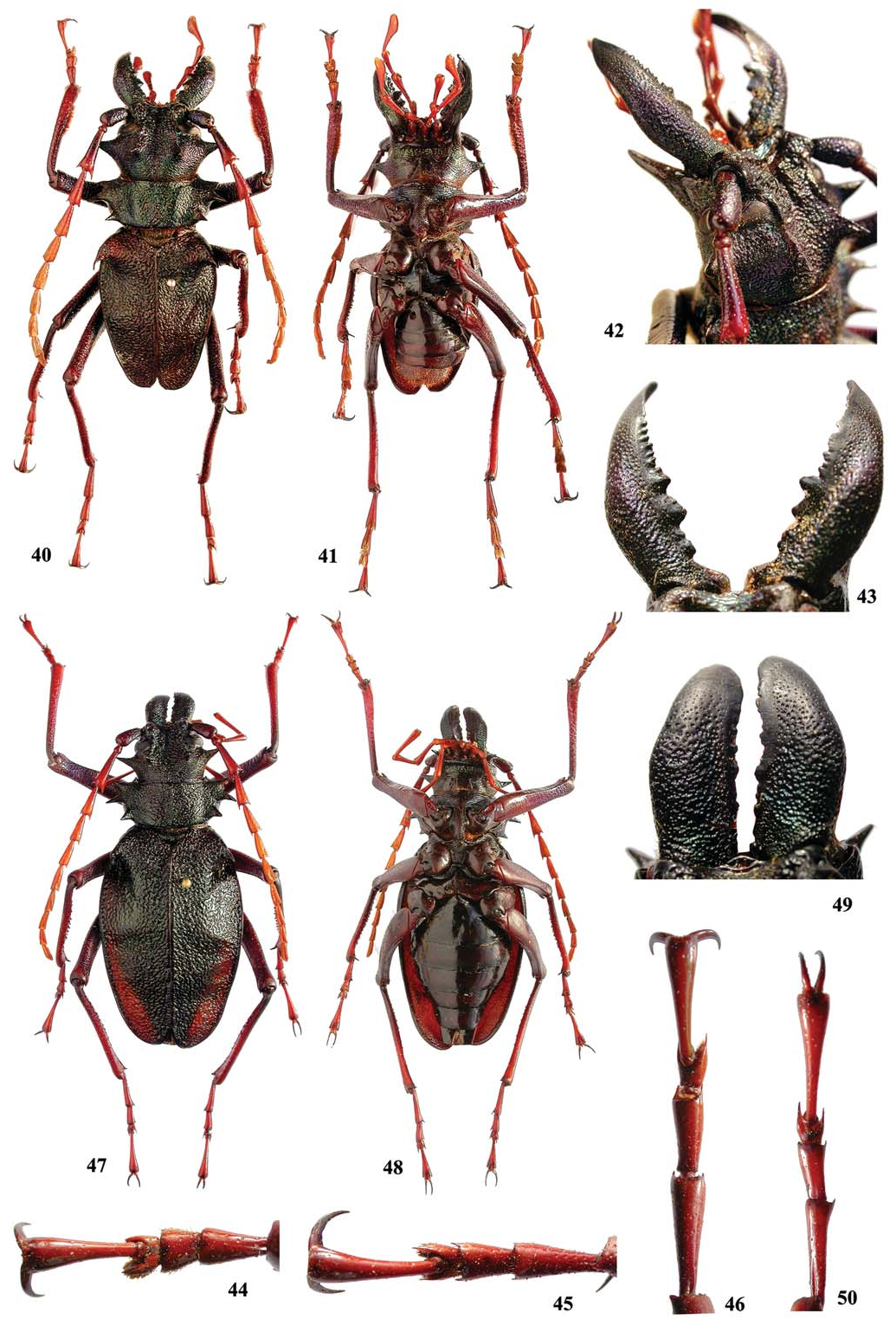
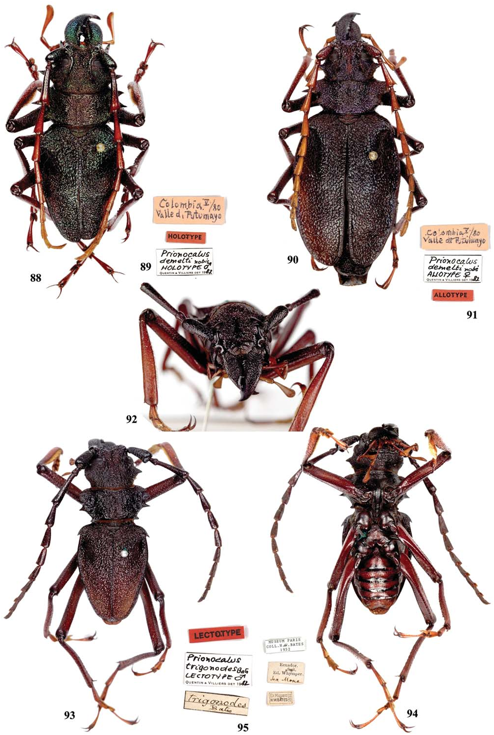
Prionacalus demelti Quentin and Villiers, 1983 ( Figs. 7, 13 View Figs , 40–50 View Figs , 88–91 View Figs )
Prionocalus demelti Quentin and Villiers 1983: 224 View in CoL , 2 figs.; Hüdepohl 1985: 122 (key); Pardo- Locarno 2006: 234.
Prionacalus demelti View in CoL ; Monné and Giesbert 1994: 15 (checklist); Monné 1995: 60 (catalogue), 2006: 83 (catalogue); Martínez 2000: 85 (distribution); Komiya 2001: 31 (figs. 6–8), 32 (key); Di Iorio 2003: 3 (distribution); Salazar 2005: 246; Arai 2005: 34, fig. 2 (male); Monné and Hovore 2005: 19 (checklist), 2006: 18 (checklist); Jeniš 2010: 16 (males, females), 17 (male), 110 (female).
Redescription. Male ( Fig. 40 View Figs ). Integument black to dark brown, usually dorsally darker; maxillary and labial palpi reddish brown; femora from brown to reddish brown, always apically blackish; tibiae from brown to reddish brown, basally blackish or dark brown; tarsi reddish brown (at least, distal 1/3 of claws always black); scape blackish to dark brown; antennomeres gradually lighter from III to V, and VI-XI with same color as V; elytra with irregular and variable reddish areas (sometimes almost absent). Head: Coarsely punctate-rugose, mainly between cephalic carinae on region of eyes and antennal tubercles; area between cephalic carinae distinctly depressed, centrally with distinct sulcus from clypeus to area of posterior ocular edge (sometimes reaching only middle of eyes). Antennal tubercle coarsely confluently punctate on base, almost smooth near apex; bases far from each other (distance between them equal to about 1.2 times tubercle width). Cephalic carinae ( Fig. 42 View Figs ) convergent from antennal tubercle to after eyes, divergent from this point to apex (sometimes subparallel), strongly elevated at apex, forming spine-like tops. Area behind lower ocular lobes with abundant small asperities. Lateral tubercle of head very large, acute at apex. Subhorizontal area of clypeus coarsely, shallowly or smoothly punctate, usually with distal margin elevated. Distal area of clypeus distinctly concave, smooth. Labrum subtriangular, apex acute or slightly rounded. Distance between upper ocular lobes 2.8–3.7 times longest width of 1 lobe. Gena ( Fig. 42 View Figs ) with long projection directed forwards (sometimes downward). Submentum depressed, coarsely, moderately abundantly punctate. Mandible ( Fig. 43 View Figs ) as long as head to slightly longer; subcylindrical on base, distinctly laminar towards apex; surface coarsely, abundantly punctate, gradually finer, sparser towards apex (smooth close to inner margin); both mandibles with inner margin pluridentate from base to distal plate, the latter also with numerous, irregular small teeth at its basal 1/2. Longest width of last maxillary ( Fig. 7 View Figs ) and labial palpomeres equal to about 0.25 times length. Antennae reaching or slightly surpassing elytral apex; scape subcylindrical, coarsely, abundantly, confluently punctate on basal 1/2, sparsely, finely punctate towards apex; antennomere III about 1.6 longer than scape; antennomeres III-X ( Fig. 13 View Figs ) with projection on both sides of apex, notably long on III-V. Thorax: Latero-anterior angles of prothorax projected forward, almost acute; latero-posterior angles of prothorax not projected sideward, rounded; lateral margin with 3 distinct teeth, the 2 anterior teeth protracted, the most anterior smaller. Pronotum with distinct depression on center of disc, sometimes longitudinally divided by slightly distinct carina; coarsely, abundantly punctate (at least partially confluent laterally). Prosternal process with rounded projection at underside of apex (sometimes slightly distinct). Elytra coarsely vermiculate throughout; apex variable: individually rounded or rounded together; with or without projection at sutural angle (occasionally, left elytron shorter than right). Apical tooth humeral projection large. Abdomen: Usually not surpassing elytral apex (sometimes slightly surpassing). Ventrites I-IV ( Fig. 41 View Figs ) very finely, sparsely punctate; ventrite V coarsely, abundantly punctate. Legs: Metafemora surpass elytral apex by about middle. Ventral sulcus of protibiae visible only between about middle and before apex. Protarsi ( Fig. 44 View Figs ) moderately elongate and narrow; protarsomeres I-III with denticle at each apex. Mesotarsi ( Fig. 45 View Figs ) slender; apex of mesotarsomeres I-III with distinct spine at each apex. Metatarsi ( Fig. 46 View Figs ) distinctly slender; apex of metatarsomeres I-III spiny.
Female ( Fig. 47 View Figs ). Dorsally blackish, ventrally dark brown; distal area of ventrites blackish; scape brownish; antennomeres reddish brown. Mandibles ( Fig. 49 View Figs ) laminar; inner margins pluridentate. Antennae reaching distal 1/3 of elytra. Longest width of last maxillary and labial palpomeres about 0.20 times length. Cephalic carinae as in males. Distance between upper ocular lobes 3.0 times longest width of 1 lobe. Lateral tubercle of head as in male. Prothorax as in males. Abdomen surpassing or not elytral apex. Ventrites I-IV almost impunctate ( Fig. 48 View Figs ); ventrite V very finely, sparsely punctate. Metatarsi ( Fig. 50 View Figs ) shorter than in male.
Dimensions. Male/female. Total length (including mandibles) = 39.0–57.0/63.0–64.0 mm; prothoracic length = 5.5–7.0/ 6.8–7.4 mm; prothoracic width between apices of anterior angles = 8.3–12.3/ 10.5–12.0 mm; prothoracic width between apices of posterior angles = 8.0–14.4/12.0– 12.5 mm; humeral width = 13.4–18.6/20.0– 20.5 mm; elytral length = 19.7–25.0/ 35.5–37.3 mm.
Geographical Distribution. Colombia.
Material Examined. COLOMBIA: Female , XII.1991, [no collector indicated] ( ZKCO) ; male, VII.1992, [no collector indicate] ( ZKCO) . Huila: Gigante , 1 male, 1 female, 1974, (no collector indicated) ( ZKCO) ; 1 male, 1976, (no collector indicated) ( ZKCO) , 1 male, 1 female, [no date indicated], local collector ( MPCO) ; male, XI.1978, [no collector indicated] ( DHPC) . Boyacá: near Muzo , male, V.1983, [no collector indicated] ( ZKCO) . Yari : Caqueta, 2 males, I.1982, (no collector indicated) ( ZKCO) .
Types and Type Locality. Prionocalus [ sic] demelti was described based on a holotype male ( Figs. 88, 89 View Figs ) and an allotype female ( Figs. 90, 91 View Figs ), both from Colombia ( Putumayo River Valley), deposited at MNHN. Additionally , there were four paratypes ( three males, one female) deposited in Carl von Demelt Collection (currently deposited at SMNS) .
Remarks. Prionacalus demelti resembles Psalidognathus superbus Fries, 1833 by its antennomeres which are distinctly spiny at apex, and by its long genal projection. Those features make this species one of the more easily recognizable Prionacalus species.
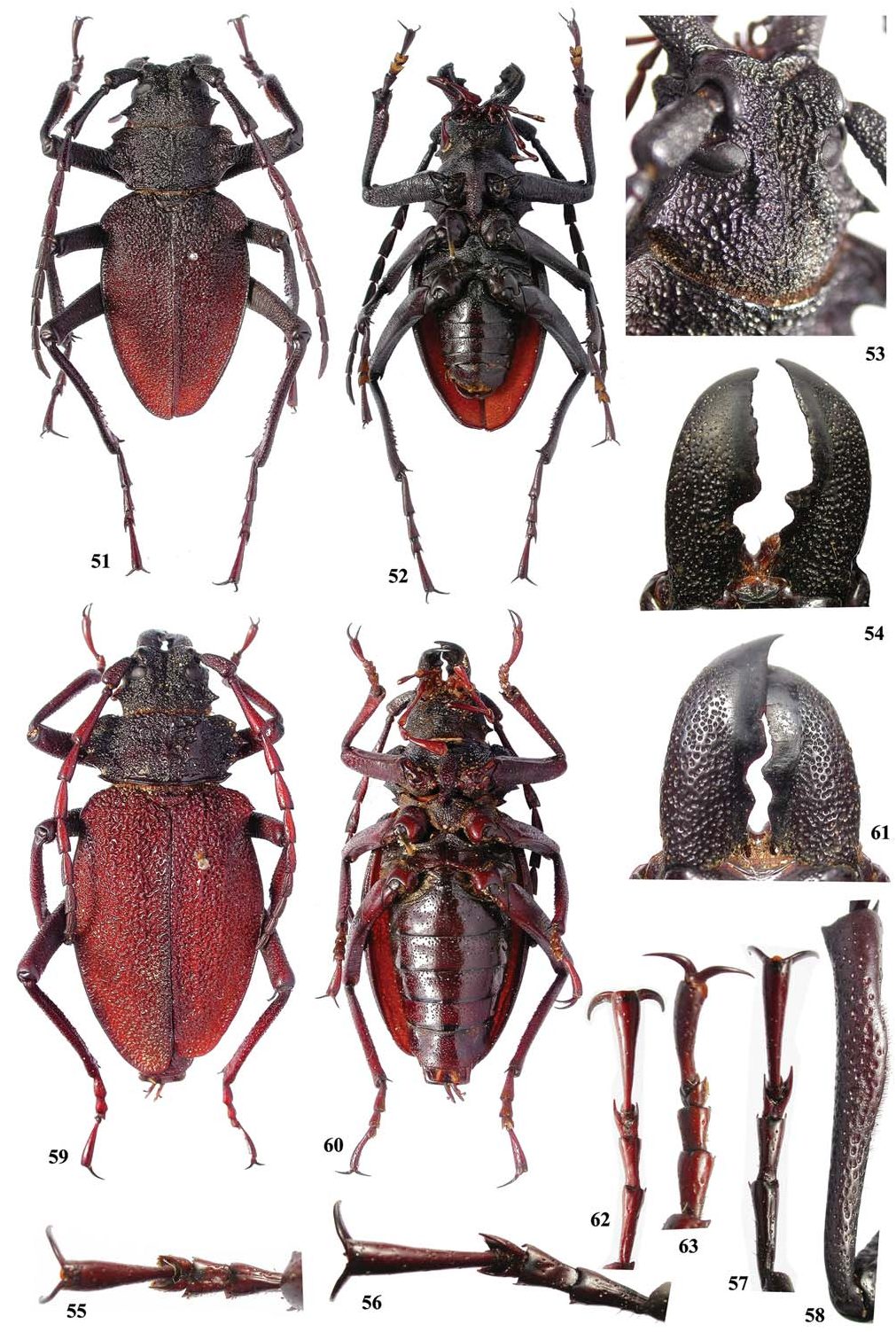
Prionacalus whymperi Bates, 1892 ( Figs. 6, 14 View Figs , 51–63 View Figs , 92–95 View Figs , 96–102 View Figs )
Prionocalus Whymperi Bates 1892: 36 , 1 fig; Campos, 1921: 87, fig. 16.
Prionocalus whymperi View in CoL ; Quentin and Villiers 1983: 223 (revalidation, lectotype).
Prionacalus whymperi View in CoL ; Monné and Giesbert 1994: 15 (checklist); Monné 1995: 61 (catalogue), 2006: 84 (catalogue); Komiya 2001: 31, 32 (key); Di Iorio 2003: 3 (distribution).
Psalidognathus ( Prionocalus) Buckleyi ; Lameere 1910: 376 (part), 1913: 65 (catalogue, part), 1919: 121, pl. 6, fig. 2 (part).
Prionocalus giovannii Hüdepohl 1985: 117 View in CoL , 4 figs. New synonymy.
Prionacalus giovannii View in CoL ; Monné and Giesbert 1994: 15 (checklist); Monné 1995: 60 (catalogue), 2006: 83 (catalogue); Komiya 2001: 32 (key); Monné and Hovore 2005: 19 (checklist), 2006: 18 (checklist).
Prionacalus giovanni View in CoL ; Di Iorio 2003: 3 (distribution).
Prionocalus trigonodes Bates 1892: 37 View in CoL ; Whymper 1892: 10; Kolbe 1902: 481; Campos 1921: 87; Quentin and Villiers 1983: 224 ( lectotype, synonymy); Hüdepohl 1985: 121. New synonymy.
Psalidognathus ( Prionocalus) trigonodes View in CoL ; Lameere 1910: 377.
Redescription. Male ( Fig. 51 View Figs ). Head, mandibles, and thorax blackish (sometimes prosternal process reddish brown); maxillary and labial palpi brown to reddish (usually slightly darker on basal segments); scape and pedicel blackish; antennomere III entirely blackish to entirely dark brown (sometimes dark brown only distally); antennomeres IV-XI dark brown (sometimes slightly reddish); elytra dark brown on basal 1/3, gradually reddish towards apex; ventrites dark brown, with distal margin blackish (sometimes, ventrite V almost entirely reddish, except on distal margin); profemora dark brown; meso- and metafemora reddish on basal 1/3, dark brown on remaining surface (occasionally almost entirely dark brown); protibiae dark brown on basal 1/3, gradually reddish towards apex (sometimes reddish only on distal 1/3); meso- and metatibiae brown on base, slightly reddish towards apex (occasionally entirely dark brown); tarsi entirely dark brown to entirely reddish (sometimes dark brown with some parts reddish), except claws that are blackish. Head: Coarsely scabrous between apex of cephalic carinae and prothorax (sometimes only punctate near prothorax), vermiculate between cephalic carinae, coarsely punctatevermiculate between upper ocular lobes and prothorax; area between cephalic carinae distinctly depressed, centrally with distinct sulcus from clypeus to area after antennal tubercles (sometimes from clypeus to almost apex of cephalic carinae); distal margin of vertex (close to abrupt declivity) centrally distinctly emarginate. Antennal tubercle coarsely punctate-vermiculate on base, gradually finer towards impunctate area of apex; bases near each other (distance between them about 0.15 times width or slightly narrower). Cephalic carinae ( Fig. 53 View Figs ) distinct, elevated from base to apex (usually less so between antennal tubercle and base of eye); convergent from antennal tubercle to little after eyes, subparallel towards apex; apex not or slightly elevated, not forming a triangular tubercle, but usually wider at this area. Area behind lower ocular lobes variable: not finely striate, and with abundant small asperities; finely striate near prothorax, and with abundant asperities near eyes. Lateral tubercle of head small to moderately large, rounded at apex ( Fig. 96 View Figs ) to distinctly acute ( Fig. 51 View Figs ). Subhorizontal area of clypeus finely, coarsely, abundantly punctate (occasionally with 1 tubercle on central area). Distal area of clypeus distinctly concave, entirely smooth to slightly rugose at base. Labrum distinctly concave, usually with central longitudinal sulcus, and apex centrally emarginate. Distance between upper ocular lobes 2.3–2.6 times longest width of 1 lobe. Gena rounded at apex ( Fig. 53 View Figs ), without downward projection. Submentum depressed, laterally confluently, coarsely punctate, remaining surface coarsely vermiculate, or abundantly punctate, or almost smooth. Mandibles ( Fig. 54 View Figs ) 0.8–0.9 times head length; inner side laminar; surface coarsely, abundantly punctate, gradually finer towards apex (smooth near inner margin); left mandible with 1 distinct tooth on basal 1/2 (sometimes another small one nearer base); right mandible with 2 jointly protracted teeth near base (posterior tooth smallest), and another large tooth close to the base of distal plate; distal plate of left mandible wider than that of right mandible. Longest width of last maxillary ( Fig. 6 View Figs ) and labial palpomeres almost equal to 0.4 times length. Antennae not or slightly surpassing elytral apex; scape slightly enlarged from base to apex, coarsely, confluently punctate; antennomere III usually distinctly dentate on inner side of apex, rounded or slightly dentate on outer margin, about 1.6 times longer than scape; antennomere IV dentate on both sides of apex (sometimes only on outer side); antennomeres V-X usually slightly projected on both sides of apex. Thorax: Latero-anterior angles of prothorax slightly projected forward, rounded (occasionally somewhat acute); latero-posterior angles of prothorax distinctly projected sideward (tooth-like), acute to moderately rounded at apex; lateral margin with 3 distinct teeth (anterior one distinctly smaller to subequal to middle tooth). Pronotum with a distinct, transverse depression on distal 1/2; centrally with longitudinal carina from near base to apex; coarsely punctate-vermiculate, usually with smooth, narrow band on base; anterior margin distinctly emarginate centrally. Prosternal process with spiniform projection at underside of apex. Elytra coarsely vermiculate throughout; apices individually to jointly rounded (sometimes moderately truncate). Apical tooth of humeral projection small to very large. Abdomen: Not surpassing elytral apex. Ventrites ( Fig. 52 View Figs ) moderately coarsely, abundantly punctate (mainly I-IV laterally and V throughout), or distinctly sparsely punctate on ventrites I-V (mainly centrally) and V abundantly punctate. Legs: Apex of metafemora surpasses elytral apex by about 0.2 times length of metafemora. Ventral sulcus of protibiae distinct only from middle to near apex (usually present after basal 1/3, but not distinctly marked); protibiae enlarged near middle ( Fig. 58 View Figs ). Protarsi ( Fig. 55 View Figs ) moderately short and wide; protarsomeres I-III with small denticle at apex. Mesotarsi ( Fig. 56 View Figs ) slender; apex of mesotarsomeres I-III with distinct denticle at apex. Metatarsi ( Fig. 57 View Figs ) distinctly slender; apex of metatarsomeres I-III spiny (mainly III).
Female ( Fig. 59 View Figs ). Head and prothorax dark brown, with some parts blackish; mandibles blackish; abdomen entirely dark brown or reddish brown with apex blackish; scape dark brown; pedicel and antennomeres III-XI reddish brown. Inner margin of mandibles ( Fig. 61 View Figs ) laminar; inner margins similar to those in males but with only 1 tooth between base and distal plate. Antennae slightly surpassing middle of elytra. Longest width of last maxillary and labial palpomeres about 0.35 times length. Cephalic carinae as in males, or distinctly divergent after eyes. Distance between upper ocular lobes 2.6–2.8 times longest width of 1 lobe. Lateral tubercle of head as in male, with the same variations. Prothorax as in males (in 2 females examined, anterior lateral tooth smaller than middle tooth). Prosternal process as in males ( Fig. 14 View Figs ). Abdomen distinctly surpassing elytral apex. All ventrites ( Fig. 60 View Figs ) moderately coarsely, sparsely punctate. Meso- and metatarsi shorter than in male; tarsomeres slender and elongate ( Fig. 62 View Figs ) or thick and short ( Fig. 63 View Figs ).
Dimensions. Male/female. Total length (including mandibles) = 44.5–58.3/51.6–53.0 mm; prothoracic length = 6.3–8.6/8.0– 8.4 mm; prothoracic width between apices of anterior angles = 9.1–12.7/ 12.0– 12.2 mm; prothoracic width between apices of posterior angles = 10.4–13.0/ 12.5–14.9 mm; humeral width = 16.0–21.3/ 19.5–20.5 mm; elytral length = 25.533.3/32.0– 33.5 mm.
Geographical Distribution. Ecuador.
Material Examined. ECUADOR: 2 males, 2 females, [no other data], Fritsche V. col. ( ZMHB) ; male, VI.1982, J. P. Marechal col. ( ZKCO) . Cotopaxi: 2500 m , 1 male, 1 female, III.1985, P. Arnaud col. ( MPCO) . Pichincha: female, IV.2001, [no collector indicated] ( ZKCO) ; 2 males, 1 female, XII.2001, [no collector indicated]; Los Alpes ( 2000 m), male, 12.VII.1935, Schultze- Rhonhof S. G. col. ( ZMHB) ; Santo Domingo de los Colorados , male, VII.2001, [no collector indicated] ( ZKCO) .
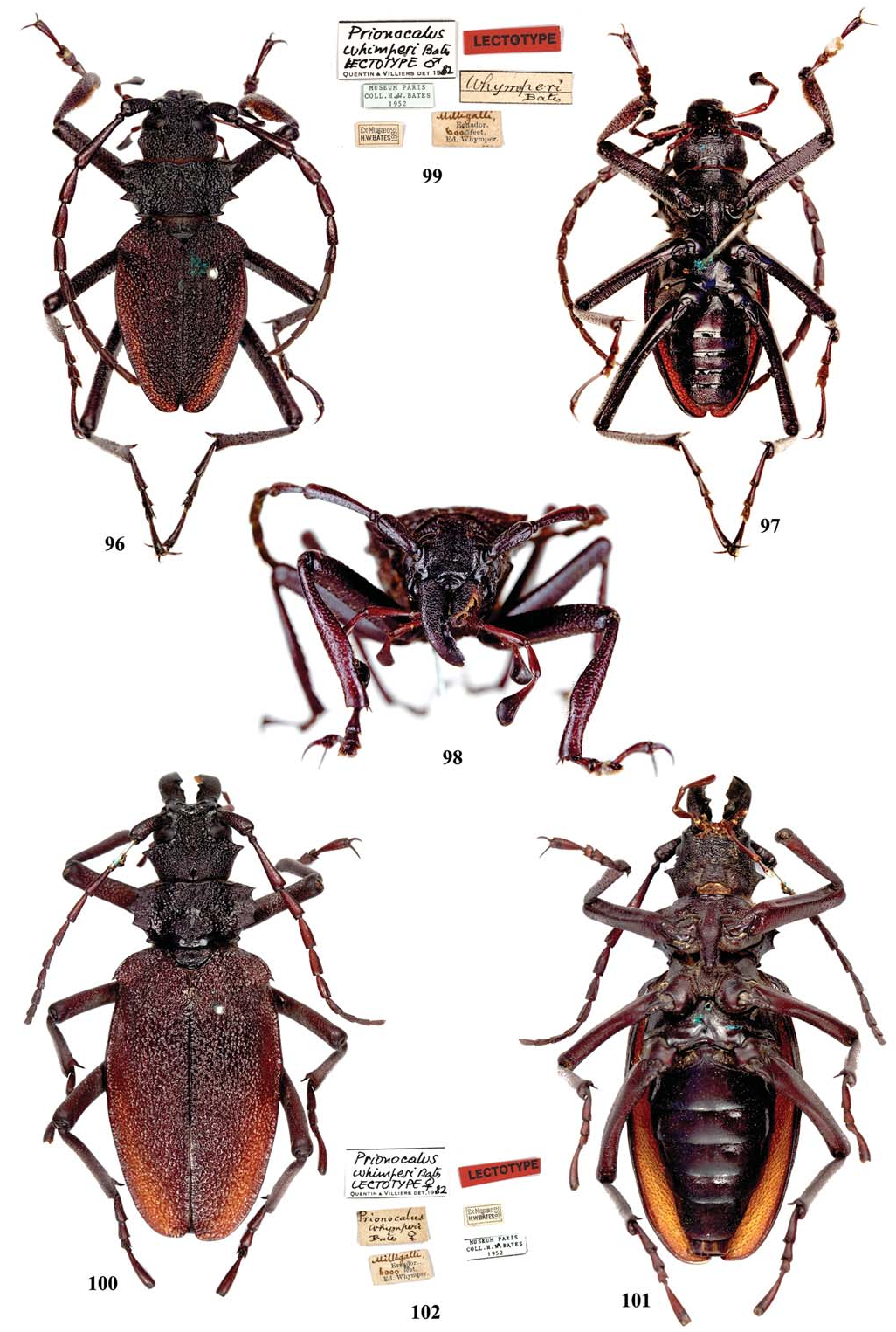
Types and Type Localities. Of Prionacalus whymperi ( Figs. 96–102 View Figs ): Described from two males and one female, all from “Milligalli ( 6200 feet).” According to Myers (1969): “…from Milligalli, Ecuador, a locality at about 1900 meters elevation on the western slopes of Cerro Corazon (Andrade Marin, 1931, p. 30).” The type locality is located in the province of Pichincha. The syntypes are deposited at MNHN .
Quentin and Villiers (1983) stated (translation): “Among the three syntypes, we designate a LECTOTYPE male with 38 mm length (without mandibles) and with the following labels: “Milligalli, Ecuador. 6000 feet. Ed. Whymper.”, “Ex Musaeo H. W. Bates, 1892 ”, “Museum Paris, Coll. H. W. Bates, 1952”, “ Whymperi Bates ”. – LECTOTYPE female with 63 mm length (without mandibles) with the same labels and: “ Prionocalus Whymperi Bates, ♀ ”. – One PARALECTOTYPE male from same place.” According to the ICZN in effect at that time ( ICZN 1964) and currently ( ICZN 1999), those designations are invalid, because a species can only have one lectotype: “74.1. Designation of a lectotype. A lectotype may be designated from syntypes to become the unique bearer of the name of a nominal speciesgroup taxon and the standard for its application;” and “74.5. Lectotype designations before 2000. - In a lectotype designation made before 2000, either the term “ lectotype ”, or an exact translation or equivalent expression (e.g. “the type”), must have been used or the author must have unambiguously selected a particular syntype to act as the unique name-bearing type of the taxon.”
We designate here as the lectotype for P. whymperi the specimen ( Figs. 96–98 View Figs ) designated by Quentin and Villiers (1983) as the lectotype male. The specimen has the following labels ( Fig. 99 View Figs ):
1. White [Handwritten]: whymperi / Bates;
2. White: Milligalli [Handwritten] / Ecuador. (Printed) / 6000 [Handwritten] feet [Printed] / Ed. Whymper [Printed];
3. White [Printed]: Ex-Musaeo H. W.Bates / 1892;
4. White [Printed]: Museum Paris / Coll. H. W. Bates / 1952
5. White: Prionocalus [ sic] whimperi [ sic] Bates [Handwritten] / Lectotype ♂ [Handwritten] / Quentin and Villiers det. 19 [Printed] 82 [Handwritten];
6. Red [Printed]: Lectotype
7. Red and yellow [Printed; added by us]: LECTOTYPE / Prionacalus whymperi
Although Bates (1892) recorded “ 6200 feet,” the type label indicates the elevation as either 600 or 6,000 ft. We can never know which is the true value recorded since the pin was inserted exactly where the last zero would be placed.
Of Prionocalus [ sic] giovannii : Holotype and paratype males, from Ecuador ( Province of Pichincha, Santo Domingo de los Colorados ), deposited at ZSMC .
Of Prionocalus [ sic] trigonodes ( Figs. 93–95 View Figs ): The number of specimens originally included in the description is controversial. Bates (1892) recorded: “Hab. La Mona (under 200 feet), one ♂. Taken also by Mr. Buckley on his last journey.” However, he also indicated: “Long. 27–37 milim. ♂.” Quentin and Villiers (1983) designated the lectotype for the species (translation): “We designated as LECTOTYPE a male with 37 mm long (without mandibles), with the following labels: “ Ecuador, …feet. Ed. Whymper, La Mona”, “Ex Museum H. W. Bates”, “ trigonodes Bates ”, “Museum of Paris, Coll. H. W. Bates”.” La Mona is located in the province of Los Rios, Ecuador. The species was likely described based on a single specimen. According to Whymper (1892): “Amongst the few species secured on the first day’ s journey, there have been found an undescribed Ant ( Camponotus ), a Bug ( Pnohirmus ), and two Beetles ( Epitragus and Prionocalus [ sic]). These are described and figured in the Supplementary Appendix with is published simultaneously with this volume. The Prionocalus [ sic] that is described by Mr. H. W. Bates under the name P. trigonodes was picked up close to La Mona.” Based on this information, we believe that Bates had only a single specimen when he described the species (indicating it was the holotype). Thus, the designation of a lectotype by Quentin and Villiers (1983) was in error, based on the incorrect information provided by Bates (1892).
Remarks. As mentioned above, Lameere (1910) considered P. whymperi as a synonym of P. buckleyi (translation): “I have not seen the type of P. Whymperi Bates ; the species is established on a male slightly differing from P. Buckleyi , according to Bates himself, but also on a female with elytra notably surpassing elytral apex. I have before me a male of P. Buckleyi that also has this feature, but I cannot see more than an individual variation.”
Quentin and Villiers (1983) revalidated the species (translation): “The study of the types of Bates, preserved in the Paris Museum, showed us that Lameere ( loc. cit.), who had not seen them, committed various mistakes that we rectify as follows [translation]: “Lameere ( loc. cit.: 731) had arbitrarily placed this species in synonymy of P. buckleyi Waterhouse. Instead , we consider it as valid [NEW COMBINATION]. They are distinguished by the following characters: P. whymperi Bates : 1. Anterior tibiae of male 6 times longer than wide, dilated at middle, and then narrowed at apical fourth; setae of inner face starting after basal fourth; 2. Tarsi slender in both sexes, second mesotarsomere about as long as wide at its apex; 3. Elytra covering the abdomen. P. buckleyi Waterhouse : 1. Anterior tibiae of male 7 times longer than wide, enlarged from base to the apex; setae of inner face extending on entire length, from the base; 2. Tarsi thicker; second mesotarsomere approximately one and a half time longer than wide at its apex; 3. Elytra usually leaving visible the apex of abdomen.” We agree with Quentin and Villiers (1983). Prionacalus whymperi is not a synonym of P. buckleyi , and thus, not a synonym of P. iphis . The main character separating P. whymperi from P. iphis is the shape of the sulcus on the inferior side of the protibiae, starting far from the base in the former and near the base in the latter.
Although Hüdepohl (1985) was aware of the work by Quentin and Villiers (1983), he excluded P. whymperi in his key to the species of Prionocalus [ sic] without explanation. Prionacalus whymperi was listed as a valid species in Monné (1995, 2006). However, it was considered a synonym of P. buckleyi in Monné and Giesbert (1994), Monné and Hovore (2005, 2006), and it was omitted in Jeniš (2010).
Based on the specimens examined, original descriptions, and photographs of the types, we conclude that P. giovannii is a junior synonym of P. whymperi . It is strange that Hüdepohl did not observe the information from Quentin and Villiers (1983) on the protibiae of P. whymperi : “dilated at middle…setae of inner face starting after basal 1/4.” This information would have shown that the protibiae in the types of P. whymperi are exactly as in the holotype of P. giovannii . This feature and the shape of the first spine on the lateral margin of the prothorax can be used to distinguish P. whymperi from the other species of the genus.
As mentioned above, Quentin and Villiers (1983) considered P. trigonodes another synonym of P. buckleyi (translation): “Lameere ( loc. cit., Revision: 732), without seeing the type, assigned to this species erroneous characters based on specimens wrongly identified in the British Museum. In fact, trigonodes corresponds to small individuals of buckleyi and the name cannot be maintained.” The short description of P. trigonodes does not allow it to be differentiated from P. buckleyi . However, examining the detailed photographs of the lectotype male, we conclude that it is equal to P. whymperi and not to P. buckleyi (= P. iphis ). Although the protibiae ( Fig. 92 View Figs ) are not distinctly enlarged from the middle, they are somewhat slender, and the ventral sulcus does not start very near the base. In addition, the prosternal process has a distinct, spiniform projection on the underside of the apex, a feature that was not observed in any specimen of P. iphis . Also, although Quentin and Villiers (1983) had affirmed that P. trigonodes agrees with small specimens of P. buckleyi , the length of the holotype (their lectotype), 37 mm, is about as long as the lectotype of P. buckleyi (18 lines). One English line corresponds to about 2.12 mm ( 2.1166 mm). Thus, the lectotype male of P. buckleyi is about 38.2 mm in length.
Both P. whymperi and P. trigonodes were recorded as having been described by Bates in 1891. However, as mentioned above, Whimper (1892) affirmed: “…in the Supplementary Appendix which is published simultaneously with this volume.” Thus, it is possible to infer that the date on the Supplementary Appendix was pre-dated. In other words, it was printed first in 1891 but not distributed until 1892.
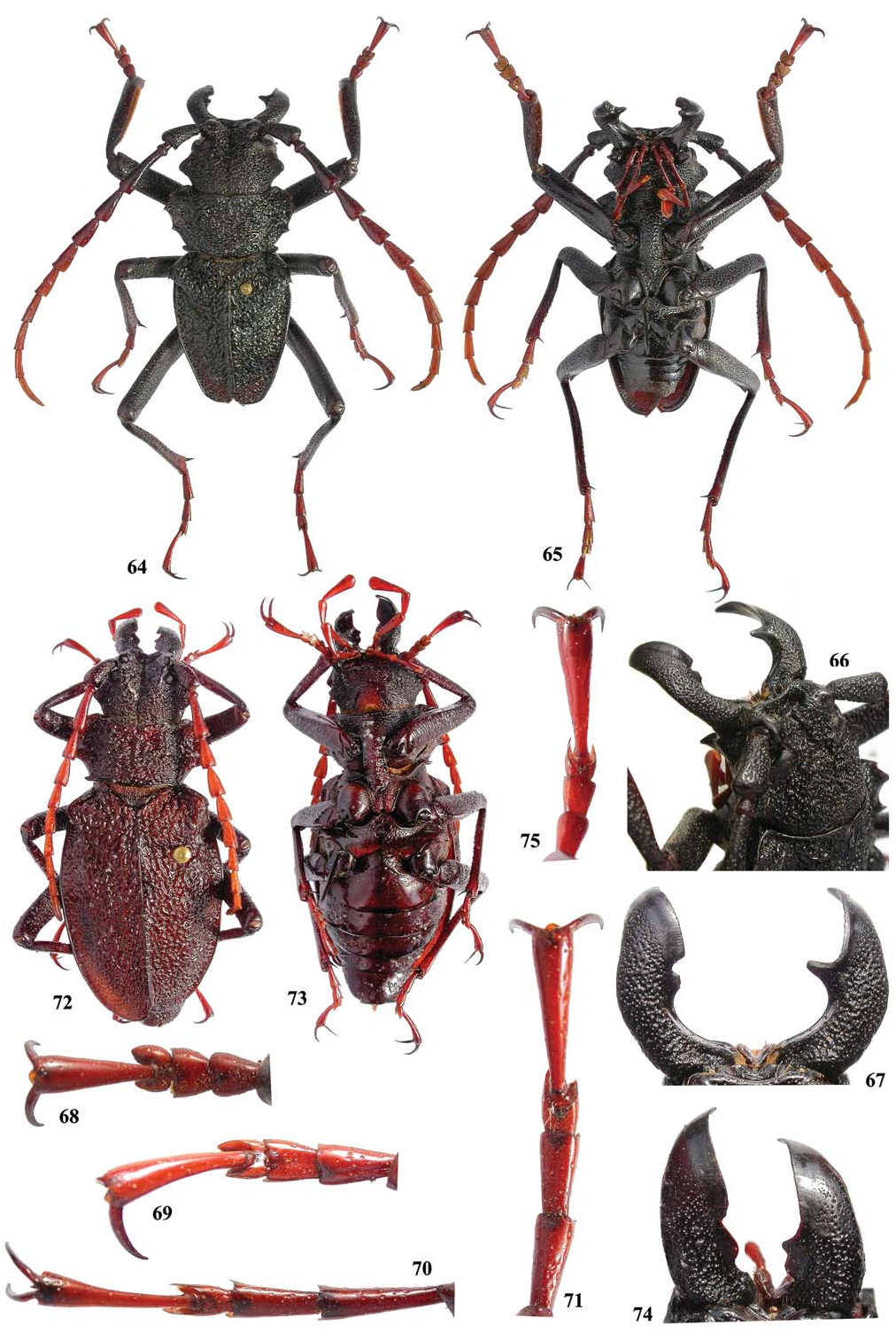
Prionacalus woytkowskii ( Heyrovsky, 1960) ( Figs. 3, 9, 10 View Figs , 64–75 View Figs )
Psalidognathus ( Prionocalus) woytkowskii Heyrovsky 1960: 162 View in CoL , 3 figs.
Prionocalus woytkowskii View in CoL ; Quentin and Villiers 1983: 225 (key); Hüdepohl 1985: 121.
Prionacalus woytkowskii View in CoL ; Monné and Giesbert 1994: 15 (checklist); Monné 1995: 61 (catalogue), 2006: 84 (catalogue); Komiya 2001: 29, 30 (figs. 2, 3), 32 (key); Di Iorio 2003: 3 (distribution); Arai 2005: 34, fig. 3 (male), 4 (female); Monné and Hovore 2005: 19 (checklist), 2006: 18 (checklist); Jeniš 2010: 16 (males, females), 17 (male), 124 (male), 125, female; Monné et al. 2012: 7 (checklist).
Prionacalus buckleyi View in CoL ; Jeniš 2001: 27 (fig. 91/55).
Prionacalus giovannii View in CoL ; Jeniš 2010: 19 (male, female), 114 (male), 115 (female); Monné et al. 2012: 7 (checklist).
Redescription. Male ( Fig. 64 View Figs ). Integument black (sometimes with small dark brown areas); maxillary and labial palpi reddish brown; tibiae reddish on distal fourth (sometimes almost from basal 1/3); tarsi from brown to reddish brown, except small dark area at apex and blackish claws; pedicel and antennomere III blackish to dark brown, except distal 1/6 of antennomere III reddish; basal 3/4 of antennomere IV brown or dark brown, distal 1/4 from brown to reddish; antennomere V brown to reddish; antennomeres VI-XI reddish. Head: Coarsely punctate-vermiculate, mainly between cephalic carinae; area between cephalic carinae distinctly depressed, centrally with distinct sulcus from clypeus to area of posterior ocular edge. Antennal tubercle coarsely punctatevermiculate on base, becoming smoother towards apex (only with very sparse coarse punctures); bases moderately far from each other (distance between them equal to almost each width). Cephalic carinae ( Fig. 66 View Figs ) slightly distinct by the coarse sculpture of dorsal surface of head but usually more or less recognizable; subparallel from base to apex. Area behind lower ocular lobes with abundant small asperities. Lateral tubercle of head large, rounded at apex ( Fig. 64 View Figs ). Subhorizontal area of clypeus smooth, usually centrally with 2 small callosities. Distal area of clypeus distinctly concave, smooth. Labrum distinctly concave (occasionally slightly concave), apex centrally emarginate or not. Distance between upper ocular lobes 2.6–3.0 times longest width of 1 lobe. Gena acute at apex ( Fig. 66 View Figs ), with short sideward projection (sometimes moderately truncate and not sideward projected). Submentum depressed, transversely sulcate (sometimes vermiculate), finely to moderately coarsely punctate (punctures moderately sparse; occasionally absent). Mandibles ( Fig. 67 View Figs ) 0.7–0.8 times head length; inner margin subcylindrical at basal 1/3; surface coarsely, abundantly punctate on basal 1/3, gradually finer, sparser towards apex (smooth near inner margin); left mandible with 1 distinct tooth near base of distal plate; right mandible with 1 large tooth close to base of distal plate (in small specimens, usually with another smaller tooth jointly protracted); distal plate of left mandible wider than that of right mandible. Longest width of last maxillary ( Fig. 3 View Figs ) and labial palpomeres about 0.45 times length. Antennae slightly to distinctly surpassing elytral apex; antennomeres moderately slender ( Fig. 10 View Figs ) to wide ( Fig. 9 View Figs ); scape slightly to distinctly enlarged from base to apex, coarsely, confluently punctate, except on distal 1/5 which is almost smooth; antennomere III rounded on both sides of apex, about 1.4 times longer than scape; antennomeres IV-X dentate on outer apex, rounded to angulate on inner apex. Thorax: Lateroanterior angles of prothorax slightly projected forward, rounded; latero-posterior angles of prothorax distinctly projected sideward (tooth-like), moderately rounded at apex; lateral margin with 3 distinct teeth (anterior tooth smallest). Pronotum with distinct depression on center of disc (occasionally slightly marked); coarsely punctatevermiculate (laterally less so), occasionally with small, impunctate, shiny callosity on each side of anterior third; anterior margin distinctly emarginate medially. Elytra very coarsely vermiculate throughout; apex dentate (occasionally not) at sutural angle (frequently, left elytron shorter than right one). Apical tooth of humeral projection small to very large. Abdomen: Not surpassing elytral apex. Ventrites I-IV finely, sparsely punctate (laterally slightly more abundantly) ( Fig. 65 View Figs ); ventrite V abundantly, moderately coarsely punctate. Legs: Apex of metafemora surpassing elytral apex by about 1/2 length of metafemora (occasionally, about 0.4 times). Ventral sulcus of protibiae distinct from near base to near apex. Protarsi ( Fig. 68 View Figs ) moderately short and wide; protarsomeres I-III with small denticle at apex. Mesotarsi ( Fig. 69 View Figs ) slender; mesotarsomeres I-III with distinct, apical denticle (mainly on III). Metatarsi moderately ( Fig. 71 View Figs ) to distinctly ( Fig. 70 View Figs ) long and slender; apex of metatarsomeres I-III spiny.
Female ( Fig. 72 View Figs ). Integument dark brown; mandibles blackish; scape brown; antennomeres III-XI reddish; elytra with or without some irregular reddish areas; color of palpi and legs as in males. Inner margin of mandibles ( Fig. 74 View Figs ) laminar; inner margin of left mandible as in male, but with tooth wider; inner margin of right mandible with 2 teeth jointly protracted, close to the distal plate. Antennae slightly surpassing middle of elytra (sometimes almost reaching distal 1/3). Longest width of last maxillary and labial palpomeres about 0.3 times length. Cephalic carinae as in males. Distance between upper ocular lobes equal to about 3.0 times longest width of 1 lobe. Lateral tubercle of head as in male. Prothorax as in males. Abdomen distinctly surpassing elytral apex. All ventrites ( Fig. 73 View Figs ) finely, sparsely punctate. Meso- and metatarsi ( Fig. 75 View Figs ) shorter than in male.
Dimensions. Male/female. Total length (including mandibles) = 29.8–48.3/ 38.6–52.1 mm; prothoracic length = 5.3–8.6/6.0– 7.8 mm; prothoracic width between apices of anterior angles = 7.5–11.8/ 7.9–10.5 mm; prothoracic width between apices of posterior angles = 8.1–12.5/ 7.9–11.8 mm; humeral width = 11.0–17.8/ 13.4–16.9 mm; elytral length = 15.9–23.4/24.0–29.0 mm.
Geographical Distribution. Peru ( Heyrovsky 1960), Bolivia (Monné 2006), and Argentina ( Di Iorio 2003).
Material Examined. PERU: Acomayo , male, III.1984 [no collector indicated] ( ZMHB) . Ucayali: Pucallpa , male, 12.XII.1984, [no collector indicated] ( DDPC) ; female, 15.XII.1984, [no collector indicated] ( DDPC) . Huanuco: male, IX.1984, local collector ( MPCO) ; Cordillera of Carpish , male, 01.X.1950, [name of collector unreadable] ( EMEC) ; female, II.1986, [no collector indicated] ( DDPC); ( 2,800 m) , 2 males, II.2009, local collector ( MPCO) ; male, local collector ( MPCO); ( Carpish Pass; 2600 m) , 3 males, III.1983, [no collector indicated] ( DHPC) ; Tingo María , 1 male, 1 female, XII.2003, local collector ( MPCO); ( 680 m) , 1 male, 1 female, 04.V.1973, [no collector indicated] ( SNSD) , 1 male 1 female, 10.VI.1974, (no collector indicated) ( ZKCO) . Junín: Satipo , female, VII.1991 [no collector indicated] ( DDPC) , 5 males, I.1990 (no collector indicated) ( ZKCO) . Pasco: Oxapampa , male, [data unreadable] ( IRSN) . San Martín: Uchiza ( Huallaga River ), 20 males, 2 females, 11–20.I.1985, (no collector indicated) ( ZKCO) .
Types and Type Locality. Holotype, allotype, and two paratypes from Peru ( Mount Carpish, 2,813 m, between Huánuco and Tingo María, Andes ). Heyrovsky did not record the sex of the types. The holotype appears to be a male. The types are deposited in the Leo Heyrovsky Collection (currently at NMPC) .
Remarks. H ü d e p o h l (1 9 8 5) s t a t e d t h a t P. woytkowskii was a likely a synonym of P. trigonodes . However, he did not comment on the affirmation by Quentin and Villiers (1983), who said that P. trigonodes is a synonym of P. buckleyi . Also, Komiya (2001) considered P. woytkowskii and P. trigonodes as synonyms, following Hüdepohl (1985). Hüdepohl (1985) and Komiya (2001) included those species in their keys, following the couplet “head without clear cephalic carinae.” However, the lectotype male of P. trigonodes has distinct cephalic carinae. We examined an aberrant specimen with elytra not entirely vermiculate, but agreeing in all other characters with the types.


No known copyright restrictions apply. See Agosti, D., Egloff, W., 2009. Taxonomic information exchange and copyright: the Plazi approach. BMC Research Notes 2009, 2:53 for further explanation.
|
Kingdom |
|
|
Phylum |
|
|
Class |
|
|
Order |
|
|
Family |
|
|
Genus |
Prionacalus atys White, 1850
| Santos-Silva, Antonio, Komiya, Ziro & Nearns, Eugenio H. 2013 |
Prionacalus gunteri
| Jenis 2010: 16 |
Prionacalus giovannii
| Jenis 2010: 19 |
Prionacalus giovanni
| Di Iorio 2003: 3 |
Prionacalus casicus
| Komiya 2001: 29 |
Prionacalus buckleyi
| Jenis 2001: 27 |
Prionacalus demelti
| Jenis 2010: 16 |
| Salazar 2005: 246 |
| Arai 2005: 34 |
| Di Iorio 2003: 3 |
| Komiya 2001: 31 |
| Martinez 2000: 85 |
| Monne 1995: 60 |
Prionacalus whymperi
| Di Iorio 2003: 3 |
| Komiya 2001: 31 |
| Monne 1995: 61 |
Prionacalus giovannii
| Komiya 2001: 32 |
| Monne 1995: 60 |
Prionacalus woytkowskii
| Jenis 2010: 16 |
| Arai 2005: 34 |
| Di Iorio 2003: 3 |
| Komiya 2001: 29 |
| Monne 1995: 61 |
atys
| Komiya 2001: 32 |
| Hudepohl 1985: 121 |
simonsi
| Hudepohl 1985: 121 |
demelti
| Hudepohl 1985: 122 |
giovannii Hüdepohl 1985: 117
| Hudepohl 1985: 117 |
woytkowskii
| Hudepohl 1985: 121 |
Psalidognathus ( Prionocalus ) woytkowskii
| Heyrovsky 1960: 162 |
Psalidognathus ( Prionocalus ) atys
| Duffy 1960: 15 |
| Blackwelder 1946: 555 |
Psalidognathus ( Prionocalus ) simonsi
| Blackwelder 1946: 555 |
Psalidognathus ( Prionocalus )
| Lameere 1913: 65 |
Psalidognathus ( Prionocalus )
| Lameere 1913: 66 |
| Lameere 1910: 377 |
Psalidognathus ( Prionocalus )
| Lameere 1910: 378 |
Psalidognathus Wallisi Taschenberg 1870: 191
| Lameere 1910: 378 |
Prionacalus atys
| Lameere 1910: 378 |
Psalidognathus ( Prionocalus )
| Lameere 1910: 376 |
Psalidognathus ( Prionocalus ) trigonodes
| Lameere 1910: 377 |
Gunteri
| Lameere 1910: 376 |
| Gahan 1894: 221 |
Prionocalus Whymperi Bates 1892: 36
| Campos 1921: 87 |
| Bates 1892: 36 |
trigonodes
| Hudepohl 1985: 121 |
| Campos 1921: 87 |
| Kolbe & Prionacalus Emmae & Berliner Entomologische Zeitschrift 1902: 481 |
| Bates 1892: 37 |
Psalidognathus ( Prionocalus ) cacicus
| Gilmour 1954: 43 |
| Blackwelder 1946: 555 |
| Lameere 1913: 66 |
| Lameere 1910: 378 |
| Reiche 1850: 265 |
Psalidognathus cacicus
| Reiche 1850: 249 |