
Microporella arctica Norman, 1903
|
publication ID |
https://doi.org/ 10.1080/00222930802126904 |
|
persistent identifier |
https://treatment.plazi.org/id/03A787D8-190C-FFAD-FE53-FD6F1958FAA8 |
|
treatment provided by |
Felipe |
|
scientific name |
Microporella arctica Norman, 1903 |
| status |
|
Microporella arctica Norman, 1903 View in CoL
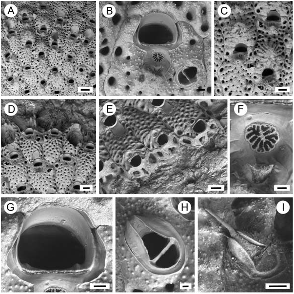
( Figures 2A–I View Figure 2 )
Microporella arctica Norman 1903, p. 105 View in CoL .
Microporella ciliata ( Pallas, 1766) Kluge 1962, p. 518 View in CoL , fig. 361; Gostilovskaya 1978, p. 214, fig. 135; Kluge 1972, p. 629, fig. 361.
Microporella svalbardensis Kuklinski and Hayward 2004, p. 82 View in CoL ; fig. 2.
Type material
Lectotype (chosen here): NHM 11.10.1.1244a (bleached specimen), Vadsö , Finmark, Norway (ca 70 ° N, 30 ° E), Norman Collection GoogleMaps . Paralectotype: NHM 11.10.1.1244b (unbleached specimen), details as for lectotype.
Other material
NHM 11.10.1.1245, Spitsbergen, Norman Collection, ex F. Smitt. NHM 11.10.1.1246, ‘‘Valorous’’ Expedition 1875, Greenland, 57 fathoms, Norman Collection. NHM 2003.3.10.7, Kongsfjorden, West Spitsbergen (79 ° 03.59N, 11 ° 39.49E), 10 m depth, 6 August 2001, P. Kuklinski Collection (holotype of M. svalbardensis ). NHM 2008.1.10.8-9, Kongsfjorden, West Spitsbergen (79 ° 01.89N, 11 ° 49.89E), 12 m depth, 3 August 2002, P. Kuklinski Collection (paratypes of M. svalbardensis ). ZI 7/5997, north coast of Murmansk, Barents Sea, dredge 12, 1895, collected by Knipovitz.
Description
Colony encrusting, developing extensive unilaminar sheets. Pore chambers with two or three small windows opening on the distal wall and one on the distolateral wall. Ancestrula not observed.
Autozooids rounded rhombic in frontal outline, separated by deep grooves, 0.52– 0.68 mm long (average 50.59 mm, n 520) by 0.22–0.49 mm wide (average 50.38 mm, n 520). Frontal shield rugose, thickly calcified, granular, perforated by numerous large pseudopores; nodular calcification developing between pseudopores as frontal shield thickens during ontogeny; areolae numbering about 14–16 per zooid, larger than pseudopores, subrounded to longitudinally elliptical. Primary orifice wider (0.12–0.16 mm, average 50.14 mm, n 520) than long (0.09–0.12 mm, average 50.10 mm, n 520), more or less semicircular, proximal border straight with a distinct condyle in each proximolateral corner, teeth lacking. Oral spines numbering two to four, basally articulated, disposed around the distal and lateral borders of the orifice, usually broken-off and overgrown in older autozooids. Secondary orifice in older zooids with distal edge formed by frontal shield calcification of succeeding zooid. Ascopore large, subcircular or crescentic, divided by thin radial septa, usually with a distinct tongue hanging downwards from the distal edge; situated close to the proximal rim of the orifice, generally set within a concave area of smooth calcification extending distally around proximal and lateral borders of orifice; large umbo developing on the proximal edge of the smooth area and becoming especially prominent in later ontogeny in certain zooids. Basal walls with uncalcified windows. Ovicell hyperstomial, prominent, wider (0.34–0.42 mm, average 50.37 mm, n 57) than long (0.26–0.37 mm, average 50.31 mm, n 57), frontal surface granular, developing a stout median umbo and sometimes radial ridges, with distinct marginal pores but imperforate centrally.
Avicularia adventitious, single (unpaired), absent in about 50% of autozooids, 0.09–0.12 mm long (average 50.11 mm, n 515) by 0.05–0.06 mm wide (average 50.06 mm, n 519), located on the right or left side of the autozooid level with the ascopore or just proximal to it, directed laterally or distolaterally; opesia semielliptical; rostrum triangular, acute to the frontal plane of the autozooid, tip channelled, crossbar calcified, straight; mandible setiform, extending beyond the tip of the rostrum for a distance equivalent to about length of rostrum.
Distribution
Confirmed records of M. arctica are from northern Norway, the Russian coast of the Barents Sea, Svalbard and Greenland ( Norman 1903; Kuklinski and Hayward 2004), i.e. the European sector of the Arctic. The species has also been reported from the ‘‘American Arctic’’ ( Osburn 1936) and the Kodiac Shelf, Alaska ( Cuffey and Turner 1987) but these occurrences need confirmation.
Remarks
Important distinguishing features of this species are the large ascopore subdivided by narrow partitions that radiate from a tongue projecting from the distal margin ( Figure 2F View Figure 2 ), the concave area of smooth calcification extending around the ascopore and bordering the proximal and lateral edges of the orifice, the umbo proximal of the ascopore, and the granular calcification of the frontal shield.
While most of the material in the Norman Collection labelled as M. arctica does belong to this species, there is one batch (‘‘Valorous’’ Expedition 1875, Greenland, 57 fathoms) containing a mixture of M. arctica and M. klugei , proving that the two Arctic species of Microporella can exist in sympatry.
Microporella svalbardensis Kuklinski and Hayward, 2004 View in CoL is here synonymized with Microporella arctica Norman, 1903 View in CoL . All important morphological details (orifice shape, ascopore, avicularia, ovicell) of M. svalbardensis View in CoL are similar to the type specimen of M. arctica View in CoL . On average, zooidal dimensions are slightly larger in the type material of M. svalbardensis View in CoL than in M. arctica View in CoL , but still fall within the range seen in the type of M. arctica View in CoL . Such variation in size could be due to differences in seawater temperatures between collection sites, as is seen in other cheilostome species (e.g., Silén and Harmelin 1976). The type material of M. arctica View in CoL was collected in Finmark in northern Norway, while that of M. svalbardensis View in CoL came from farther north in West Spitsbergen where sea temperatures are lower, consistent with the larger zooid size.
Microporella klugei View in CoL sp. nov
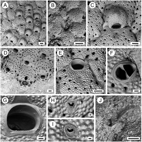
( Figures 3A–J View Figure 3 )
Microporella ciliata var. arctica Kluge 1962, p. 519 View in CoL , fig. 362; Gostilovskaya 1978, p. 215, fig. 136; Kluge, 1975; p. 631; fig. 362.
Microporella arctica View in CoL – Soule et al. 2005; fig. 1.
Type material
Holotype: NHM 2008.02.06.12, Hollender bukta, Isfjorden (77 ° N, 15 ° E), West Spitsbergen, on Chlamys islandica shell, 22 August 2000, collected by P. Kuklinski, dredge. GoogleMaps
Paratype: NHM 2008.02.06.13–15, details as for holotype.
Other material
NHM 2008.02.06.16, ‘‘Valorous’’ Expedition 1875, Greenland, 57 fathoms, Norman collection. ZI 1/3427, Spitsbergen, Green Harbour 1837, determined by G.A. Kluge, Kluge collection.
Diagnosis
Microporella with encrusting colony; autozooids large, averaging 0.79 mm long by 0.58 mm wide, frontal shield pustulose; areolae and pseudopores morphologically similar; orifice averaging 0.12 mm long by 0.15 mm wide, without teeth or condyles; oral spines lacking; ascopore small, reniform or crescentic with short, tooth-like spines, situated about one orifice length proximally of orifice, set within narrow concave area of smooth calcification, umbo often developing just proximal of ascopore; ovicell with median umbo and pores around distal and later edges only; avicularia lacking in the great majority of autozooids, single when present, situated level with the ascopore or just proximal to it, directed laterally or distolaterally, causing bulge in outline of autozooid, rostrum arch-shaped, the distal tip rounded and not channelled, mandible extending beyond tip of rostrum for a distance equivalent to about twice length of rostrum.
Description
Colony encrusting, developing extensive unilaminar sheets. Pore chambers with two or three windows opening on the distal wall and one larger more elongate window on each distolateral wall. Ancestrula damaged in available material,?tatiform, longer than wide.
Autozooids rounded rhombic in frontal outline, separated by deep grooves, 0.62–0.99 mm long (average 50.79 mm, n 520) by 0.34–0.85 mm wide (average 50.58 mm, n 520). Frontal shield thickly calcified, pustulose, the pustules smaller around the ascopore; pseudopores scattered, lacking between ascopore and orifice; areolae averaging about 12 per zooid, the same size as or slightly larger than pseudopores, subcircular or elliptical. Orifice wider (0.14–0.17 mm, average 50.15 mm, n 520) than long (0.10–0.13 mm, average 50.12 mm, n 520), more or less semicircular but widest somewhat distally of proximal margin, proximal border straight, above a slightly convex inner rim, without condyles or teeth. Oral spines not observed. Ascopore small, reniform or crescentic, margin minutely crenulate with short tooth-like spines projecting into lumen; situated proximally of the orifice by about same distance as orifice length, set within a narrow concave area of smooth calcification sometimes prolonged proximally; often a small umbo develops on frontal shield proximal to the ascopore, especially prominent in later ontogeny. Ovicell hyperstomial, prominent, almost as long (0.35–0.45 mm, average 50.40 mm, n 520) as wide (0.36–0.47 mm, average 50.39 mm, n 520); opening high, arch-shaped or rounded; frontal surface coarsely pustulose, developing a median umbo, with pores around the edge but imperforate centrally.
Avicularia adventitious, rare, present in a small minority of autozooids, single (not paired), 0.10–0.15 mm long (average 0.13 mm, n 55) by 0.08–0.10 mm wide (average 50.09 mm, n 55), situated on the right or left level with the ascopore or proximal to it, directed laterally or distolaterally, causing bulge in outline of autozooid; opesia semielliptical; rostrum arch-shaped, the distal tip rounded and not channelled, acute to the frontal plane of the autozooid, crossbar calcified, a median indentation present on proximal side of at least some examples; mandible setiform, extending beyond the tip of the rostrum for a distance equivalent to about twice length of rostrum.
Distribution
This is an Arctic circumpolar species recorded from Point Barrow, Alaska ( Soule et al. 2005), Greenland and Spitsbergen (see previous section).
Etymology
Named for G.A. Kluge (1870–1952), the Russian bryozoologist responsible for the most important monograph on Arctic bryozoans.
Remarks
Microporella klugei View in CoL sp. nov. was described by Kluge (1962, 1975) as Microporella ciliata var. arctica ( Norman 1903) View in CoL . However, M. klugei View in CoL differs in many respects from both Microporella arctica View in CoL and M. ciliata View in CoL . The zooids of M. klugei View in CoL are larger than those of both M. ciliata View in CoL and M. arctica View in CoL . The proportional size of the orifice to the zooid as a whole is smaller in M. klugei View in CoL than the other species described in this account. Furthermore, M. klugei View in CoL lacks oral spines in all post-ancestrular zooids and the orifice has neither teeth nor condyles ( Figure 3G View Figure 3 ). Avicularia are rare and when present are small in proportion to the autozooid bearing them and, unlike M. arctica View in CoL and M. ciliata View in CoL , have rostra with rounded, unchanneled tips ( Figure 3F View Figure 3 ). The ascopore of M. klugei View in CoL is small, crescentic and situated more proximally on the frontal shield than that of M. arctica View in CoL (compare Figure 2F View Figure 2 with Figures 3H and I View Figure 3 ).
Among species of Microporella View in CoL described from other regions of the globe, M. klugei View in CoL resembles M. diademata ( Lamouroux, 1825) View in CoL from New Zealand (see Gordon 1989) but this latter species has larger avicularia located more centrally on the autozooid and about 6 oral spines compared to none in M. klugei View in CoL . Oral spines are also present in another similar species, M. ventricosa Canu and Bassler, 1929 from the Philippines, and this species also has minute teeth along the proximal edge of the orifice.
No known copyright restrictions apply. See Agosti, D., Egloff, W., 2009. Taxonomic information exchange and copyright: the Plazi approach. BMC Research Notes 2009, 2:53 for further explanation.
|
Kingdom |
|
|
Phylum |
|
|
Class |
|
|
Order |
|
|
Family |
|
|
Genus |
Microporella arctica Norman, 1903
| Kuklinski, Piotr & Taylor, Paul D. 2008 |
Microporella svalbardensis
| Kuklinski P & Hayward PJ 2004: 82 |
Microporella ciliata ( Pallas, 1766 )
| Gostilovskaya MG 1978: 214 |
| Pallas PS 1962: 518 |
Microporella ciliata var. arctica
| Gostilovskaya MG 1978: 215 |
| Kluge GA 1962: 519 |
Microporella arctica
| Norman A 1903: 105 |