
Dorcopsistrongylus supriyatnai, Purwaningsih & Dewi & Smales, 2019
|
publication ID |
https://doi.org/10.11646/zootaxa.4565.3.10 |
|
publication LSID |
lsid:zoobank.org:pub:3B766431-5C26-40DD-91D2-931912CFF463 |
|
DOI |
https://doi.org/10.5281/zenodo.5922226 |
|
persistent identifier |
https://treatment.plazi.org/id/03A82709-FFA3-FFE0-9BE0-FCECAF563F59 |
|
treatment provided by |
Plazi |
|
scientific name |
Dorcopsistrongylus supriyatnai |
| status |
sp. nov. |
Dorcopsistrongylus supriyatnai n. sp.
( Figs. 16–28 View FIGURES 16–25 View FIGURES 26–28 )
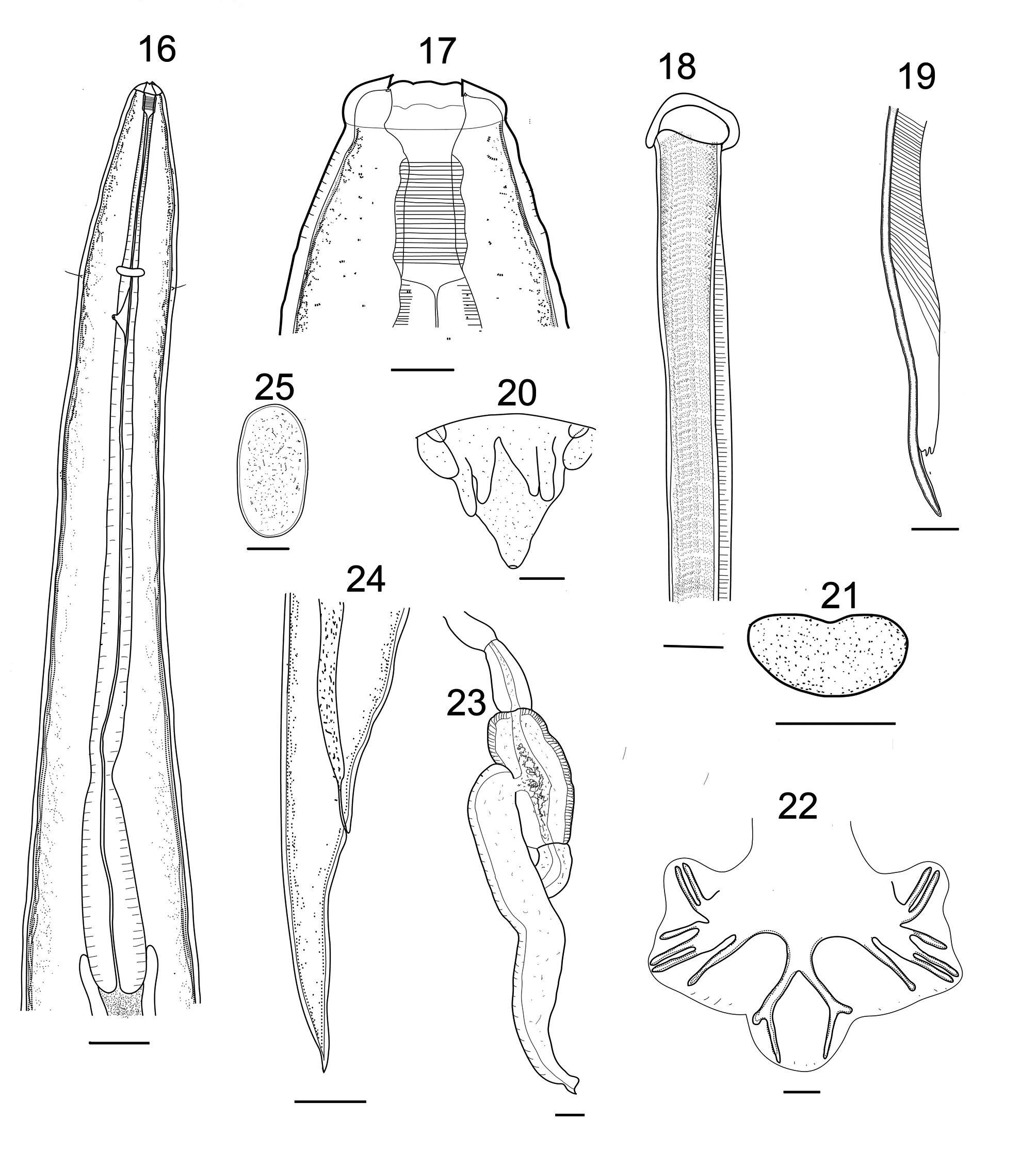
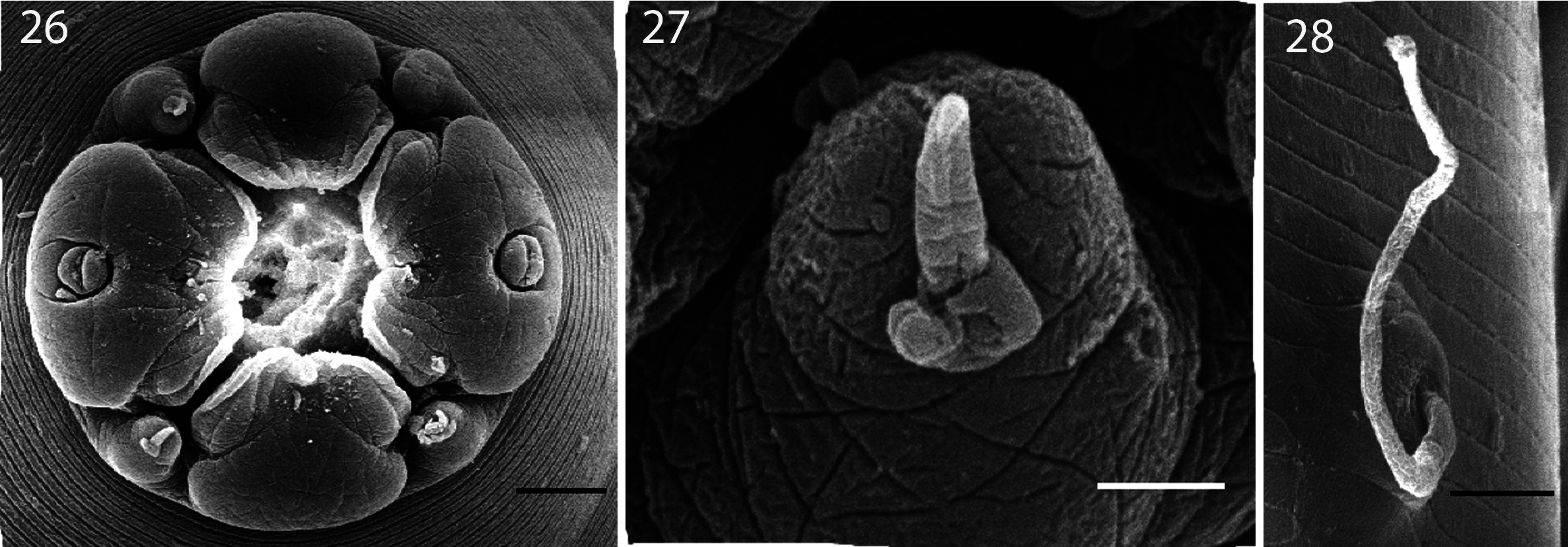
Description. General: Body small, attenuated posteriorly, cuticle with fine transverse striations. Anterior end with cephalic collar, without fine longitudinal striae, bearing 4 submedian papillae, each with single seta, between elements of the labial crown ( Fig. 27 View FIGURES 26–28 ). Mouth opening circular, labial crown divided into 4 lip-like elements, anterior end of each ridged, amphids, 1 each on base of lateral lip ( Fig. 26 View FIGURES 26–28 ). Lateral labial crown elements larger than dorsal and ventral elements. Buccal capsule cylindrical, thick walled, lining with prominent transverse striations ( Fig. 17 View FIGURES 16–25 ), longer than deep. Anterior end of buccal capsule with labial collar, lacking fine longitudinal striae. Oesophagus about 15.6% body length, lining strongly sclerotized, cylindrical with elongated terminal bulb following slight constriction ( Fig. 16 View FIGURES 16–25 ). Nerve ring at anterior extremity, deirids setate ( Figs. 16 View FIGURES 16–25 , 28 View FIGURES 26–28 ), posterior to nerve ring, excretory pore posterior to deirids. Anterior end of intestine without diverticula.
Male (n=12): Body length 9.50 (8.70–12.30) mm; width 473 (365–530). Buccal capsule 58 (50–70) long, 19 (15–23) wide. Oesophagus 1,485 (1,370–1,710) long, bulb 217(180–240) long. Nerve ring 338 (230–400), deirids 396 (320–670), excretory pore 487 (410–580) from anterior end, respectively. Bursal lobes not separate, dorsal lobe longest, without median cleft, ventral lobes shortest. Ventro-ventral and ventro-lateral rays apposed, reaching bursal margin, externo-lateral ray divergent, shorter than medio-lateral and postero-lateral, not reaching bursal margin, medio-lateral and postero-lateral apposed, reaching bursal margin, externo-dorsal ray arising from the lateral trunk, not reaching margin of bursa; dorsal trunk stout, bifurcating at about 1/4 its length, each branch giving off pair of short lateral branches at 2/3 its length ( Fig. 22 View FIGURES 16–25 ). Spicule slender, wider at proximal end ( Fig.18 View FIGURES 16–25 ), pointed at distal end ( Fig.19 View FIGURES 16–25 ), 1,942 (1,860–2,090) long, about 20.3% body length. Gubernaculum bean shaped, 17 long, 34 wide ( Fig. 21 View FIGURES 16–25 ). Genital cone prominent, dorsal lip conical, ventral lip with 1 pair of bifid appendages and 2 pair simple l appendages ( Fig. 20 View FIGURES 16–25 ).
Female (n=18): Body length 13.60 (8.25–19.00) mm; width 576 (400–680); buccal capsule 71 (60–78) long 38 (25–40) wide. Oesophagus 1,711 (1,180–1,950) long. Nerve ring 409 (330–580), deirids 496 (370–570), excretory pore 577 (410–630) from anterior end, respectively. Vulva 727 (440–749) from tip of tail. Ovejector with long and sinuous shaped, vagina vera 973 (850–1190) long, vestibule large, 283(260–305), sphincter short, 76 (50–98) and infundibulum not seen ( Fig. 23 View FIGURES 16–25 ). Tail tapering, with pointed tip, 349 (320–420) long ( Fig. 24 View FIGURES 16–25 ). Eggs thin-shelled, oval, 71(60–78) long, 38 (35–40) wide ( Fig. 25 View FIGURES 16–25 ).
Type host: Dorcopsis muelleri (Lesson) (Mammalia: Macropodidae ).
Type locality: Kumawa Mountains , West Papua, Indonesia (1°6 S, 130°51 E).
Site of infection: Stomach.
Type specimens: Holotype male, allotype female (MZB Na764); paratypes, 11 males and 17 females (MZBNa 765). Date of collection: November 2014. Etymology. The name of the species is given in honour of the collector of the host specimens. Remarks. These specimens belong in the genus Dorcopsistrongylus because they have a suite of characters including an external labial crown of four lip-like elements, a long cylindrical buccal capsule lined with prominent transverse striations and a long narrow oesophageal corpus with a clavate terminal bulb ( Smales, 1982). Dorcopsistrongylus supriyatnai n. sp. differs from all congeners in lacking large anteriorly directed intestinal diverticulae. The species is most similar to D. labiacarinatus in morphometrics ( Table 1), but further differs in having larger lateral than ventral and dorsal labial crown elements, not having fine longitudinal striae on the anterior end of the buccal capsule, a narrower buccal capsule, a longer vagina vera, the proportions of the ovejector, a shorter vestibule and longer sphincter, and a larger, bean shaped compared with a smaller rectangular gubernaculum.
Dorcopsistrongylus supriyatnai further differs from D. ewini in being a larger worm with a shorter buccal capsule, proportionally shorter oesophagus, shorter spicules without the proximal twist, larger gubernaculum, longer vagina vera and an ovejector with a shorter sphincter ( Table 1). Dorcopsistrongylus supriyatnai further differs from D. salawatiensis in being a larger worm with a narrower buccal capsule, proportionally shorter oesophagus and spicules, larger gubernaculum, longer vagina vera, and an ovejector with a smaller vestibule and sphincters ( Table 1). Dorcopsistrongylus supriyatnai , D. ewini and D. salawatiensis are all found in Do. muelleri from different localities in Papua, while D. labiacarinatus is found in Do. luctuosa , from Papua New Guinea.
No known copyright restrictions apply. See Agosti, D., Egloff, W., 2009. Taxonomic information exchange and copyright: the Plazi approach. BMC Research Notes 2009, 2:53 for further explanation.
|
Kingdom |
|
|
Phylum |
|
|
Class |
|
|
Order |
|
|
Family |
|
|
Genus |