
Pissodes yunnanensis Langor and Zhang, 1999
|
publication ID |
https://doi.org/ 10.1649/072.065.0215 |
|
persistent identifier |
https://treatment.plazi.org/id/03A84D5D-FFD5-FFF2-E4FF-FE34684F8398 |
|
treatment provided by |
Diego |
|
scientific name |
Pissodes yunnanensis Langor and Zhang, 1999 |
| status |
|
Pissodes yunnanensis Langor and Zhang, 1999 View in CoL ( Figs. 1–9 View Figs View Figs View Figs , 10 View Figs , 12 View Figs )
Pissodes yunnanensis Langor and Zhang View in CoL , in Langor et al. 1999: 601
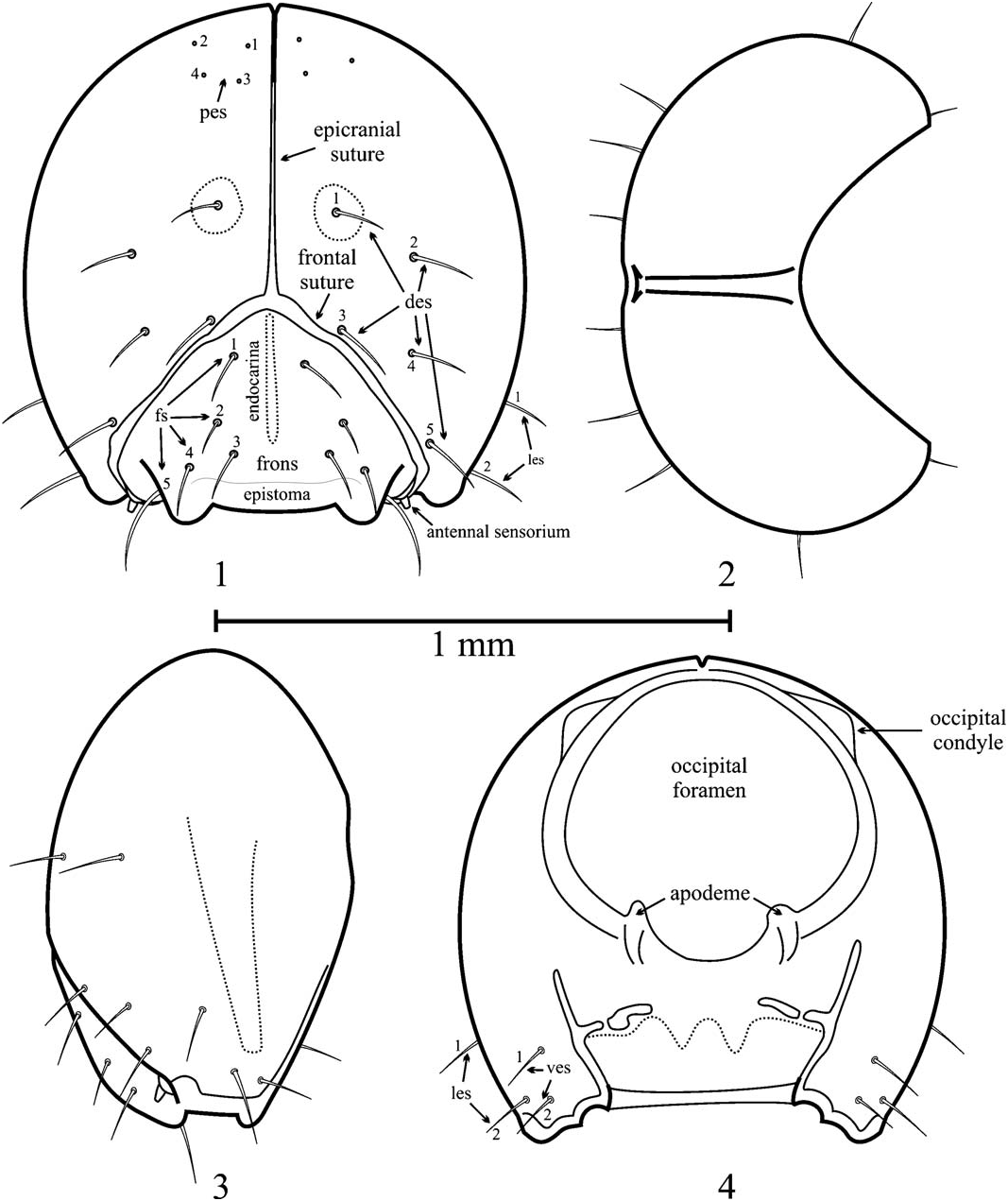
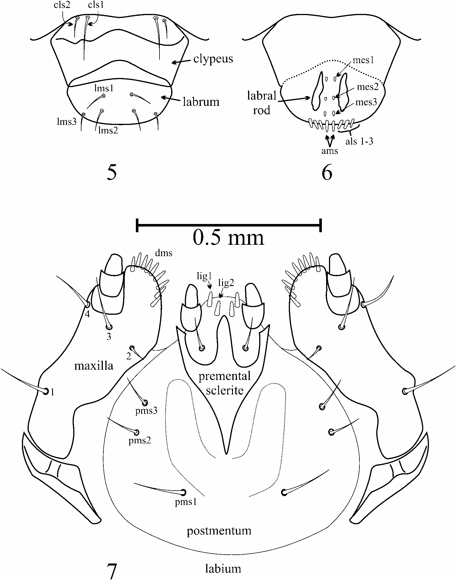
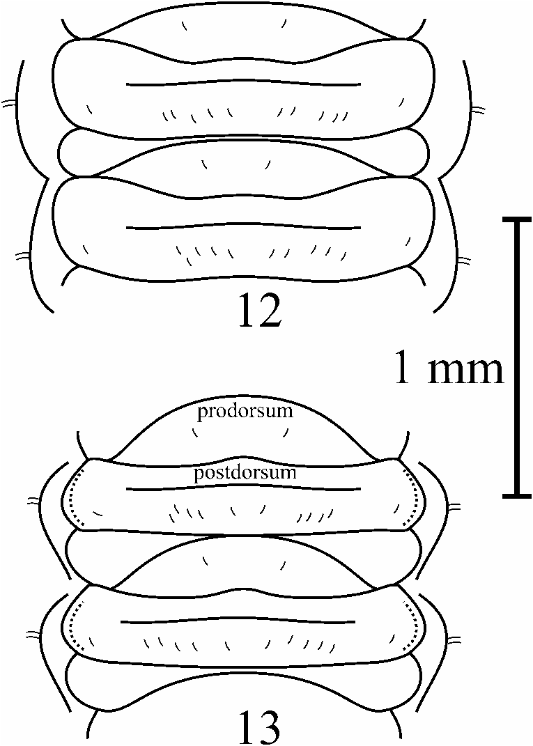
Diagnosis. Larvae of P. yunnanensis may be separated from those of other known Pissodes species by the following combination of characters: head with pale area on center of dorsal surface present only around base of des1 ( Fig. 1 View Figs ); antenna with a conspicuous, elongate sensorium, as long as wide at base or longer, with truncate apex ( Fig. 1 View Figs ); occipital foramen with ventral apodemes, curvature of foramen between apodemes discontinuous with remainder ( Fig. 4 View Figs ); most specimens with pigmentation of pronotum pale and present only as a faintly pigmented stripe near anteromedial margin to faintly pigmented on mediodorsal ¼ of sclerite, rarely absent ( Fig. 10 View Figs ); and prodorsum of abdominal segments with posterior margin medially sinuate, prodorsum arcuate ( Fig. 12 View Figs ).
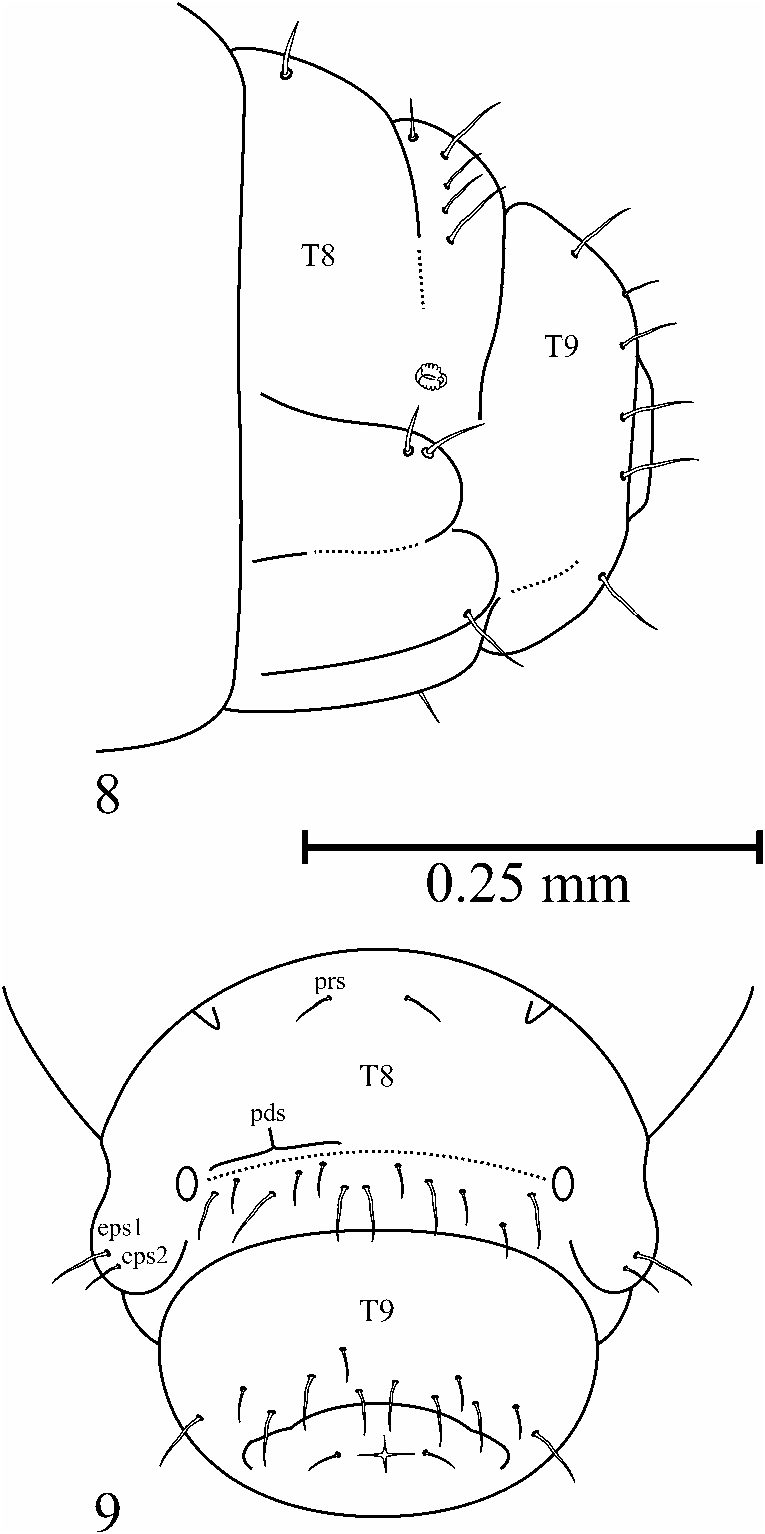
Description. Larva typical curculionid in form, slightly arcuate or comma-shaped in natural position, with free, sclerotized head. Head light brown, thorax and abdomen creamy white. Most of integument covered with fields of minute, stout microtrichia, except for head capsule, pronotum, integumental folds, posterior surface of abdominal segment IX, and around bases of setae. Color light brown; epistoma, cataphophyses, posterior 0.25 of epicranial suture, and endocarina dark brown and sclerotized; epicranial and frontal sutures clear and pale; pale markings on dorsal surface limited to small ovoid areas around des1 setal bases ( Fig. 1 View Figs ). Side of head capsule with slightly indented and unpigmented stripe from near ventral mandibular articulation 0.70–0.80 of distance to posterior margin of head; unpigmented stripe directed toward ves2 anteriorly ( Fig. 3 View Figs ). Pigmented and sclerotized bars on ventral surface moderately well developed ( Fig. 4 View Figs ). Head capsule round in shape, width/length in anterior view 0.98–1.00 ( Figs. 1, 4 View Figs ); posterior margin in lateral view with middle quarter flat to slightly concave ( Fig. 3 View Figs ). Epicranial suture continuous and distinct from occipital foramen to frontal suture; fused on posterior quarter ( Figs. 1, 2 View Figs ). Endocarina covering 0.66 of distance between epicranial suture and epistoma ( Fig. 1 View Figs ). Frons triangular; length/width = 0.65–0.70; frontal sutures weakly arcuate, anteriorly reaching antenna; antennae with sensorium elongate, slightly longer than wide, lightly pigmented, conical in shape ( Fig. 1 View Figs ). Epistoma and frontoclypeal sutures weakly arcuate ( Fig. 1 View Figs ). Occipital foramen with anterior half less concave than posterior half, widest point near anterior margin; ventral apodemes moderately well developed, foramen curvature between apodemes greater than and discontinuous with remainder of foramen ( Fig. 4 View Figs ). pes1–pes 4 in quadrate arrangement in specimens with all four present, but number of setae variable, any or all setae apparently lacking in some specimens; when present very minute or represented by clear socket; hidden by thorax when head in normal position ( Fig. 1 View Figs ). Cephalic setae: des3–des5 evenly spaced in arc basad of frontal suture; les1 about half as long as les2; setae separated by less than length of les2; les2 close to anterior margin of head ( Fig. 1 View Figs ), ves1 shorter than ves2 and both setae less than 0.66 as long as les2; ves2 near anterior margin adjacent to posterior mandibular articulation ( Fig. 4 View Figs ); fs1 near basal angle of frons, and fs2 anteriad of fs1, fs3 and fs4 midlaterally near epistoma, and fs5 anterolateral, adjacent to catapophysis; fs5 about 0.33 longer than other frontal setae ( Fig. 1 View Figs ). Clypeus width/ length = 2.0–2.3; lateral margins convex basally, straight apically; apical margin broadly concave; proximal 0.33 sclerotized and pigmented, apical 0.66 translucent, margin between these areas concave and irregular; cls1 length about 0.66 of clypeal length, cls2 length 0.5 or less than clypeal length ( Fig. 5 View Figs ). Labrum width/length about 1.2; basal margin moderately produced and convex; lms1 shorter than distance between clypeus and labrum at midline, lms2 longer than lms1, lms3 slightly shorter than lms2; apex with als1–als3 present, these plus ams minute ( Fig. 5 View Figs ). Labral rods subparallel, attached to outer labral surface about 0.33 of distance between midline and apical labral corners; mes1–mes3 present, mes1 shorter and thinner than mes2 and mes3 ( Fig. 6 View Figs ). Maxillary mala with a row of 8 or 9 dorsal setae (dms); stipes of maxilla with inner and outer margins sinuate; inner margin emarginated and unpigmented and outer margin produced into a convex protuberance, about 0.33 of distance between cardo and apex of mala; protuberance blunt tipped, with acute, unpigmented apex; stipital seta 3 and seta 4 as long as or longer than palp; seta 2 minute, seta 1 slightly longer than seta 3 and 4 ( Fig. 7 View Figs ). Premental sclerite of labium with anterior and posterior extensions well developed, the later subtriangular or acute at apex; premental seta as long as or slightly longer than labial palp ( Fig. 7 View Figs ). Ligular seta 1 and seta 2 as long as apical segment of labial palp ( Fig. 7 View Figs ). Postmentum membranous, with elongate-oval pigmented area on each side between premental sclerite and postmental setae, forming pigmented U-shaped area ( Fig. 7 View Figs ); pms 1 and pms 2 setae about 1.5 times as long as labial palp, pms 3 shorter than labial palp ( Fig. 7 View Figs ). Pronotum in most specimens with pair of lightly pigmented sclerites that cover most of pronotum in dorsal view; most specimens with pigmentation of pronotum pale and present only as a faintly pigmented stripe near anteromedial margin to faintly pigmented on mediodorsal ¼ of sclerite, rarely absent ( Fig. 10 View Figs ). Spiracular area as longest pleural seta; 3 on anterior pronotal margin, all slightly shorter than longest pleural seta; 1 on posterior pronotal margin about midway between midline and lateral pronotal margin, about as elongate oval, smooth, flat, lacking in microtrichia, bearing a spiracle and unpigmented to faintly pigmented depression; spiracle large, bicameral, oval; depression about the size of, and anteroventral to spiracle. Epipleurum forming a triangular area above pleural sclerite, continuous with pleurum. Pleurum separated from epipleurum by a broad shallow groove and from pedal area by a more distinct one. Eusternum triangular, about equilateral; surface about half that of pedal area plus pleurum; a pigmented, depressed, irregularly shaped, elongate sclerotized area at posterior apex. Pedal area irregularly oval, unpigmented, lacking microtrichia, about the same size as pleural sclerite. Pronotum with 10 dorsal setae per side; 2 medial on sclerite, about as long long as longest pleural seta; 2 pairs of 2 setae dorsal to spiracle one pair near pronotal sclerite and one near spiracle, about half as long as longest pleural seta. Pleural sclerite with ps1 and ps2 subequal and longer than other prothoracic setae. Eusternum with setae about half a long as longest pleural seta, about midway between anterior and posterior eusternal margins. Pedal area with 5 or 6 setae, in most specimens the 6 th seta displaced medially and separate from pedal sclerite. Meso- and metathorax with prodorsum lens-shaped, medial, comprising about 0.5–0.6 of width of thorax in dorsal view; postdorsum more narrow medially than laterally, about 0.5–0.6 as wide as prodorsum at middorsal line; epipleurum bearing a lightly sculptured and depressed, unpigmented, irregularly oval bare area that is lacking in microtrichia; pleurum bearing a lightly sculptured, unpigmented, irregularly oval bare area that is lacking in microtrichia; eusternum triangular, anterior margin longer than posterolateral margins, nearly as great in area as pedal area plus pleurum; pedal sclerite ovoid, unpigmented to slightly pigmented, lacking microtrichia, larger than pleural sclerite. Thoracic setae similar in thickness and pigmentation to setae of head; length of longest prothoracic setae about 0.75 longest setae of head, longest setae of meso- and metanotum slightly shorter. Meso- and metanotum with prs about 0.33 or less as long as longest epipleural seta; postdorsum with pds1 minute, pds2–pds4 progressively longer with pds4 slightly shorter than longest epipleural seta; alar area with a single seta; epipleurum with 1 or 2 setae dorsad and 1 seta venterad of pleural sclerite; dorsal and ventral setae subequal in length and usually the longest setae on the segment; when second dorsal seta is present it is shorter; pleurum with 1 seta, about as long as the longest epipleural seta; eusternum with setae minute; pedal area setation similar to prothorax. Abdominal segments I–VII with prodorsum arcuate and acute ended, medial, wider than metathoracic prodorsum ( Fig. 12 View Figs ; anterior section of postdorsum 0.25–0.33 as long as posterior section; postdorsum narrower medially than laterally, transverse fold somewhat obscure near spiracles; spiracular area keyhole-shaped, faintly sculptured, concave, microtrichia absent; spiracle about 0.6 as large as thoracic spiracle, bicameral, slightly oval, with anteroventral depression absent to present but unpigmented; epipleurum with smooth area between seta bases separate, round to oval; pleurum with smooth area between setal bases separate, round to oval; pleurum and epipleurum about the same size; eusternum delimited laterally by poorly developed oblique folds, weakly arcuate, area 3–4 times greater than pedal area; sternellum thin, posterior, arcuate, separated from eusternum by a well-developed groove. Setation of abdominal segments I–VI with prs minute, about 0.5 of dis- tance between dorsal midline and lateral margin; pds1–pds5 minute to about as long as longest epipleural seta, irregularly spaced between midline and spiracle; 1 ss present; epipleurum with eps1 0.50–0.66 as long as eps2, eps2 longest seta on segment in most specimens; pleurum with ps1 about 0.5 as long as, and ps2 about 0.7 as long as, eps2; eusternum with a row of 4 setae, minute to about 0.5 as long as eps2; pedal area with setae minute to about 0.5 as long as eps2. Setation of abdominal segment VII similar to preceding segments, except some postdorsal setae longer than epipleural setae. Abdominal segment VIII with prodorsum with prs near dorsal midline, about 0.5 as long as eps2; postdorsum less convex than preceding segments, about 0.33 as wide as prodorsum, and bearing 5 rather long pds, irregularly spaced near posterior margin, and together with the longest ss, which is shifted dorsally, and minute ss dorsal to spiracle and about 0.33 as long as eps2 ( Figs. 8, 9 View Figs ); spiracular area located near posterior margin of postdorsum ( Figs. 8, 9 View Figs ). Abdominal segment IX with dorsum separated from pleurum by shallow groove or change of convexity, or continuous; ventrolateral grooves separating dorsum and sternum well developed ( Figs. 8, 9 View Figs ). Abdominal segment X lacking microtrichia, with 1 minute seta per side ( Fig. 9 View Figs ); anus terminal, X-shaped; dorsal and lateral lobes subequal ( Fig. 9 View Figs ). Epipleurum with eps1 and eps2 longer than epipleural setae on preceding segments. Pleurum with 1 ps, about 0.66 as long as eps2. Eusternal setae minute to about 0.5 as long as eps2, arranged in pairs, one on each side. Pedal area with 2 setae, one about 0.66 as long as eps2, the other minute. Dorsum of abdominal segment IX with 6–7 setae per side, irregularly scattered on apical half, most specimens with 5 long and 1 or 2 minute setae; sternum with 4 setae, a minute pair near midline and a larger pair about midway between midline and lateral margins ( Fig. 9 View Figs ).
Material Examined. China. Guizhou Province. Haila , 26°49′N 103°46′E, iv.2003, col. Hongrui Zhang, 1 second instar, 4 third instar GoogleMaps . Kunming Province. Shongming, ex. Pinus yunnanensis , v.2003, col. Hongrui Zhang, 1 second instar, 2 third instar, 3 pupae, 2 adults . Sichuan Province. Sichang , 27°90′N 102°25′E, ex. Pinus yunnanensis , 10–20. v.1993, col. N. Jielin, 1 first instar, 10 third instar, 26 fourth instar, 26 pupae . Yunnan Province. Dashao , 25°26′N 102°56′E, 26.1 v.2003, col. Hongrui Zhang, 4 third instar, 2 fourth instar, 3 pupae GoogleMaps ; Lashi , 26°48′N 100°04′E, 11.iv.2004, col. Hongrui Zhang, 3 fourth instar, 3 pupae GoogleMaps ; Linjiang , ex. Pinus yunnanensis , v.2002, col. Hongrui Zhang, 1 third instar, 2 fourth instar, 3 pupae, 2 adults ; Songming County, ex. Pinus yunnanensis , iv.2003, col. D. Langor, 2 third instar, 3 fourth instar, 1 pupa .
15) Posterior aspect (habitus dorsal view); 16) Lateral aspect; 17) Ventral aspect (habitus posterior view).
No known copyright restrictions apply. See Agosti, D., Egloff, W., 2009. Taxonomic information exchange and copyright: the Plazi approach. BMC Research Notes 2009, 2:53 for further explanation.
|
Kingdom |
|
|
Phylum |
|
|
Class |
|
|
Order |
|
|
Family |
|
|
Genus |
Pissodes yunnanensis Langor and Zhang, 1999
| Williams, Daryl J. & Langor, David W. 2011 |
Pissodes yunnanensis
| Langor 1999: 601 |