
Anoplophrya vulgaris de Puytorac, 1954
|
publication ID |
https://doi.org/ 10.5852/ejt.2019.559 |
|
publication LSID |
lsid:zoobank.org:pub:8DC07412-8619-4A03-B524-04019880B9D6 |
|
DOI |
https://doi.org/10.5281/zenodo.3475265 |
|
persistent identifier |
https://treatment.plazi.org/id/03A86948-FFEE-A07E-FDBA-FC3F86BE5815 |
|
treatment provided by |
Plazi |
|
scientific name |
Anoplophrya vulgaris de Puytorac, 1954 |
| status |
|
Anoplophrya vulgaris de Puytorac, 1954
Figs 10 View Fig , 11 View Fig A–C
Description
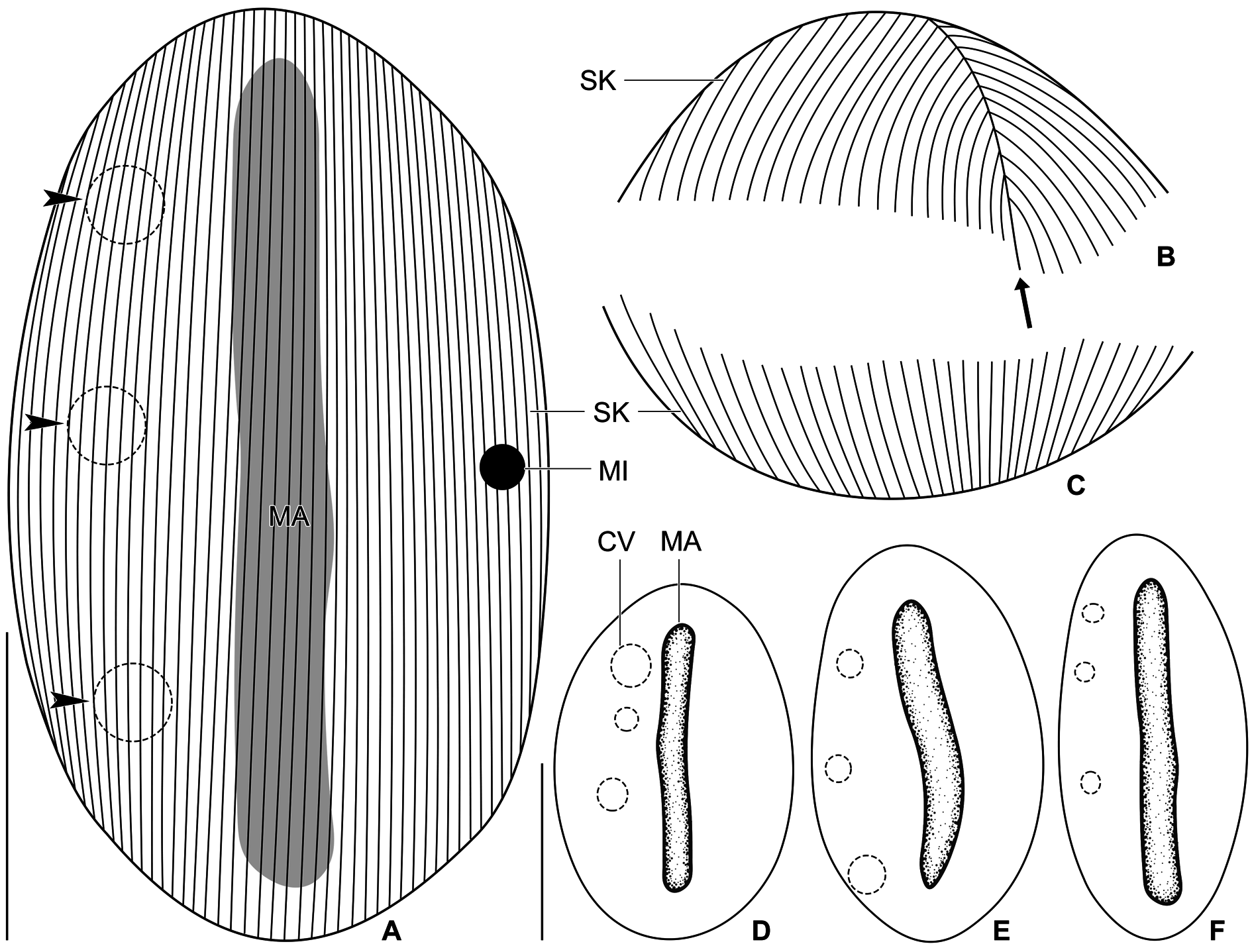
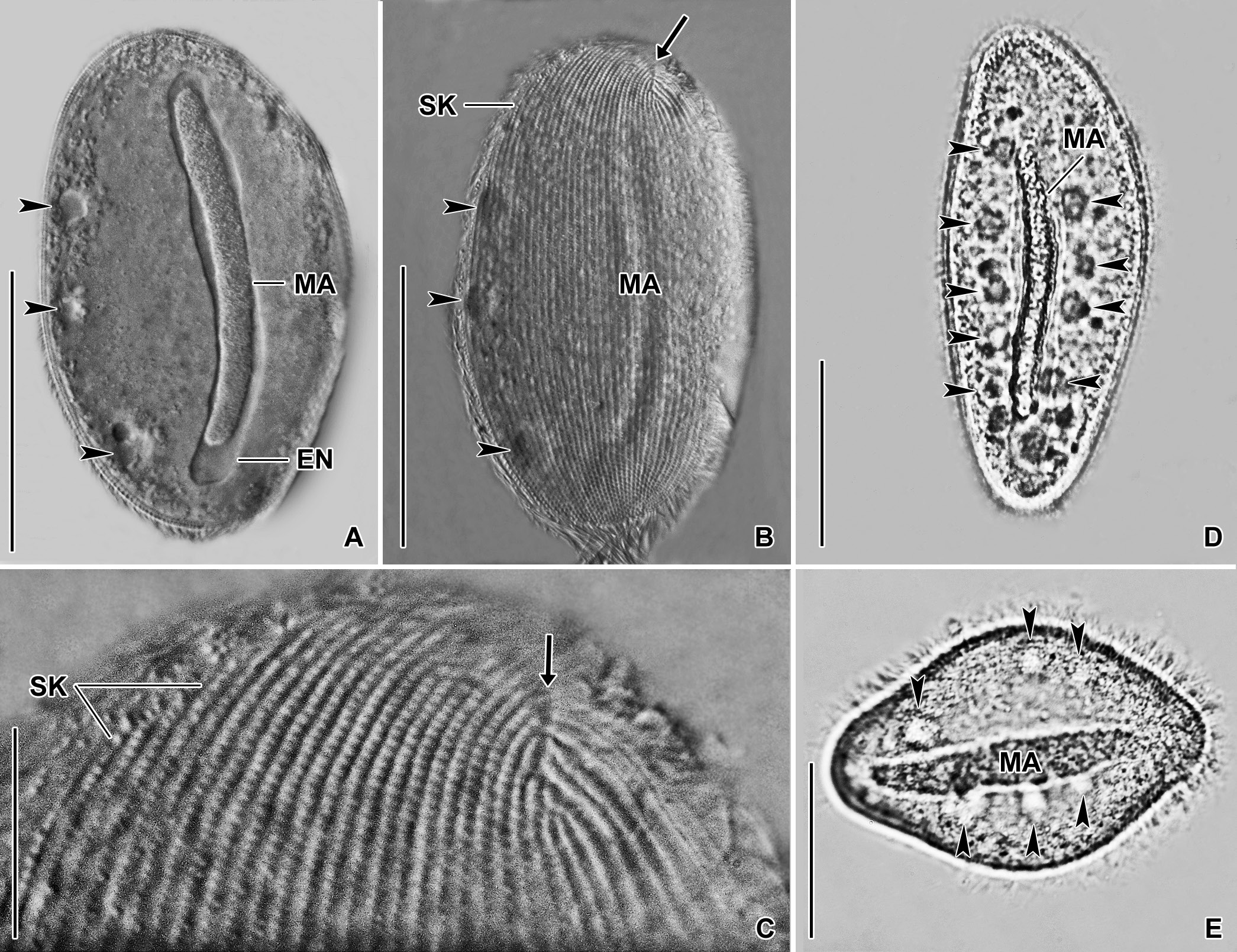
The body size is about 100–145 × 60–80 µm. The body shape is broadly elliptical to elliptical with both ends rounded. The cell is distinctly dorsoventrally flattened ( Figs 10A View Fig , D–F, 11A–B).
The nuclear apparatus is composed of a single macronucleus and a single micronucleus.The macronucleus begins about 6 µm away from the anterior body end extends through the cell’s midline into the posterior body region. The length of macronucleus varies from 80 to 110 µm and its width ranges from 9 to 18 µm. The macronuclear surface is smooth. The macronucleus diminishes in size leaving behind a conspicuous hyaline envelope in dying cells. The micronucleus is situated conspicuously far away from the macronucleus, namely, near the middle of the left body margin and opposite to the row of contractile vacuoles. The micronucleus is globular and approximately 4 µm in diameter ( Figs 10A View Fig , D–F, 11A–B).
There is a single row of contractile vacuoles arranged along the right cell margin. It is composed of three or four vacuoles being 5–9 µm across during diastole ( Figs 10A View Fig , D–F, 11A–B). The cytoplasm is colorless and studded with granules measuring approximately 0.5–1.0 µm in diameter. The cortex is semi-rigid and without specific granules. Swims moderately fast by rotation about the main body axis.
Somatic ciliature is holotrichous and composed of kineties meridionally extending over both cell sides. There are on average 34 (24–45) kineties on each body side. Individual kineties are very narrowly arranged and the interkinetal distance ranges from about 0.6 to 2.0 µm. Likewise, basal bodies are very narrowly spaced within kineties and the intrakinetal distance is ca 1 µm. There is an apical and a terminal suture at each cell pole where individual somatic kineties commence and terminate, respectively ( Figs 10 View Fig A–C, 11B–C).
Occurrence
Anoplophrya vulgaris was recorded in two species of epigeic earthworms, viz., in E. fetida and D. veneta . Ciliate 18S rRNA gene sequences originated from both hosts were identical. Host earthworms came from a compost heap in the Botanical Garden of Comenius University and at the Jakubská ulica street in Rača as well as from humous soil with a high content of decomposing plant material from a garden at the foothill of the Malé Karpaty Mts. ( Table 2 View Table 2 ). Endosymbiotic ciliates typically occurred in the middle part of the gastrointestinal tract. There were usually 10 to 15 endosymbionts per host.
No known copyright restrictions apply. See Agosti, D., Egloff, W., 2009. Taxonomic information exchange and copyright: the Plazi approach. BMC Research Notes 2009, 2:53 for further explanation.
|
Kingdom |
|
|
Phylum |
|
|
Class |
|
|
Order |
|
|
Family |
|
|
Genus |