
Ocypode sinensis Dai
|
publication ID |
https://doi.org/10.11646/zootaxa.3760.3.4 |
|
publication LSID |
lsid:zoobank.org:pub:1787FEE3-CDF8-4FA5-86E0-39FE5080CDF7 |
|
DOI |
https://doi.org/10.5281/zenodo.5680590 |
|
persistent identifier |
https://treatment.plazi.org/id/03A887A3-1A59-EA70-59F2-8B5CFC47FD94 |
|
treatment provided by |
Plazi |
|
scientific name |
Ocypode sinensis Dai |
| status |
|
Ocypode sinensis Dai View in CoL , Song & Yang, 1985
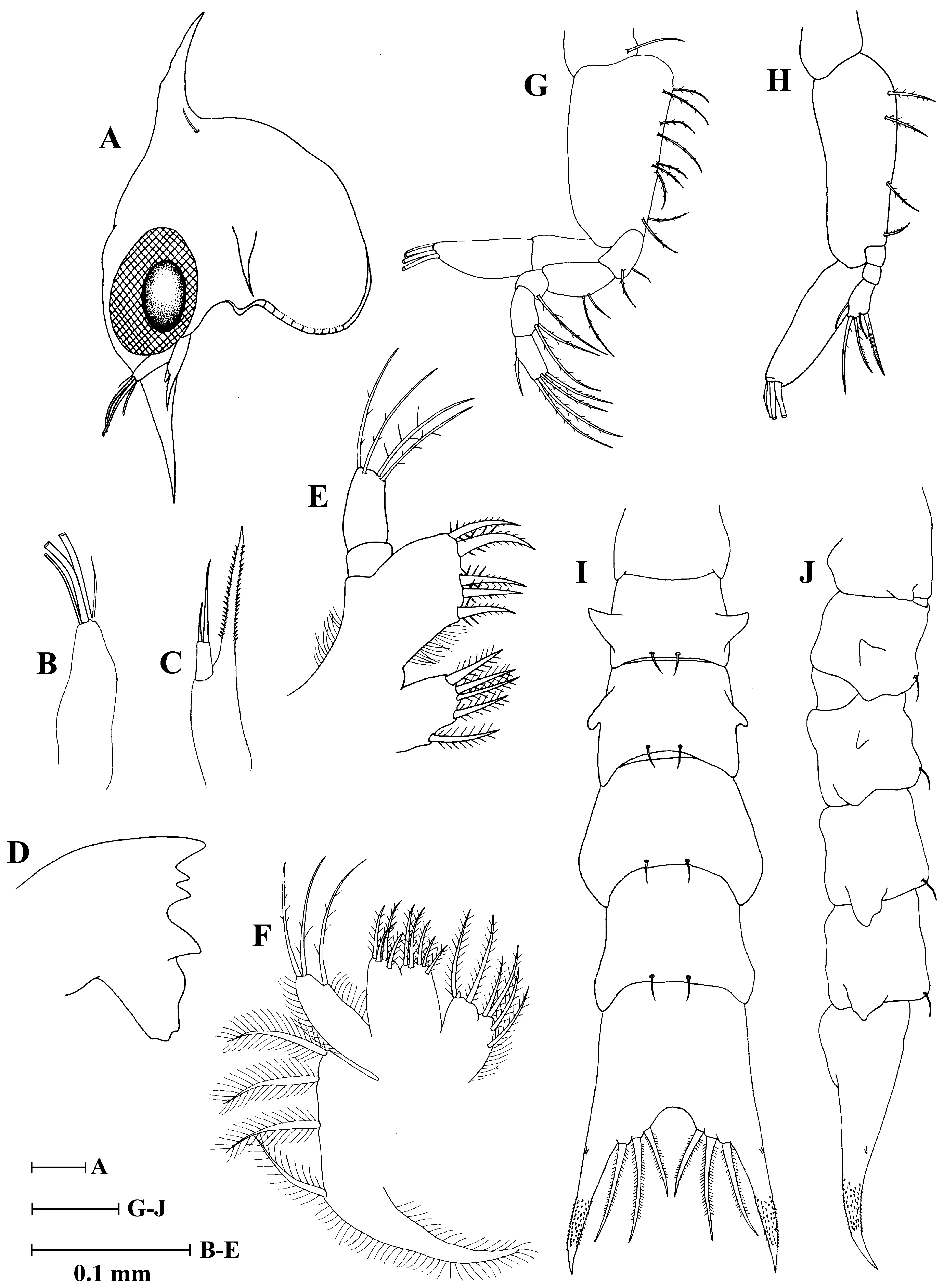
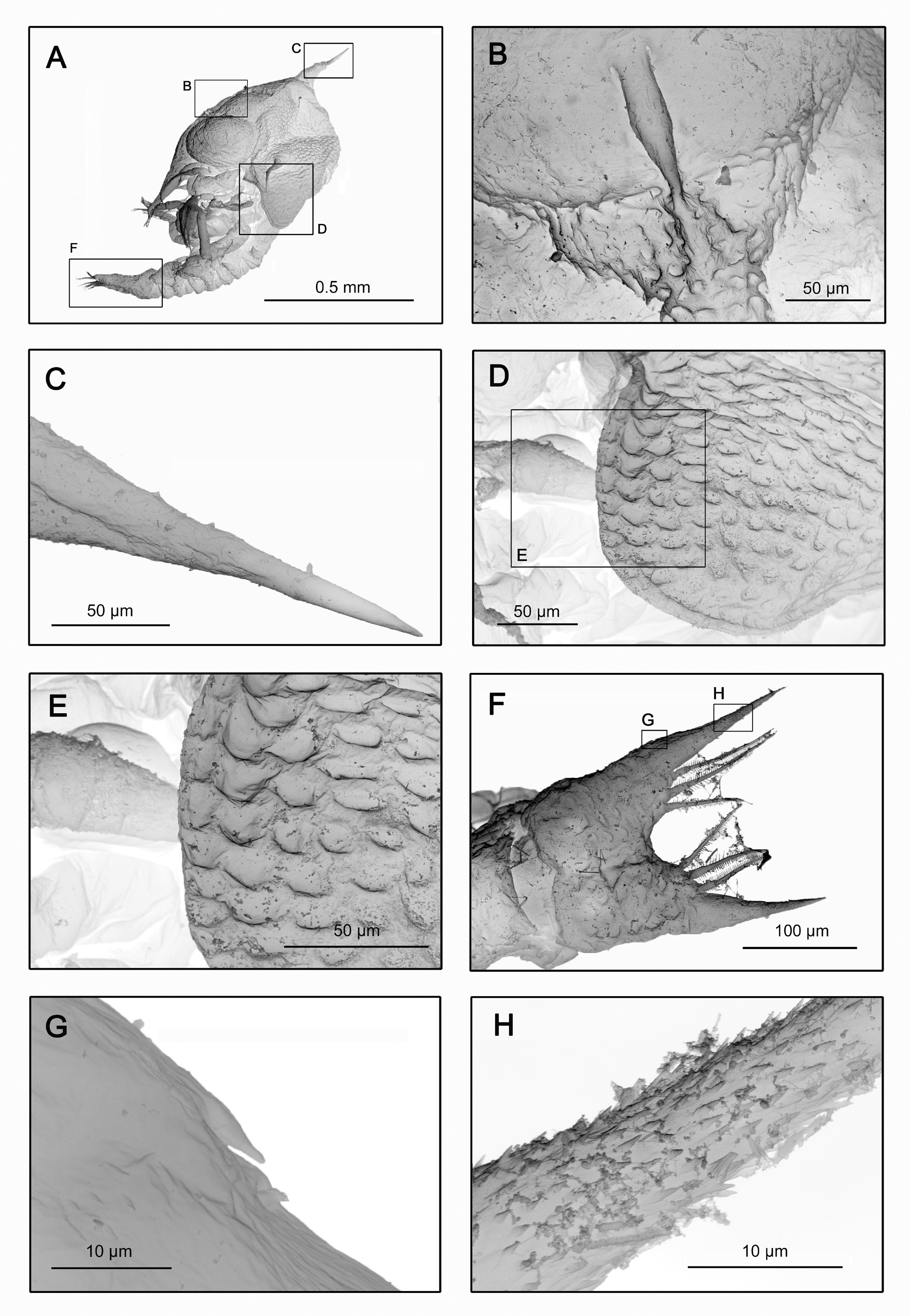
( Figs. 3 View FIGURE 3 , 7 View FIGURE 7 )
Carapace ( Figs. 3 View FIGURE 3 A, 7A–E): Pockmarked patterns on surface, bearing indistinct rectangular patterns on ventral margin ( Fig. 7 View FIGURE 7 D, E); frontal foramen present ( Fig. 7 View FIGURE 7 B); dorsal spine with some tubercles ( Fig. 7 View FIGURE 7 C), slightly straight, relatively short, approximately same length as rostral spine; rostral spine slightly longer than antennal protopod, without distal spinulation; lateral spines shorter, slightly curved downward; 1 pair of posterodorsal setae; eyes sessile.
Antennule ( Fig. 3 View FIGURE 3 B): Uniramous, endopod absent; exopod unsegmented with 3 (2 broad, 1 slender) terminal asethetascs, 1 terminal seta.
Antenna ( Fig. 3 View FIGURE 3 C): Protopod longer than antennule, bearing two rows of small lateral spines; endopod absent; exopod short, with 2 subterminal setae (1 long, almost reaching tip of protopod, 1 shorter).
Mandible ( Fig. 3 View FIGURE 3 D): Endopod palp absent.
Maxillule ( Fig. 3 View FIGURE 3 E): Epipod seta absent; coxal endite with 5 plumose setae; basial endite with 5 setal processes; endopod 2-segmented, proximal segment without setae, distal segment with 4 terminal setae; exopod seta absent.
Maxilla ( Fig. 3 View FIGURE 3 F): Coxal endite bilobed, proximal lobe with 4 setae, distal lobe with 2 setae + 1 rudimentary seta; basial endite bilobed with 5+4 setae; endopod with 1+2 terminal setae; scaphognathite margin with 4 setae, 1 long stout plumose distal process.
First maxilliped ( Fig. 3 View FIGURE 3 G): Coxa with 1 seta; basis with 9 setae arranged as 2, 2, 3, 2; endopod 5-segmented with 2, 2, 1, 2, 5 (1 subterminal + 4 terminal) setae; exopod 2-segmented, distal segment with 4 long terminal plumose natatory setae.
Second maxilliped ( Fig. 3 View FIGURE 3 H): Coxa without setae; basis with 4 setae arranged as 1, 1, 1, 1; endopod 3- segmented with 0, 0, 5 (3 subterminal + 2 terminal) setae; exopod unsegmented, distal segment with 4 long terminal plumose natatory setae.
Third maxilliped: Absent.
Pereiopods: Absent.
Abdomen ( Fig. 3 View FIGURE 3 I, J): With 5 somites; somites 2, 3 with pair of dorsolateral processes; somite 1 with rounded posterolateral processes, somites 2–3 with short posterolateral spinous processes, somites 4–5 with expanded posterolateral spinous processes; somites 2–5 with 1 pair of posterodorsal setae; pleopods absent.
Telson ( Figs. 3 View FIGURE 3 I, J, 7F–H): Telson bifid, curved upward distally; with 1 pair of minute lateral spines; generally pointed on each furca; posterior margin with 3 pairs of stout pointed spines.
No known copyright restrictions apply. See Agosti, D., Egloff, W., 2009. Taxonomic information exchange and copyright: the Plazi approach. BMC Research Notes 2009, 2:53 for further explanation.
|
Kingdom |
|
|
Phylum |
|
|
Class |
|
|
Order |
|
|
Family |
|
|
Genus |