
Meristotheca lysonensis X.-V. Nguyen, X.-T. Nguyen, Kittle & McDermid, 2022
|
publication ID |
https://doi.org/ 10.11646/phytotaxa.574.2.2 |
|
DOI |
https://doi.org/10.5281/zenodo.7383482 |
|
persistent identifier |
https://treatment.plazi.org/id/03A887A3-FF84-FFBB-1DE8-2EB7A00259C4 |
|
treatment provided by |
Plazi |
|
scientific name |
Meristotheca lysonensis X.-V. Nguyen, X.-T. Nguyen, Kittle & McDermid |
| status |
|
Meristotheca lysonensis X.-V. Nguyen, X.-T. Nguyen, Kittle & McDermid sp. nov. ( Figs. 3–14 View FIGURES 3–6 View FIGURES 7–10 View FIGURES 11–14 )
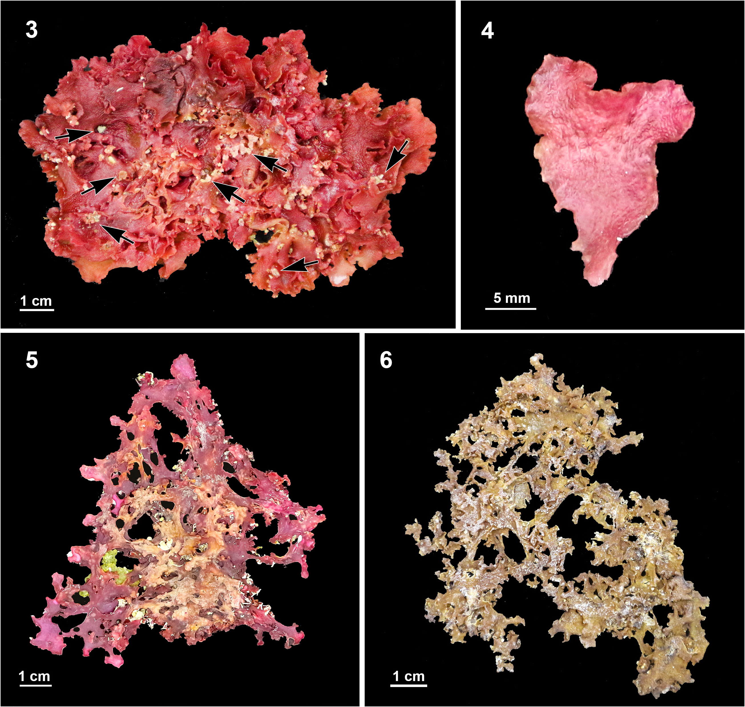
Holotype: VIETNAM. Quang Ngai: Ly Son Island ; 15.3743 ° N; 109.1329 ° E, 15 February 2021, coll. X.- V. Nguyen & X.- T. Nguyen (ION - CS 210215b, tetrasporophyte, Fig. 3 View FIGURES 3–6 ). GenBank accession number for rbc L and COI-5 P: MZ 770759 View Materials and MZ 770756 View Materials GoogleMaps
Isotypes: VIETNAM. Ninh Thuan: Thai An ; 11.5648 ° N; 109.1269 ° E, 20 February 2021, coll. X.- V GoogleMaps . Nguyen & X. - T . Nguyen (ION - CS 210301b, tetrasporophyte, Fig. 5 View FIGURES 3–6 ). GenBank accession number for rbc L and COI-5 P: MZ770761 View Materials and MZ770758 View Materials . VIETNAM. Quang Ngai: Ly Son Island ; 15.3743 ° N; 109.1329 ° E, 02 April 2003, coll. H GoogleMaps .- D. Nguyen & H.- T . Pham (ION -01011b, male plant, Fig. 6 View FIGURES 3–6 ). GenBank accession number for rbc L and COI-5 P: MZ770760 View Materials and MZ770757 View Materials
Habitat: Growing on dead corals in the mid-to lower intertidal zone where subjected to strong wave action.
Etymology: The specific epithet refers to Ly Son Island, the type locality of the new species in Viet Nam.
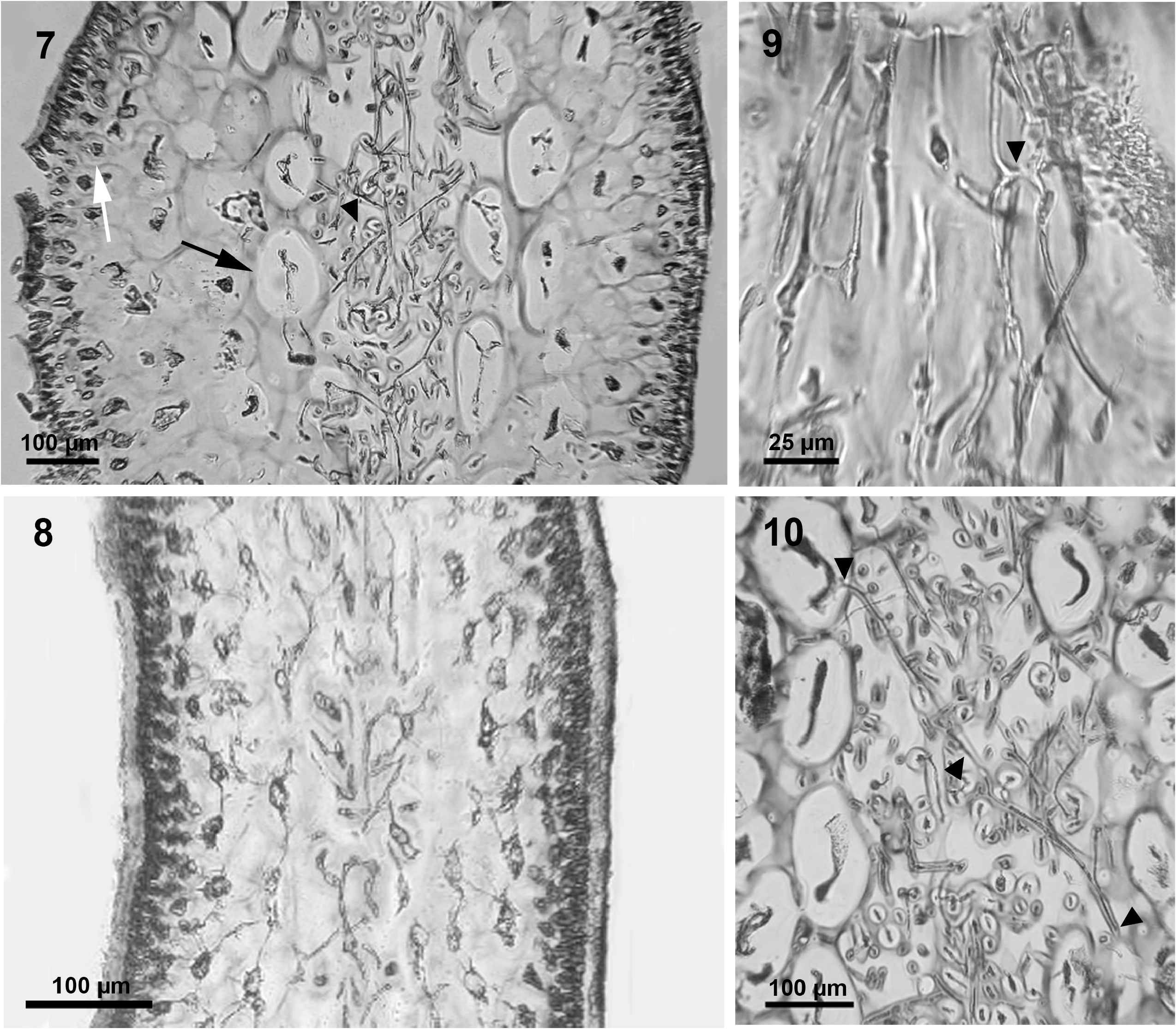
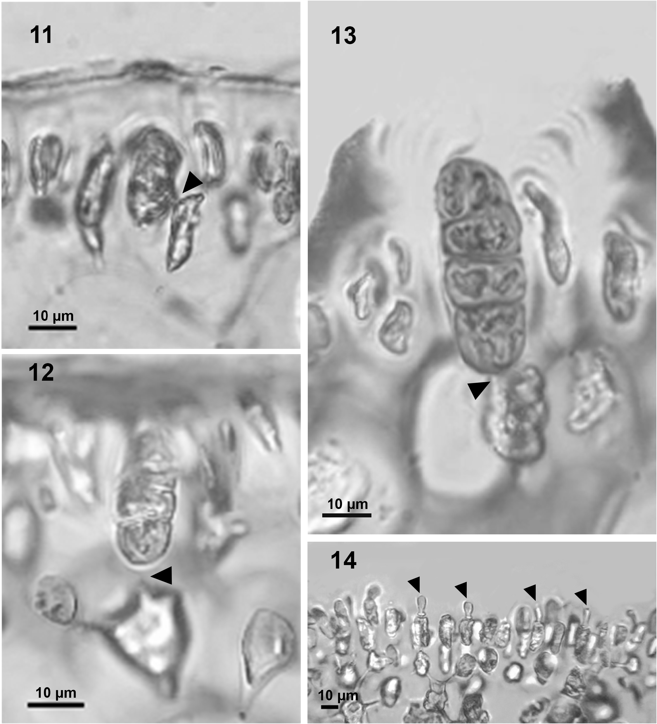
Description: Thalli prostrate or forming decumbent prostrate clumps; thallus attached to the substratum by many secondary holdfasts; thalli flattened, gelatinous in texture, deep rose-red to dark red, 10–20 cm tall with small papillae on the surface; divided into many broad segments in an irregularly dichotomous manner with rounded apices, becoming gradually narrower above and tapering below to a cuneate or sometimes cordate or heart-shaped base with a short slender stipe. Mature blades are 1–2 cm broad, margins entire at the beginning, later giving rise to irregularly branched obvious proliferations, broader or narrower intermixed; surface smooth, sometimes roughened by the presence of warty or short spinose processes ( Figs 3, 4 View FIGURES 3–6 ), pinkish or yellowish when dried ( Figs 5, 6 View FIGURES 3–6 ). Blades are 1.3–1.8 mm thick in the basal portions ( Fig. 7 View FIGURES 7–10 ), becoming progressively thinner upward, 320–450 µm thick below the apices ( Fig. 8 View FIGURES 7–10 ). Thalli are multiaxial and internally consist of a filamentous medulla. In traverse section, the outer cortex consists of 3– 4 layers of small, elongate cells, 12–20 µm in diameter, the inner cortex consists of 4–5 layers of bigger, ellipsoidal, or rounded cells, 40–140 µm in diameter. The medulla occupies about 20–30% of the blade thickness. Axial filaments run parallel to the longitudinal plane of the blade; adjacent axial filaments frequently linked by secondary pit-connections ( Fig. 9 View FIGURES 7–10 ). Crosswise filaments originate from inner cortical cells and traverse the blade to connect with inner cortical cells of the opposite side ( Fig. 10 View FIGURES 7–10 ). Tetrasporangia in scattered patches throughout the outer cortex on both dorsal and ventral sides of the entire blade except at tips and lowermost portions. Tetrasporangia are cut off from cortical cells located in the second layer of the outer cortex. In a few cases, tetrasporangial initials appear laterally, pit-connected to their parental cells ( Fig. 11 View FIGURES 11–14 ). Tetrasporangial initials are basally attached to their supporting parental cortical cells ( Fig. 12 View FIGURES 11–14 , solid triangle), and basal pit-connections remain in mature tetrasporangia. Tetrasporangia zonately divided, 10–15 µm diameter and 40–45 µm long ( Fig. 13 View FIGURES 11–14 ). Spermatangia produced from the outermost cortical cells ( Fig. 14 View FIGURES 11–14 ). Female gametophytes were not observed.
R emarks:A morphological comparison of M. lysonensis and other related species is given in Table 1 View TABLE 1 . Meristotheca lysonensis sp. nov. differs from M. coacta based on M. lysonensis’ larger size and greater blade thickness, blades that do not bear proliferations along the margins, and usually smooth blade margins. M. lysonensis clade habit is compressed to flattened. Pit-connections between tetrasporangial initials and the supporting parental cells change from lateral (young tetrasporangium) to basal (divided tetrasporangium). Meristotheca lysonensis also differs from M. procumbens in terms of thallus size and thickness, and the smaller medulla thickness in cross-sections. Position of pit-connections between tetrasporangial initials and the parental cells differ: basal position in M. lysonensis sp. nov., unlike lateral position in M. procumbens . This new species also differs from M. papulosa because M. lysonensis has many secondary holdfasts compared to single or a few erect blades arising from a discoid holdfast in M. papulosa . There are no branchlets in M. lysonensis whereas M. papulosa shows branchlets.
| V |
Royal British Columbia Museum - Herbarium |
| T |
Tavera, Department of Geology and Geophysics |
| CS |
Musee des Dinosaures d'Esperaza (Aude) |
| L |
Nationaal Herbarium Nederland, Leiden University branch |
| P |
Museum National d' Histoire Naturelle, Paris (MNHN) - Vascular Plants |
| MZ |
Museum of the Earth, Polish Academy of Sciences |
| H |
University of Helsinki |
No known copyright restrictions apply. See Agosti, D., Egloff, W., 2009. Taxonomic information exchange and copyright: the Plazi approach. BMC Research Notes 2009, 2:53 for further explanation.
|
Kingdom |
|
|
Phylum |
|
|
Class |
|
|
Order |
|
|
Family |
|
|
Genus |