
Amphicutis stygobita, Pomory, Christopher M., Carpenter, Jerry H. & Winter, John H., 2011
|
publication ID |
https://doi.org/ 10.5281/zenodo.205286 |
|
DOI |
https://doi.org/10.5281/zenodo.6183985 |
|
persistent identifier |
https://treatment.plazi.org/id/03A887C0-FF81-BF6E-B4D3-FDE7FE8AF8DF |
|
treatment provided by |
Plazi |
|
scientific name |
Amphicutis stygobita |
| status |
sp. nov. |
Amphicutis stygobita sp. nov.
Type material. Five specimens have been deposited at the Smithsonian Institution, National Museum of Natural History, P. O. Box 37012, Washington D.C., 20013-7012 including holotype ( USNM 1164447), paratype 1 ( USNM 1164451), paratype 2 ( USNM 1164452), paratype 3 ( USNM 1164453), and paratype 4 ( USNM 1164454). All five were collected by John Winter and Jerry Carpenter on 6 January 2011.
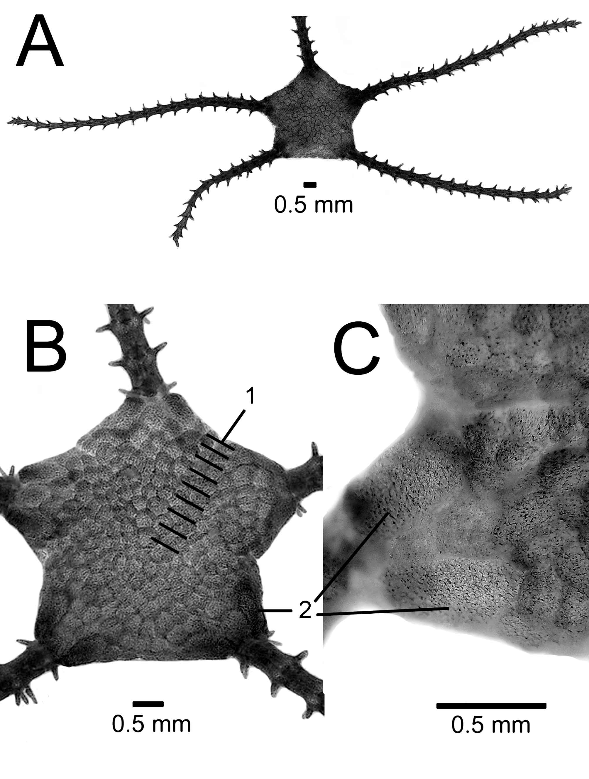
Diagnosis. Disk small, 3–4 mm in diameter, including characters of genus ( Fig. 3 View FIGURE 3 ). Arms short, 2–2.5X disk diameter. Dorsal arm plates small diamond-oval shaped, widely separated from one another by lateral arm plates. Ventral arm plates small figure-8 shaped, widely separated from one another by lateral arm plates. Lateral arm plates large, making up most of an arm segment, 2 arm spines per side. Tentacle pores large, 1 large oval tentacle scale per pore. Color in life translucent white.
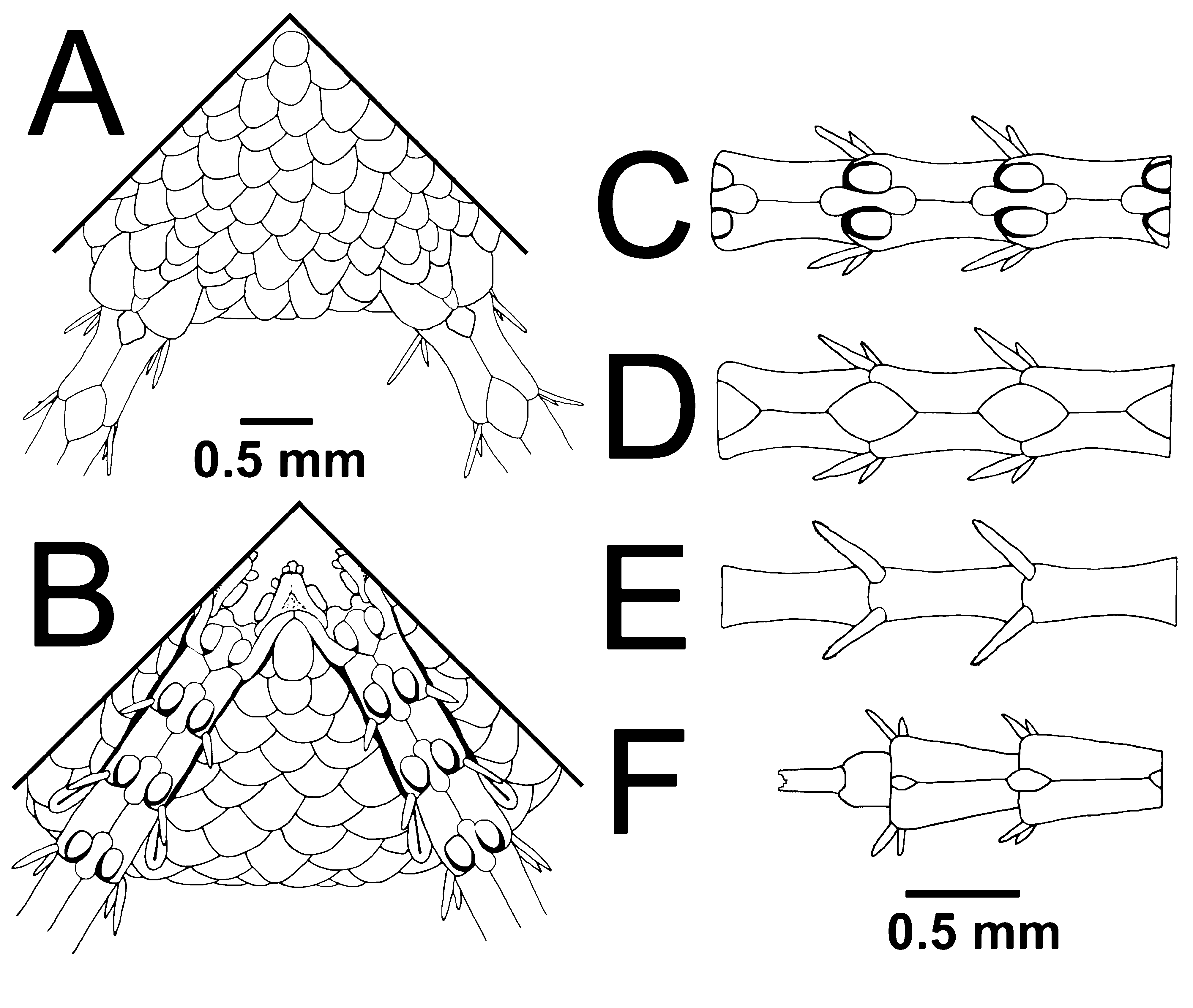
Description (holotype). Disk pentagonal to star-shaped, 3.5 mm diameter. Dorsal disk surface covered by coarse imbricating flat scales, 8–9 rows of scales from disk center to disk edge ( Fig. 4 View FIGURE 4 ). Radial shields oval, 0.4–0.5 mm long, 0.3–0.4 mm wide, divergent, separated medially along their entire length by one column of disk scales, located on disk edge, directly abut and overhang arms.
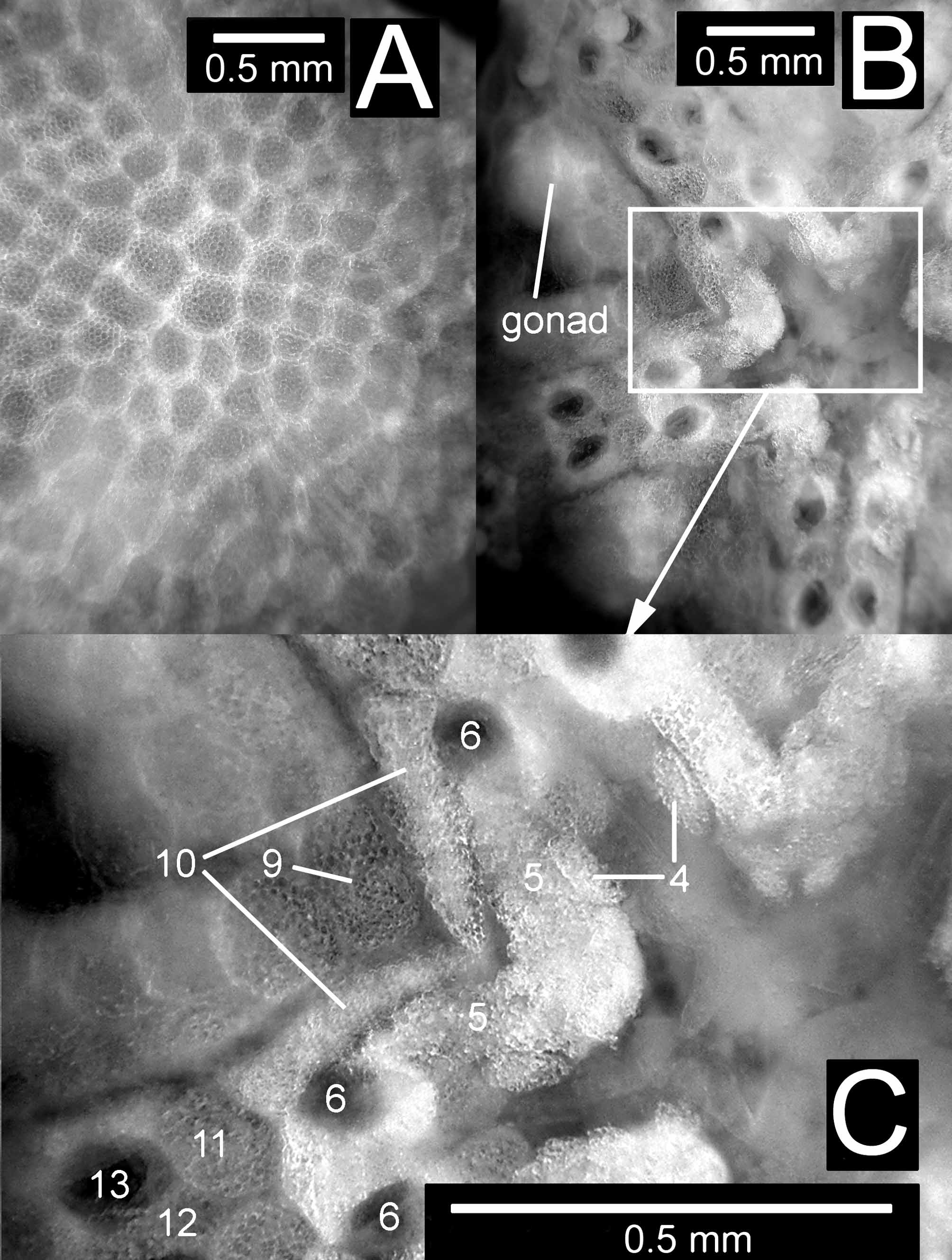
Ventral interbrachial (interradial) area covered by scales similar to dorsal disk surface, 5–6 rows of scales from disk edge to oral frame (Fig. 5). Genital slits extend about three quarters of the way to disk edge. Genital scales flat and thin marking end of genital slits, apparently no bursal sacs. Three arm segments with lateral plates distal to plate bounding end of mouth slit on ventral side of disk, first and second segments bear 0–1 spine per side, third segment bears 1–2 spines per side, tentacle pores large, 1 large oval tentacle scale per pore. Disk color translucent white, color of internal organs shows through scales in life.
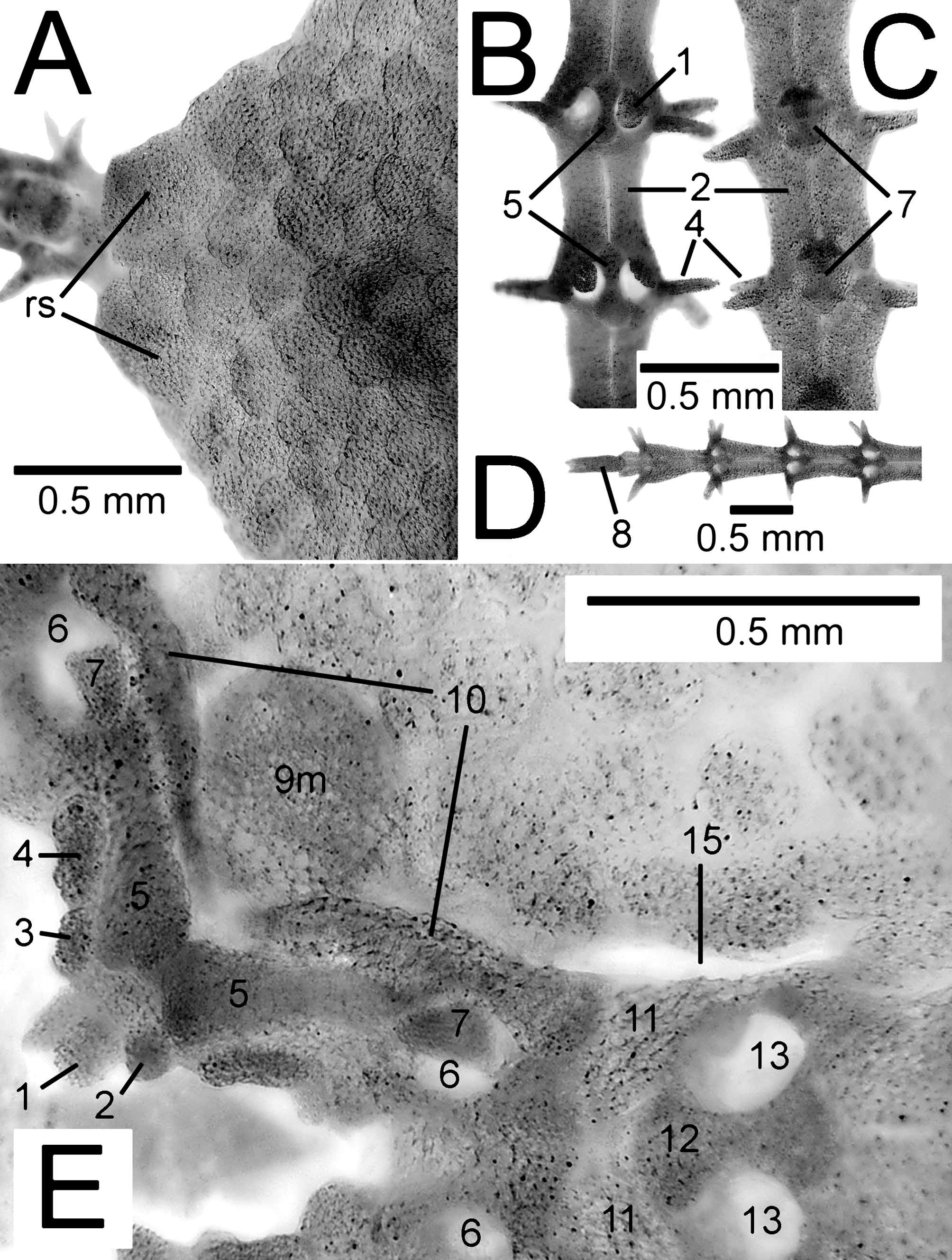
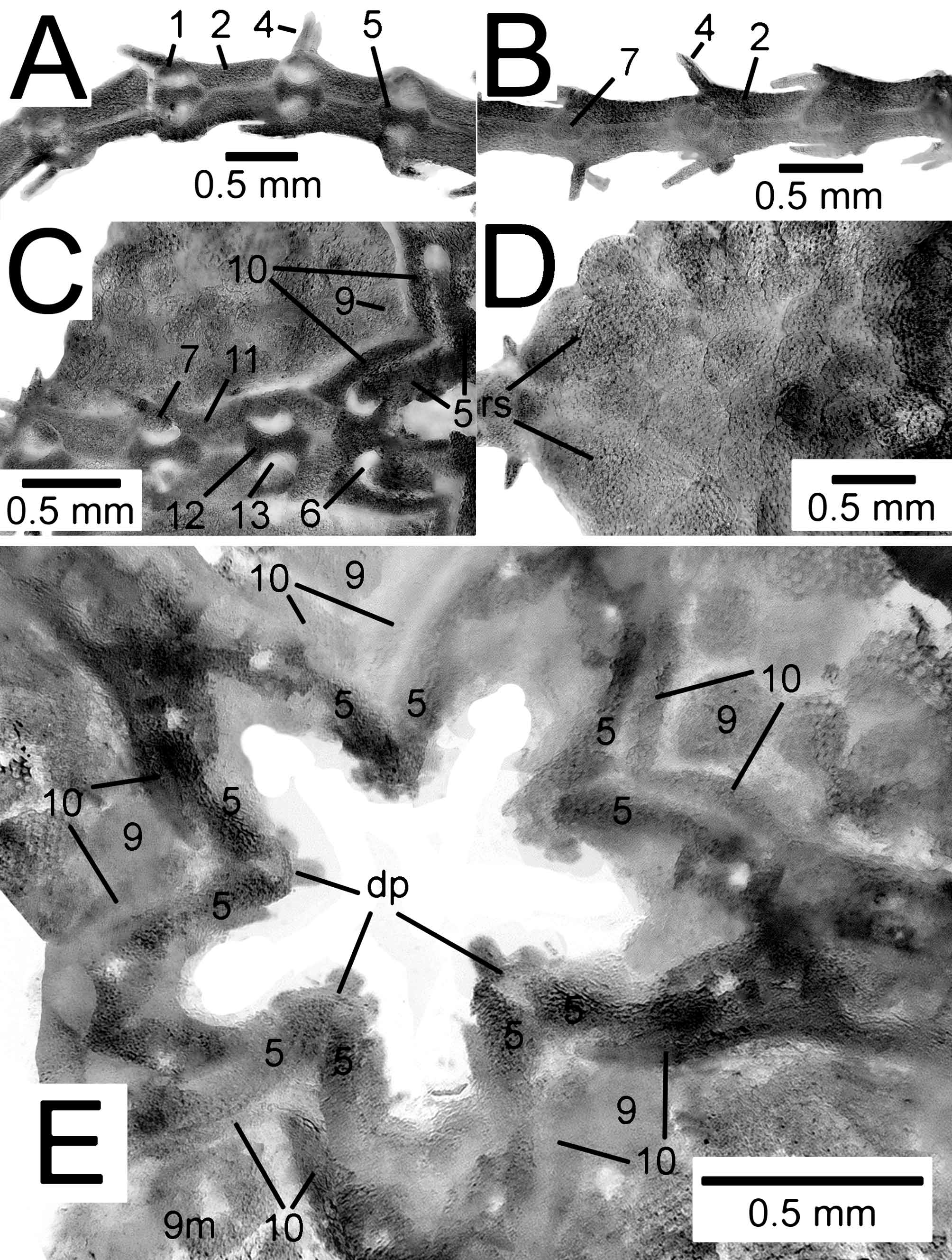
Four non-madreporite oral shields oval-tear shaped, longer than wide, thin, not much larger than and similar in appearance to ventral interbrachial scales. Madreporite oral shield slightly larger, wider, and more distinct in appearance compared to ventral interbrachial scales. Adoral shields thin, narrow, in the shape of a curved (concave proximal side, convex distal side) golf tee or tusk, pointed ends meet proximally, distal flared ends join first lateral arm plates. Adoral shields and oral shield not tightly fused together or thickened as usually seen in ophiuroids. Oral plates long, divergent, proximal end blunt-flat ended, some plates missing proximal infradental papilla. Most complete jaw with 3 oral papillae per side on oral plate; first proximal papilla set on oral plate apex, infradental, small oval shaped; second papilla set on oral plate edge in-line with first, oval shaped, similar in size to first, often missing; third distal papilla set on oral plate edge in-line with second, narrow, oblong to rectangular, 2– 3 X larger than first two papillae, closing off mouth slit with distal oral papilla on adjoining jaw. Except for the distal large papillae, no two jaws have the same papillae arrangement. Ventral tooth at the apex of each jaw broadly rounded. Distal ends of adoral shield and oral plate form border for 2nd tentacle pore of oral frame, which is outside mouth slit. Tentacle scale of 2nd tentacle pore of oral frame more triangular than oval, attached to adoral shield.
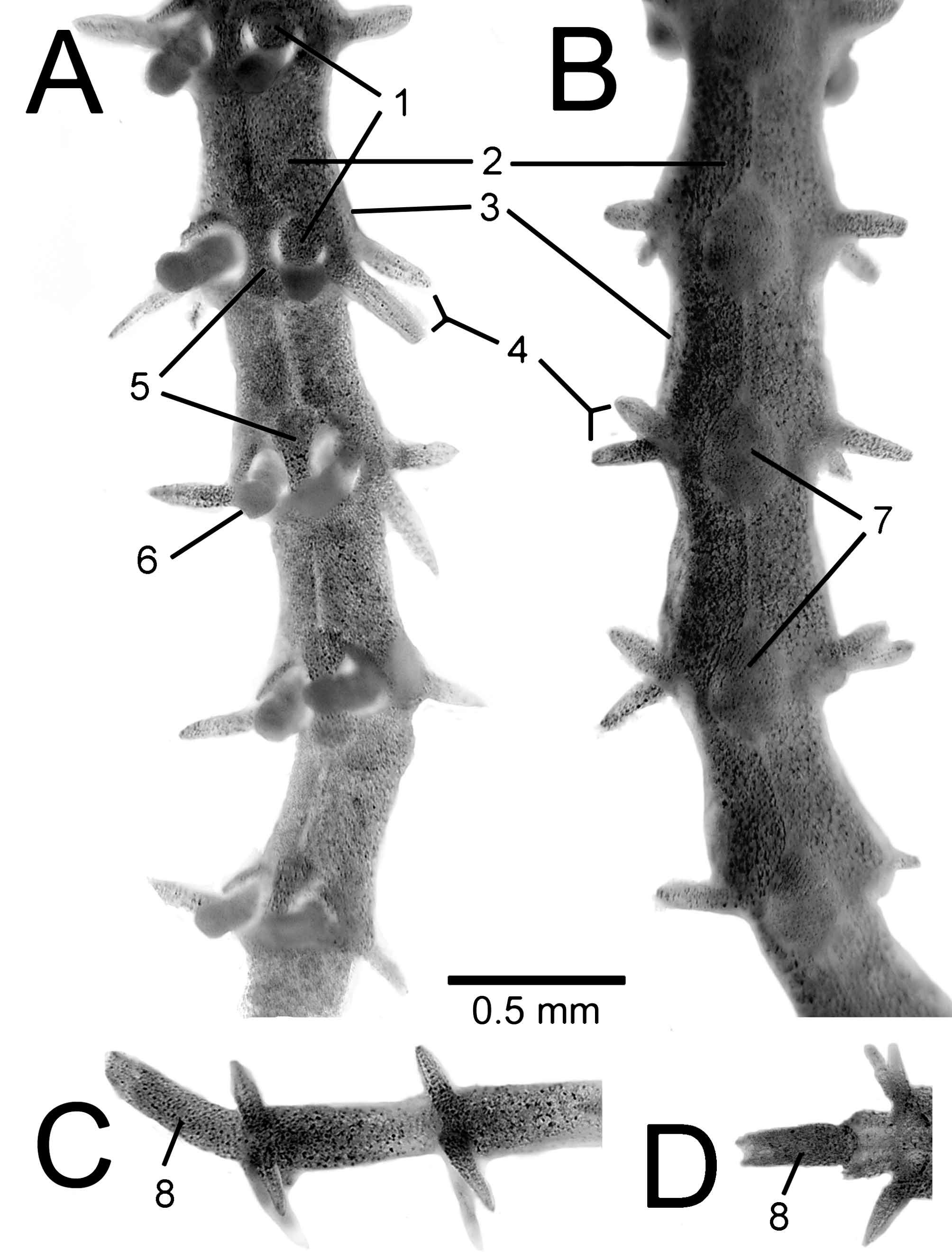
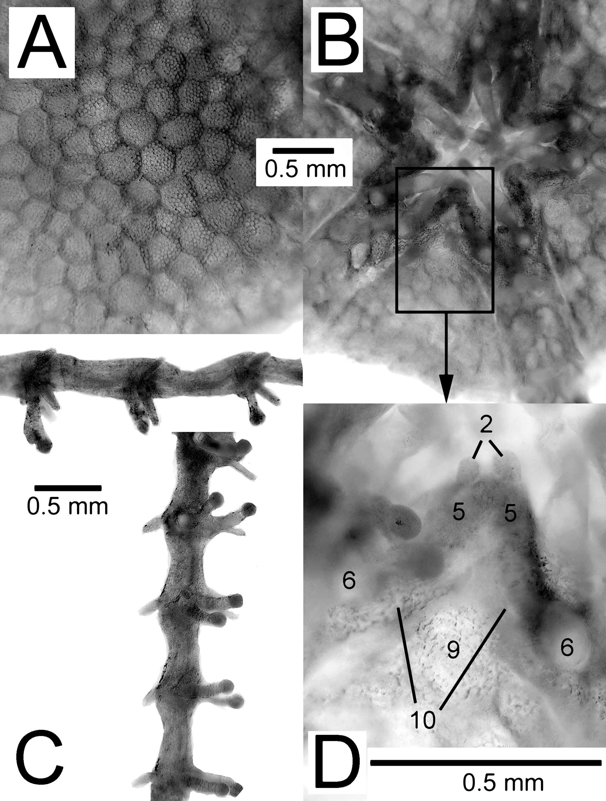
Arms short, five in number, 7–9 mm long, 2–2.5X disk diameter, number of arm segments per arm 18, 19, 14 (missing terminal plate), 20, 13 (regenerating last seven segments). Arm segments composed mainly from lateral arm plates ( Fig. 6 View FIGURE 6 ). Each lateral arm plate 0.4–0.5 mm long, 0.2–0.3 mm wide, with concave sides, more constricted in middle of segment and flared at both ends, 2 arm spines per side. Three individual arm segments on one side only have a small third spine closely associated with one of the two “regular” arm spines and are considered aberrations, possibly due to regeneration error. Arm spines one-half to three-quarters of an arm segment in length, angled at about 45o relative to one another, conical near base, flattened towards tip, irregular fine dentition on edges near tip.
FIGURE 5. Amphicutis stygobita gen. nov., sp. nov. Holotype, disk, ventral view (photos by Chris Pomory). A, interbrachial area. B, arm edge of interbrachial area. C, mouth area. 1. ventral tooth; 2. infradental papilla (variable in form and placement); 3. second oral papilla on oral plate (often missing); 4. large distal oral papilla on oral plate (least variable papilla), opercular, closing off mouth slit with distal oral papilla on adjoining jaw; 5. oral plates; 6. 2nd tentacle pore of oral frame outside mouth slit; 7. one large tentacle scale per pore; 8. 1st tube foot inside mouth slit; 9. oral shield; 10 adoral shields; 11 lateral arm plates of arm segments under disk; 12. ventral arm plate of arm segments under disk; 13. tentacle pore of arm segments under disk; 14. arm spine; 15 genital (non-bursal) slit; 16. disk scales on ventral interbrachial area; 17. genital scale.
Ventral arm plates small figure-8 shaped, 0.3 mm long near disk. Dorsal arm plates small diamond-oval shaped, 0.3 mm long, 0.17 mm wide near disk. Lateral arm plates meet medially on dorsal side of arm separating dorsal arm plates by a gap about equal to dorsal arm plate length. Lateral arm plates meet medially on ventral side of arm separating ventral arm plates by a gap about equal to ventral arm plate length. Dorsal and ventral arm plates become relatively smaller toward distal end of arm. Several arm segments before arm tip shorter with tapering proximal ends leading to narrow terminal plate. Tentacle pores large, 1 large oval tentacle scale per pore on all arm segments. Individual tube feet very large, enlarged smooth knob on end, 2–3 rings/ridges below knobbed end, rest of tube foot smooth. Arm and arm spine color translucent white. Surface of arms with thin skin raised above plates, especially noticeable in life.
Variation (paratypes). Paratype 1 ( Fig. 7 View FIGURE 7 ): a calcified specimen similar to holotype; disk diameter 3.8 mm; arms short, five in number, 8–9 mm long, number of arm segments per arm 15, 15, 15, 12 (regenerating last three segments), 2 (broken arm); oral papillae arrangement highly variable; other characters as in holotype. Paratype 2 ( Fig. 8 View FIGURE 8 ): a relatively uncalcified specimen that was dried; disk diameter 3.8 mm; arms short, five in number, 9–10 mm long, number of arm segments per arm 17, 17, 16, 14, 17; oral papillae arrangement highly variable and many missing due partly to uncalcified state; gonads were visible through disk before drying; other characters as in holotype. Paratype 3 ( Fig. 9 View FIGURE 9 ): a relatively uncalcified specimen that was not dried; disk diameter 3.3 mm; arms short, five in number, 6–7 mm long, number of arm segments per arm 14, 14, 14, 14, 11 (arm tip regenerating); oral papillae arrangement highly variable and many missing due partly to uncalcified state; other characters as in holotype. Paratype 4 ( Fig. 10 View FIGURE 10 ): a calcified specimen with four missing arms that was dried and dissected for viewing of internal frame; disk diameter 3.8 mm; arms short, five in number, 8 mm long, number of arm segments per arm 15, other arms broken after 1–2 segments; oral papillae arrangement highly variable; other external characters as in holotype; internal frame with small dental plates, long narrow oral plates without enlarged lateral wings, no bursal sacs, genital plate and scale not strongly associated with arm, genital plate-radial shield connection by single large condyle and socket respectively.
Biology/Ecology. Individuals were found crawling on the bottom of a shallow pool in Bernier Cave. A quantitative survey has not been made, but 15 individuals were collected on 6 January 2011 and several others were seen along the edge of the shallow pool. The cave water is hyposaline at 25–28ppt and appears to be partitioned from nearby aquatic habitats. The cave entrance is only ~ 20 m from the northeast arm of Great Lake, which is strongly hypersaline at ~70ppt near the cave. It appears meteoric water or water from a subsurface freshwater lens infiltrates Bernier Cave, so that the shallow-water environment may never reach total marine salinity of 35ppt. Other caves on San Salvador have consistent salinity readings over time (Carpenter personal observation). This isolation may partially explain the apparent absence of the new species from other caves, such as Lighthouse Cave, on San Salvador (Carpenter personal observation). Since no large direct connection exists with the ocean to allow easy entry of marine organisms, we suggest the new species completes its life cycle in the cave and is endemic to Bernier Cave. Specimens spawned during transport following collection and eggs started to develop, but stopped at an early stage.
During observation in culture brittle stars ignored or moved away from finely ground Tetra-Min fish food (a food source readily eaten by several species of ophiuroid, Pomory personal observation) and other particulate food sources that were offered possibly indicating the new species may utilize dissolved organic matter leaching off cave detritus (bat guano, leaf debris, or algae which collects on the bottom of the cave). The new species may also feed on bacteria growing on cave detritus. A stomach is present and can readily be seen through both the ventral and dorsal side of the disk. Most ophiuroids move by bending the arms, rather than podial walking as is common in asteroids; however, the new species mainly uses podial walking for locomotion. This may be a function of the short arms and the relatively large podia.
Etymology. Specific epithet stygobita is derived from the word stygobite, meaning an aquatic cave-dwelling organism.
| USNM |
Smithsonian Institution, National Museum of Natural History |
No known copyright restrictions apply. See Agosti, D., Egloff, W., 2009. Taxonomic information exchange and copyright: the Plazi approach. BMC Research Notes 2009, 2:53 for further explanation.