
Haasea Verhoeff, 1895
|
publication ID |
https://doi.org/ 10.11646/zootaxa.4798.1.1 |
|
publication LSID |
lsid:zoobank.org:pub:4830ED2F-DB55-468E-97D4-2A278CBA6CE0 |
|
persistent identifier |
https://treatment.plazi.org/id/03A90C3B-153D-FF82-FF24-7298FE7BFD5C |
|
treatment provided by |
Plazi |
|
scientific name |
Haasea Verhoeff, 1895 |
| status |
|
Genus Haasea Verhoeff, 1895 View in CoL
Xiphogona Cook, 1895: 3 View in CoL (junior objective synonym)
Xiphigona Cook, 1895 View in CoL —misspelled by Hoffman (1980: 130) (junior objective synonym)
Orobainosoma Verhoeff, 1897: 135 View in CoL (junior objective synonym)
Rhopalogona Silvestri, 1898: 666 View in CoL (junior objective synonym)
Deuterohaasea Verhoeff, 1898: 226 View in CoL (junior objective synonym)
Orobainosoma (Brachybainosoma) Verhoeff, 1899: 128 (junior subjective synonym)
Heterohaasea Verhoeff, 1900: 362 (junior objective synonym)
Orobainosoma (Histrosoma) Strasser, 1973a: 419 (junior subjective synonym)
Type species: Craspedosoma flavescens Latzel, 1884 accepted as Haasea flavescens ( Latzel, 1884) View in CoL [by subsequent designation of Cook, 1895 in Cook & Collins (1895: 3)]
Diagnosis: A genus of Haaseidae with leg-pair 10 in males reduced in size compared to other walking legs (vs. robust in Hylebainosoma and Xylophageuma , an exception is H. nontronensis also with a reduced leg-pair 10 but this species differs from Haasea by numerous other gonopod and vulvar structures). Medial sternal process of the anterior gonopodal block bilobed distally (vs. not bilobed in Hylebainosoma and Xylophageuma ). Angiocoxites sigmoid in lateral or mesal view (vs. mainly straight in Hylebainosoma and Xylophageuma , an exception is H. gulickai Tajovský, Mock & Papáč, 2014b with sigmoid angiocoxites, but this species differs from the other haaseids by the absence of ommatidia). Additional gonopodal processes mainly strongly curved posteriad in lateral or mesal view (vs. mainly straight in Hylebainosoma and Xylophageuma ). Vulval bursae mesally completely merged.
Short description: Body in adults with 28 or 30 segments (including telson); 6.5–12 mm long.
COLORATION ( Fig. 1 View FIGURE 1 ). Variable, from poorly pigmented to light or darker brown. Most of species with yellowish-brown body.
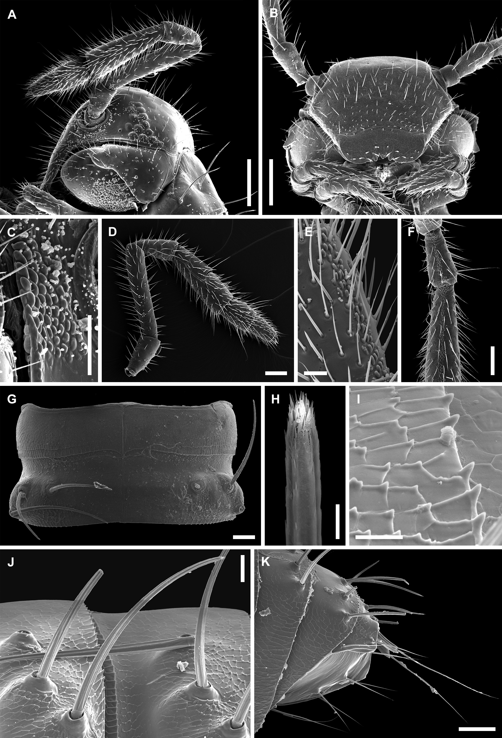
HEAD ( Fig. 2 View FIGURE 2 A–F). Setose, frontal side flattened in males ( Fig. 2A View FIGURE 2 ), slightly convex in females. Labrum with three medial teeth ( Fig. 2B View FIGURE 2 ). Antennae elongated, slender, with dorsomedial and ventromedial lines of small papillae-like outgrowths ( Fig. 2 View FIGURE 2 D–F). Lateral to antennal sockets a group of similar papillae-like outgrowths present ( Fig. 2C View FIGURE 2 ). Ommatidia brownish to black arranged in triangle, number from 9–16 ( Fig. 2A View FIGURE 2 ).
BODY ( Figs 1 View FIGURE 1 , 2 View FIGURE 2 G–K). With lateral humps. Macrochaetae long and trichoid, present on large knobs. Posterior metazonal margins with small, triangular denticles. Collum and telson not modified.
LEG- PAIRS 1 AND 2 ( Fig. 3E View FIGURE 3 ). In both sexes with tarsal combs; prefemora with several long and robust setae; femora and postfemora each with a group of several long and robust setae.
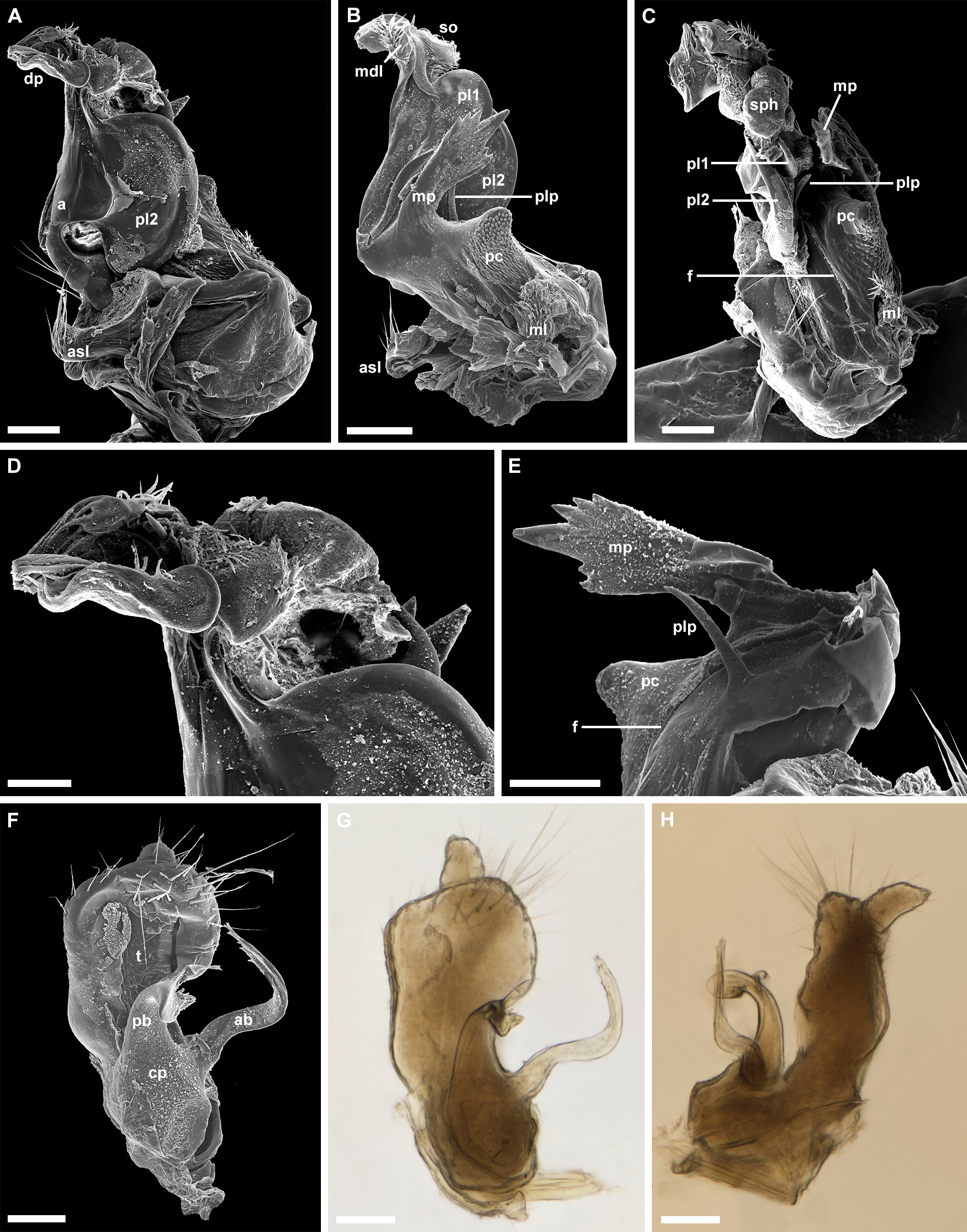
MALE SEXUAL CHARACTERS ( Fig. 3 View FIGURE 3 E–G). Leg-pair 2 with genital openings on coxae ( Fig. 3E View FIGURE 3 ). Leg-pairs 3–7 enlarged. Distal half of tarsi 3–7 with ventral papillae ( Fig. 3F View FIGURE 3 ). Leg-pair 10 significantly smaller than the other walking legs; with coxal sacks and with a robust, blunt coxal process ( Fig. 3G View FIGURE 3 ). Leg-pair 11 with coxal sacks, no other modifications.
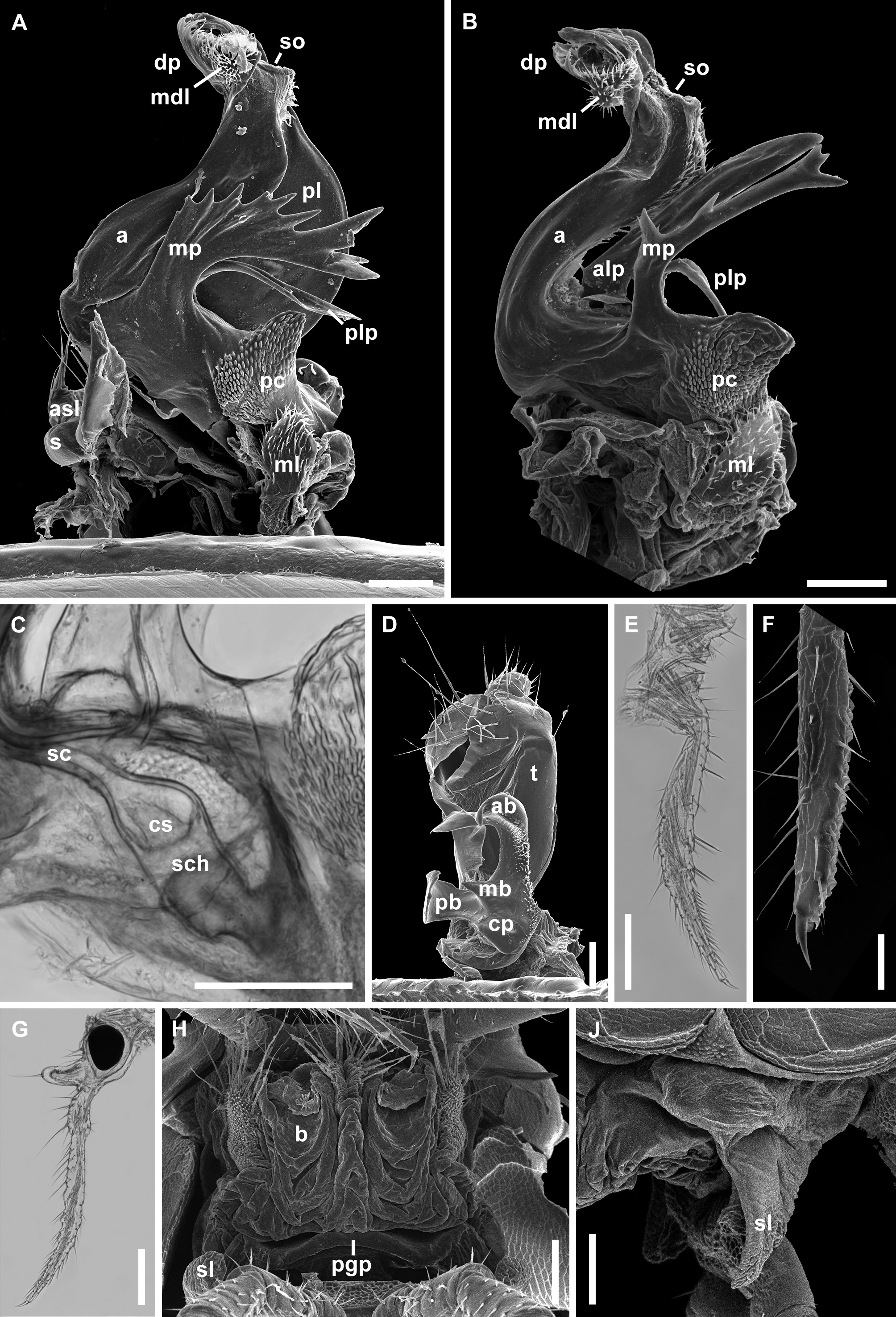
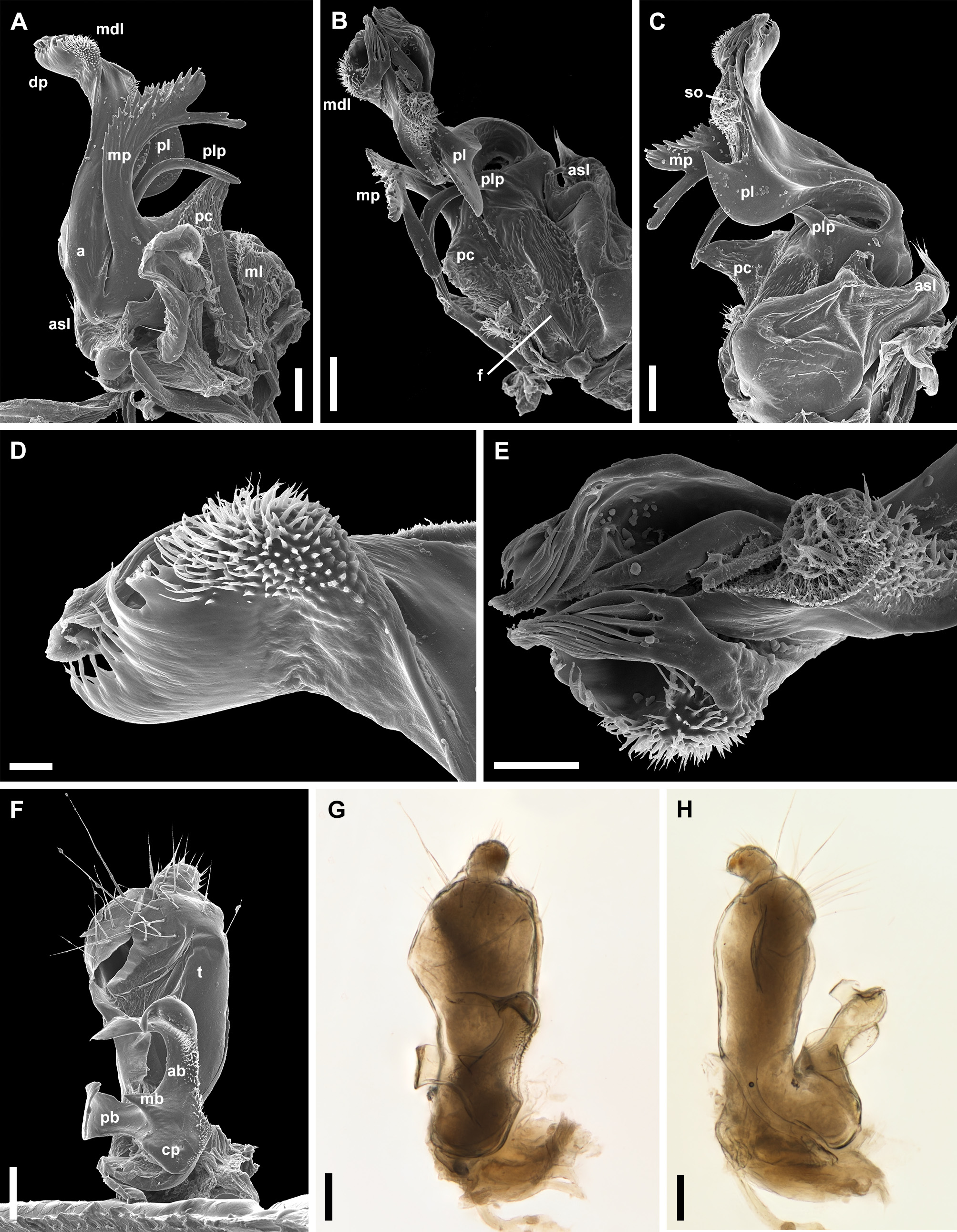
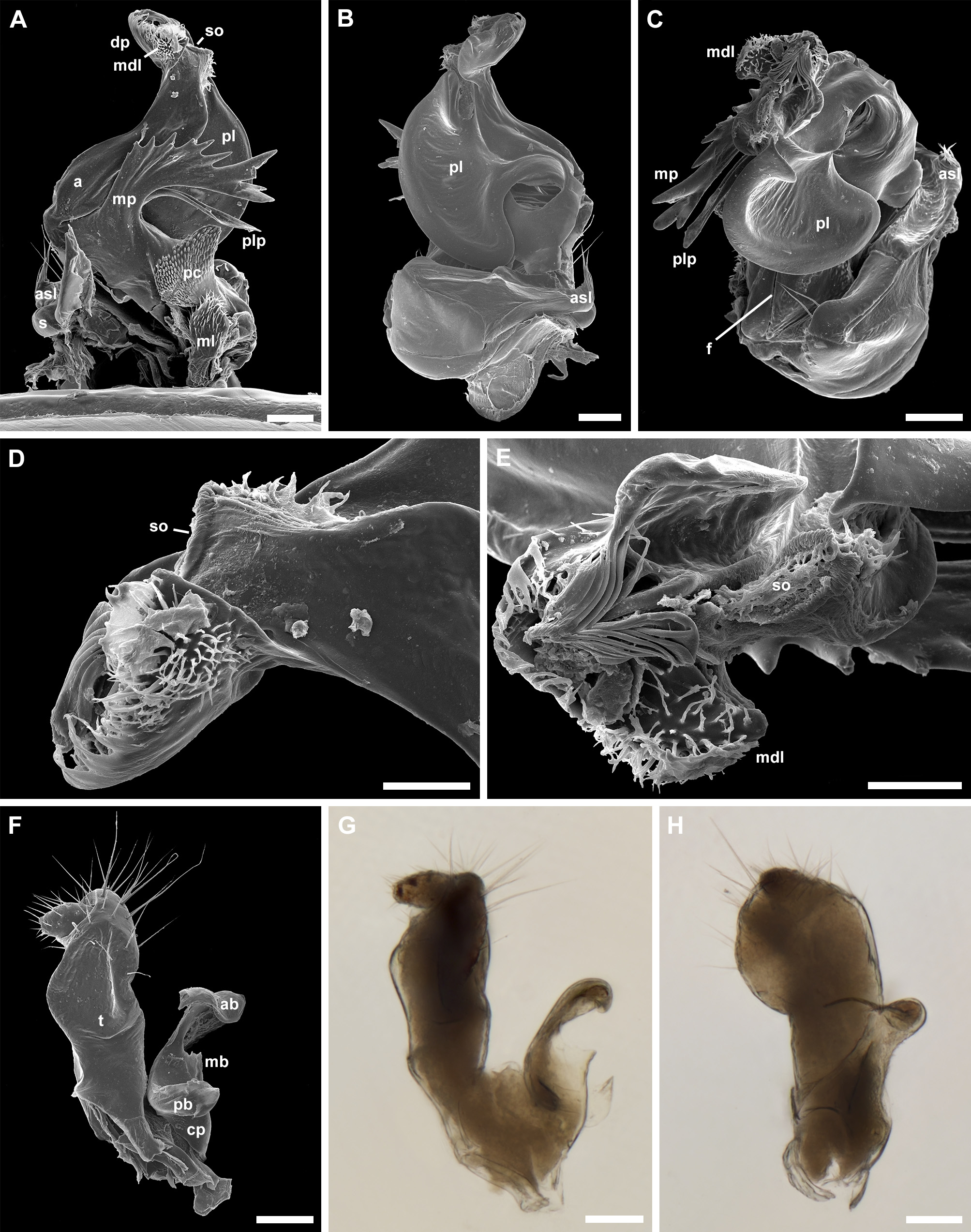
ANTERIOR GONOPODS ( Fig. 3 View FIGURE 3 A–C) Sternum (s) wide, with medial sternal process (msp, see Figs 25D View FIGURE 25 and 34C View FIGURE 34 ) bilobed distally and anterior sternal lobe (asl) with several long setae, on both lateral sides. Long setae (1–3) also present on antero-paramedial and lateral parts of s. Angiocoxites (a) erected, sigmoid in lateral or mesal view, characterized by the presence of a deep, posterior and longitudinal furrow (f, see Figs 9B View FIGURE 9 , 14C View FIGURE 14 , 18C, E View FIGURE 18 or 34D), extending from the base to the distal part, dividing superficially posterior portions of the angiocoxite into the mesal and lateral sides. Angiocoxites supplied with a few additional processes or extensions on both mesal and lateral sides. Mesal process (mp) always present, more or less well-developed, with or without additional teeth. Apart from mp, two more structures are always present on mesal side, viz., papillated cone (pc) and “ Opuntia -like” membranous lobe (ml) which might be a colpocoxite. Both structures more or less identical in all species. Laterally, two processes present, viz., anterior lateral process (alp) in only a few species and posterior lateral process (plp) in all species. The distal half of a bends anteriad. The most distal part (dp) complex, composed of structures with “hairs” or fimbriaelike outgrowths. In some species, this part has a mesodistal lobe (mdl) sometimes with fimbriae. Posteriorly, dp has a seminal opening (so) surrounded by minute fimbriae-like outgrowths. Below so, posterior lamella pl (sometimes also pl1, pl2) present, simple or bilobed. Inside a, (?) seminal canal (s c) is visible, starts from the basal half and opens distally in so. The most basal part of sc enlarged, in form of seminal chamber (sch) and ends with a cone structure (cs) covered with cilia or sensilla.
POSTERIOR GONOPODS ( Fig. 3D View FIGURE 3 ). With reduced sternum (s, see Fig. 26J View FIGURE 26 ). Telopodites (t) well-developed, robust, with two podomeres, covered by long setae. Coxal processes (cp) present mesally, with two or three branches (ab, mb, pb).
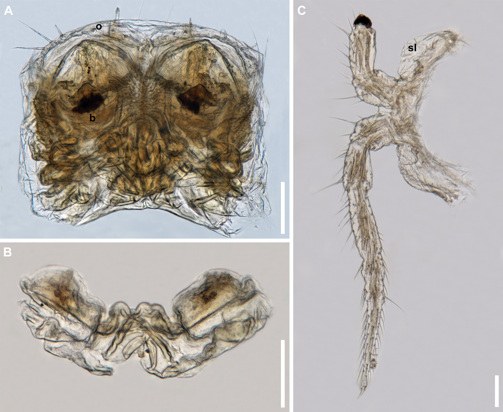
LEG- PAIR 3 IN FEMALES ( Fig. 3 View FIGURE 3 H–J). With lateral sternal lobes (sl).
VULVAE ( Fig. 3H View FIGURE 3 ). Operculum (o, see Figs 35G View FIGURE 35 and 37A View FIGURE 37 ) membranous, with serrated edge and with a row of long setae. Bursae (b) merged mesally with bulbous structure, long setae and triangular denticles present on mesal and lateral parts. Postgenital plate (pgp) present, with lateral lobes and medial lobe. For vulvae details see Kurnik (1988: 234, taf I, 246–251).


No known copyright restrictions apply. See Agosti, D., Egloff, W., 2009. Taxonomic information exchange and copyright: the Plazi approach. BMC Research Notes 2009, 2:53 for further explanation.
|
Kingdom |
|
|
Phylum |
|
|
Class |
|
|
Order |
|
|
Family |
Haasea Verhoeff, 1895
| Antić, Dragan & Akkari, Nesrine 2020 |
Xiphigona
| Hoffman, R. L. 1980: 130 |
Orobainosoma (Histrosoma)
| Strasser, K. 1973: 419 |
Heterohaasea
| Verhoeff, K. W. 1900: 362 |
Orobainosoma (Brachybainosoma)
| Verhoeff, K. W. 1899: 128 |
Rhopalogona
| Silvestri, F. 1898: 666 |
Deuterohaasea
| Verhoeff, K. W. 1898: 226 |
Orobainosoma
| Verhoeff, K. W. 1897: 135 |
Xiphogona
| Cook, O. F. & Collins, G. N. 1895: 3 |