
Myrmozercon brevipes Berlese, 1902
|
publication ID |
https://doi.org/ 10.1051/acarologia/20152151 |
|
persistent identifier |
https://treatment.plazi.org/id/03A98790-FF85-FFF8-2EF0-FE4A0D03F823 |
|
treatment provided by |
Marcus |
|
scientific name |
Myrmozercon brevipes Berlese, 1902 |
| status |
|
Myrmozercon brevipes Berlese, 1902
Myrmozercon brevipes Berlese 1902: p. 700 .
Myrmozercon brevipes: Berlese 1904: p. 313 (female chelicera), p. 314 (male chelicera), p. 445 (redescription). Table 19, Figs. 169-172.
Material examined — Four females and one male ( HNHM). Greece, Aetolia-Acarnania peripheral unit, Akarnanika Mts, Trifos, small artificial pond and its shore vegetation S of the village, 38°48.396’ N, 21°05.650’ E, 330 m, 6 May 2011, nest of Tapinoma erraticum Latreille , coll. J. KontschAEn, D. MurAEnyi, T GoogleMaps . Szederjesi and Zs. UjvAEri.
Five females, one male ( HNHM) and three females ( QM). Greece, Ionian Islands, Lefkada peripheral unit, Rahi, 38°43.363 N, 20°41.404 E, 50 m, 06 May 2011, nest of T. erraticum , coll. J. KontschAEn, D. MurAEnyi, T GoogleMaps . Szederjesi and Zs. UjvAEri.
Diagnosis (based on female) — Dorsal shield densely hypertrichous, setal length 20 – 40; posterior margin of shield rounded, not truncated; soft cuticle posterior to shield hypertrichous. Sternal shield horseshoe-shaped (inverted U-shape); setae st4 present; epigynal shield sculpturing lineatereticulate, shield bearing setae st5 only; one pair of metapodal shields present; anal shield peltate. Palp setal count (trochanter to tibia) 1-5-5-11; subcapitular groove with 8 rows of denticles, with one large denticle per row; palp coxal seta absent. Chelicerae highly modified, terminating in small membranous flange; fixed digit absent. All legs short, leg I distinctly shorter (280 – 295) than all other legs (leg II 370 – 410; leg III 385 – 425; leg IV 410 – 445); setal counts (coxa to tibia) for legs I-IV: 2-4-8-8-8, 2-4-8-8- 7, 2-5-6-10-9, 2-5-8-8-7; all leg setae simple.
Description — Female ( Figs. 1-6 View FIGURE View FIGURE View FIGURE View FIGURE View FIGURE View FIGURE ; n = 12).
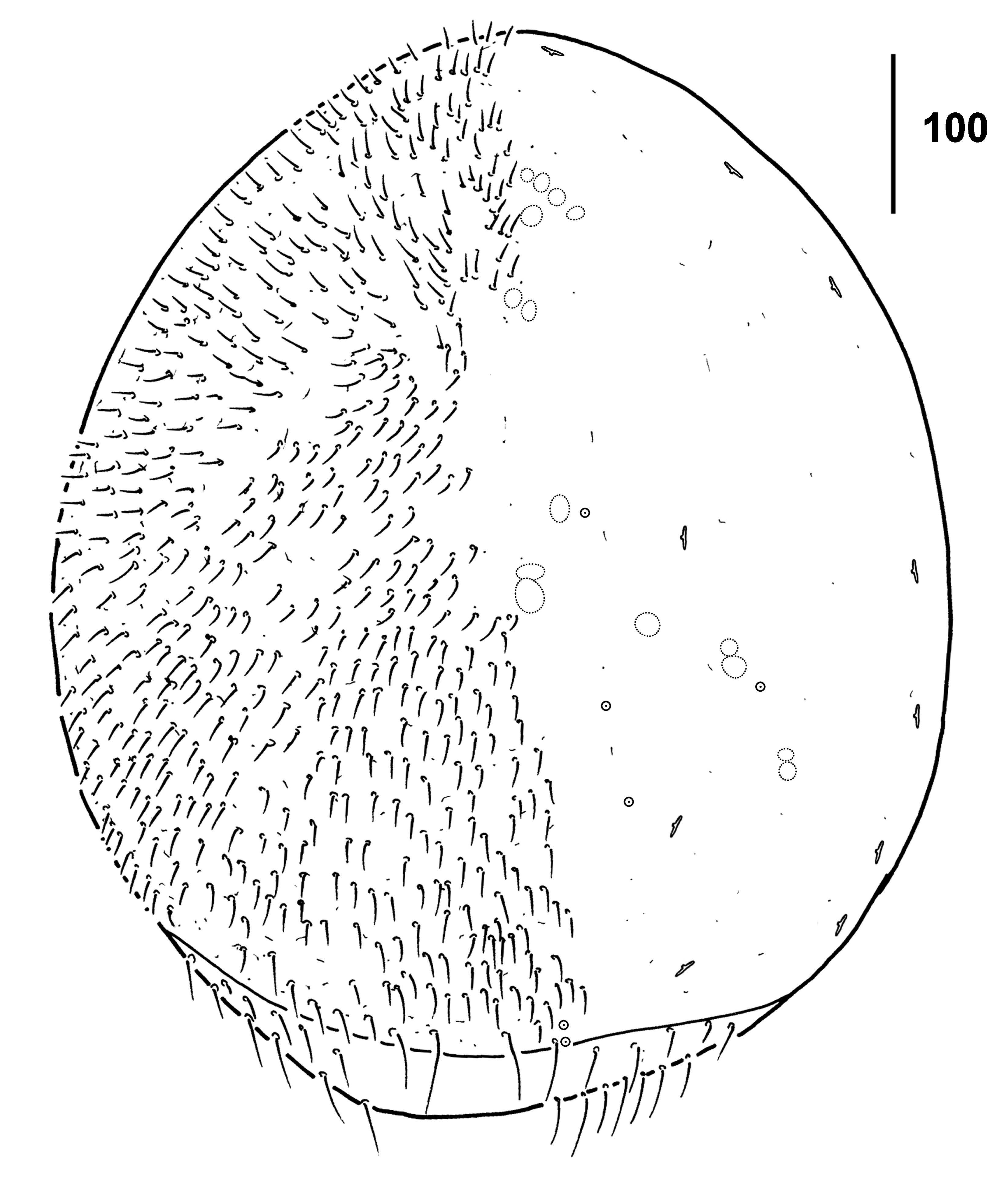
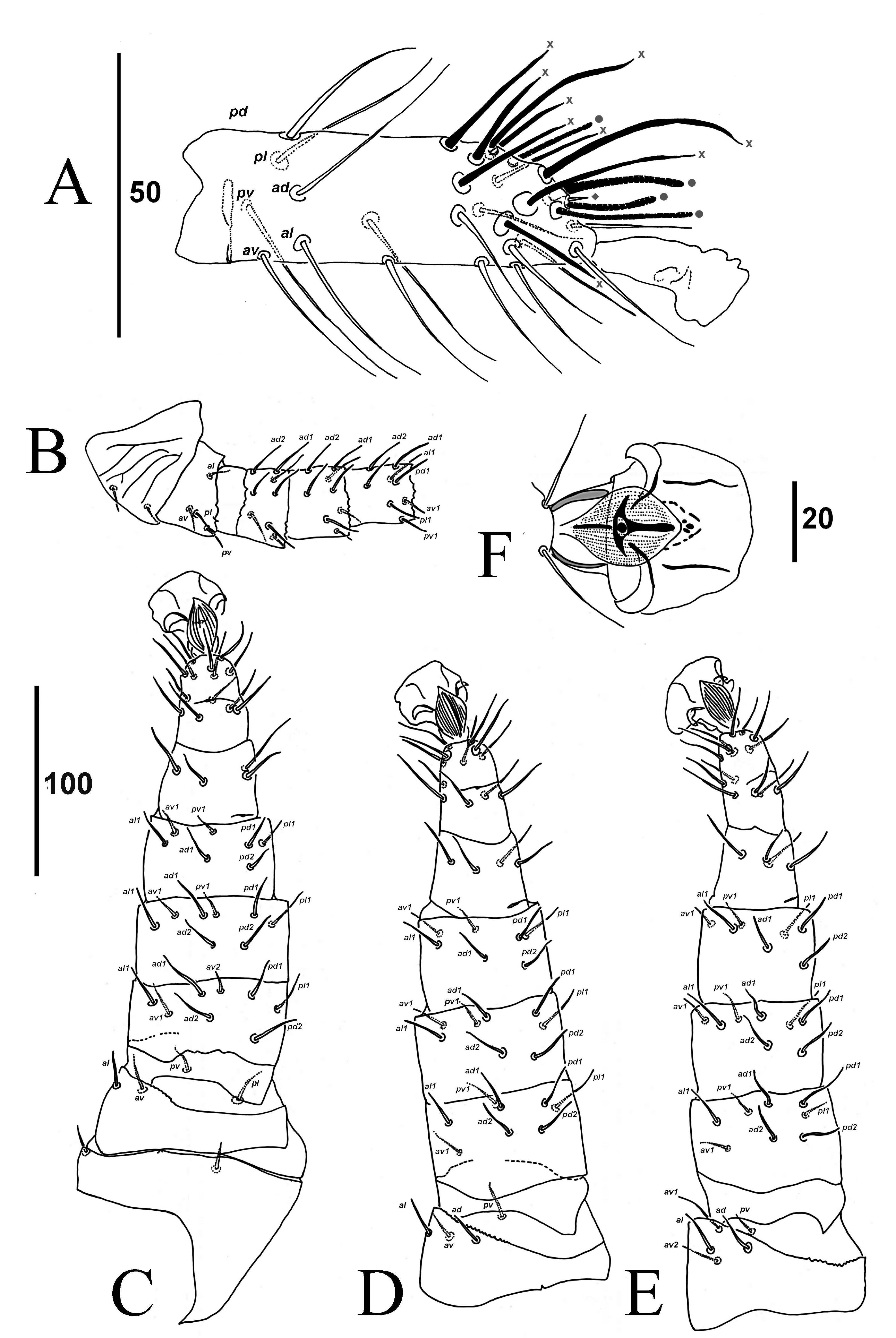
Dorsal idiosoma ( Figs. 1 View FIGURE , 2A View FIGURE ) — Dorsal shield length 640 – 760, width 550 – 640, subcircular, cuticle smooth, with several hundred fine setae, length 20 – 40. Most marginal setae short; dorsal marginal setae become longer posteriorly (longest 50 – 75) on shield and soft cuticle. Dorsal shield with 16 pairs of dal platelet); B – Tritosternum. F – Ambulacrum III.
visible pore-like structures (ten slit-like lyrifissures; all others rounded, surrounded by lacunae). Sigillae small arranged in medial groups on podontal region; groups on opisthonotal region more lateral.
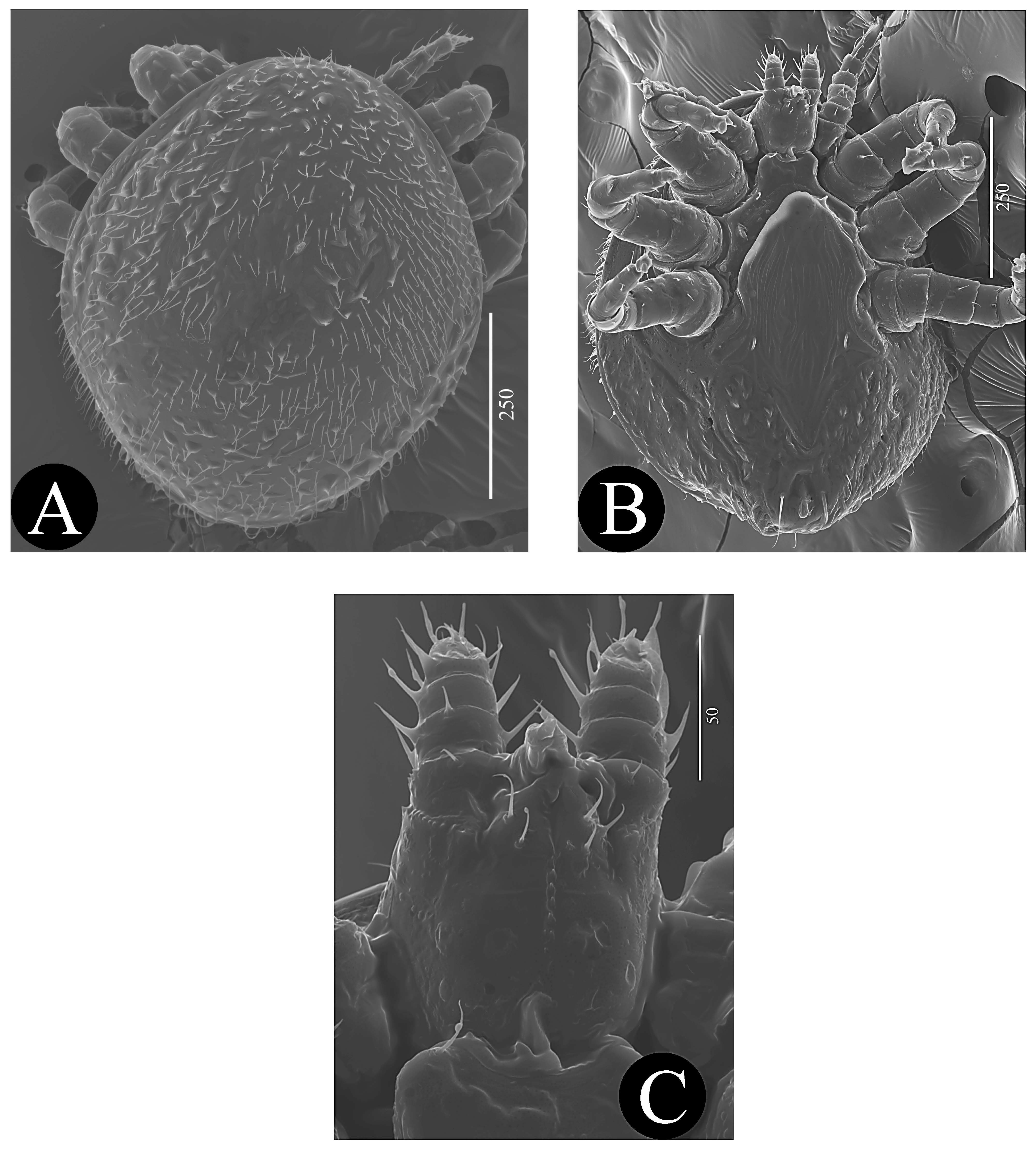
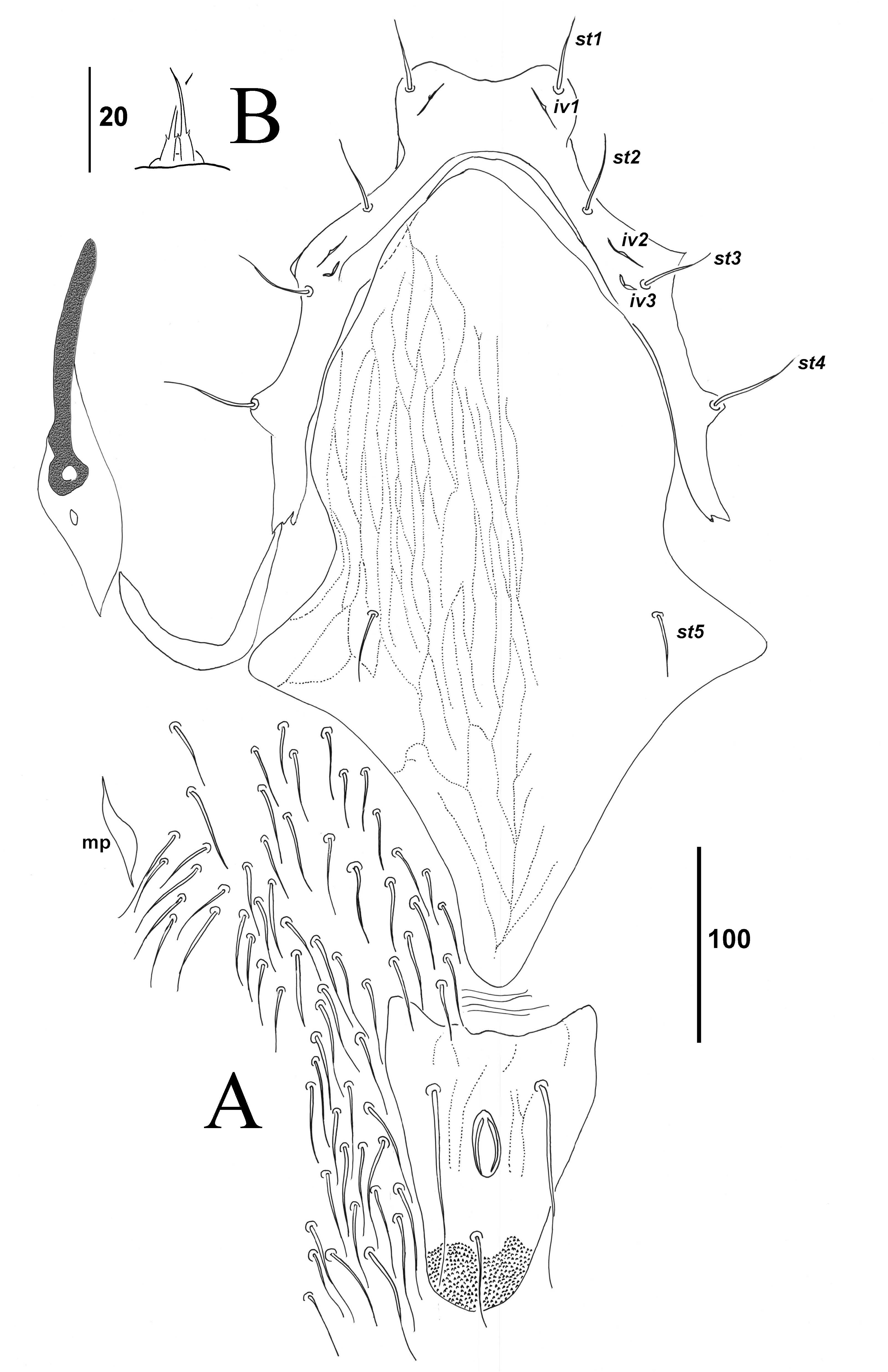
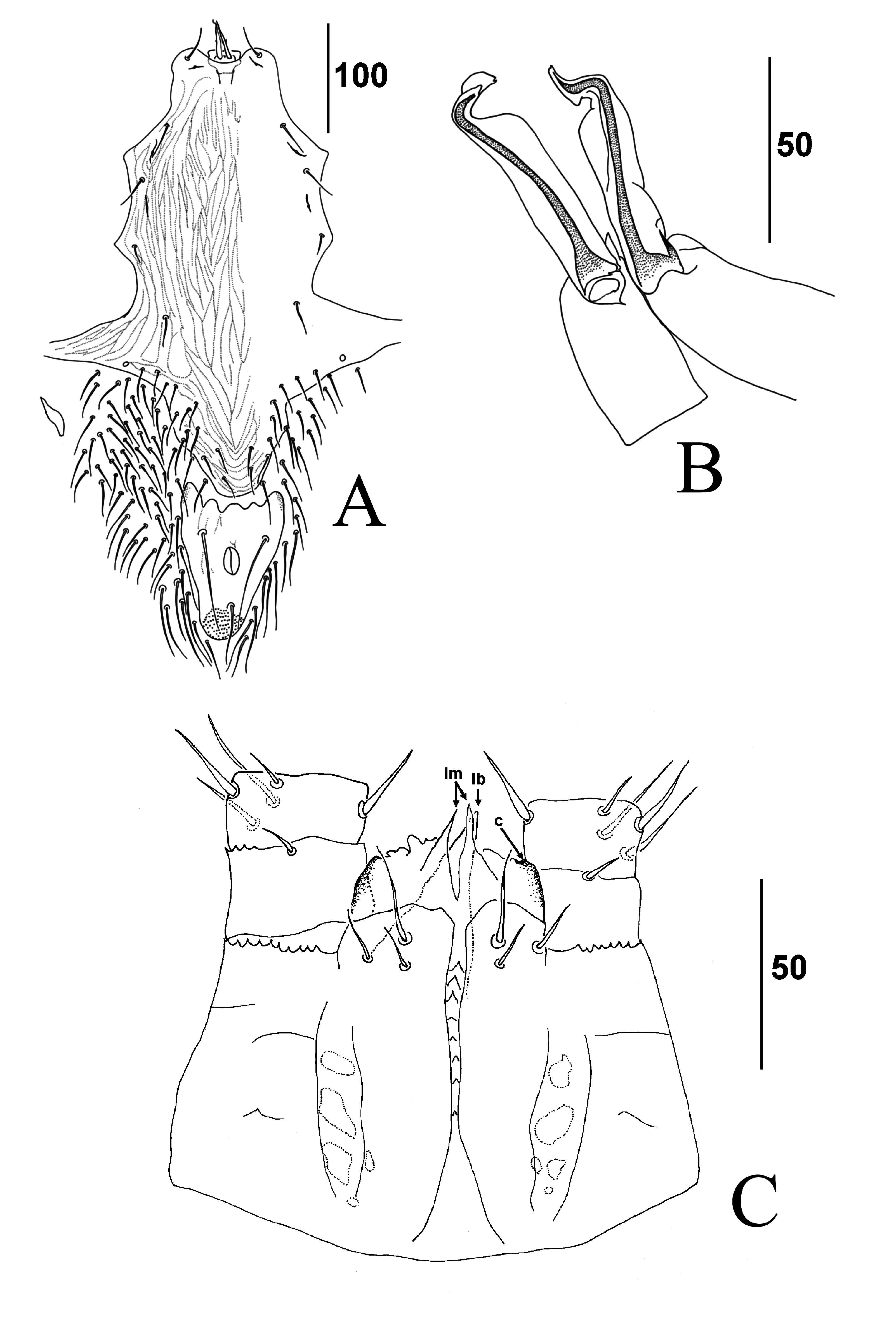
Ventral idiosoma ( Figs. 2B View FIGURE , 3A View FIGURE ) — Tritosternum ( Fig. 3B View FIGURE ) with short laciniae, length 40 – 45, unfused, laciniae each with a pair of tiny spines 14 – 16 from base, base hyaline, evanescent. Presternal area membranous, presternal shields absent. Sternal shield narrow, highly concave, horse-shoe shaped, medial length 30 – 34, maximum length 240 – 260, length to seta st4 160 – 185, width at level of setae st2 125 – 140, width at level of st4 250 – 270, ratio medial length / length to st4 0.2; sternal shield smooth, bearing smooth slender setae st1-4 (39, 35 – 37, 38 – 39, 43 – 45, respectively) and lyrifissures iv1- 3. Endopodal shields absent, or reduced and fused to exopodal shield around posterior margin of coxa IV. Epigynal shield (full length 410 – 420; 180 – 195 from st5 to posterior tip) with well-defined broadly rounded anterior margin, with longitudinal reticulation, pointed posteriorly, maximum width 265 – 270; setae st5 30 – 35; lyrifissures iv5 not present on shield (nor detected in soft cuticle). Metapodal platelets elongate, weakly formed (55 – 63 × 12 – 14). Peritremal shield small, smooth, free posteriorly and surrounding only posterior half of peritreme, not fused to dorsal shield, bearing one gland pore; peritreme extending to level of anterior coxa III. Anal shield peltate (length 150 – 170 × width 105 – 110), shield with few longitudinal reticulations, with post-anal seta (36 – 37) shorter than para-anal setae (100 – 105); cribrum a dense field of spicules extending from just behind post-anal seta to posterior margin; sigillae absent. Soft cuticle hypertrichous, with 350 – 400 setae, setal length 30 – 45.
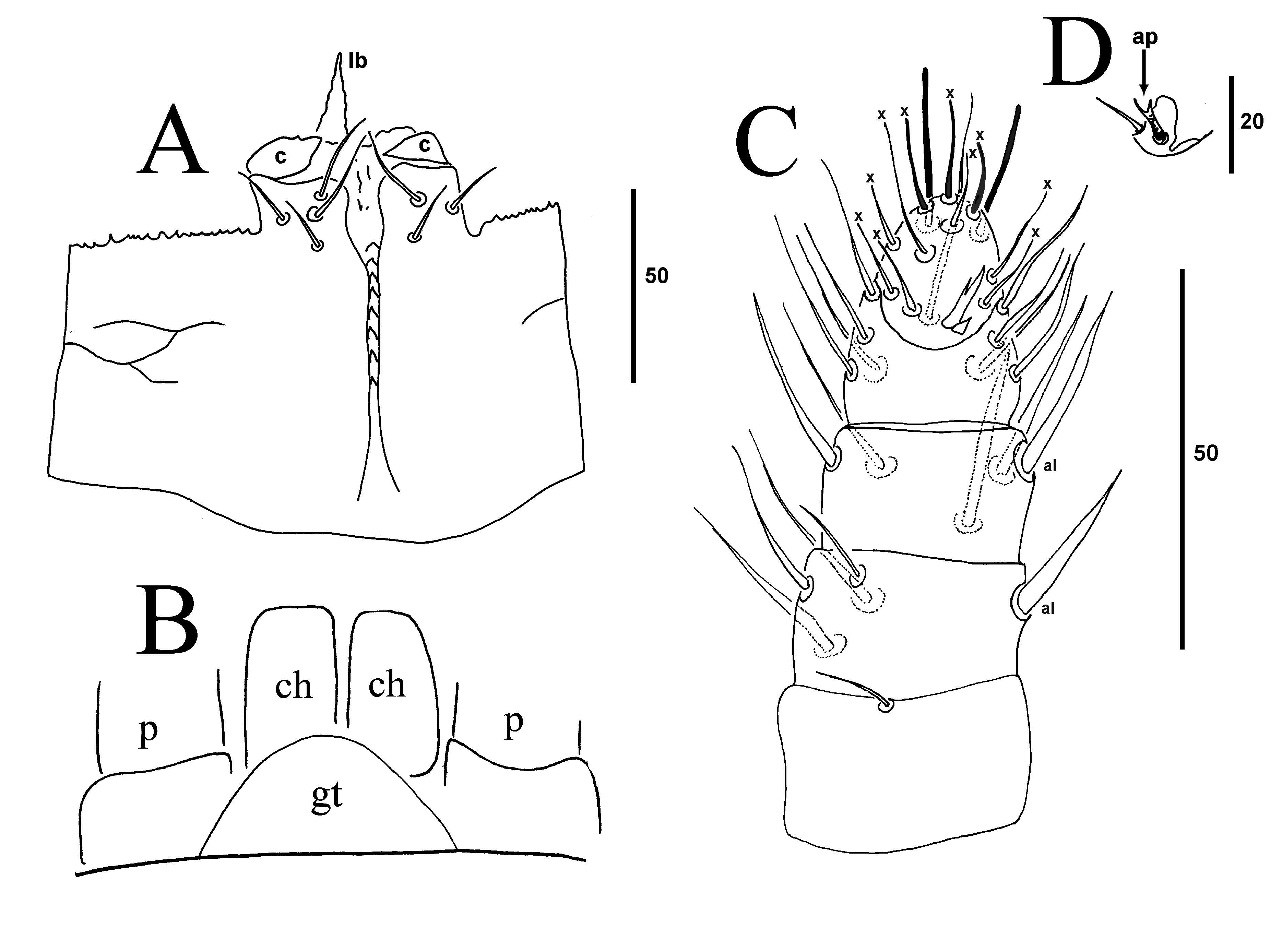
Gnathosoma ( Figs. 2C View FIGURE , 4 View FIGURE ) — Gnathotectum ( Fig. 4B View FIGURE ) with anterior margin smooth, rounded. Subcapitulum ( Fig. 4A View FIGURE ) with eight deutosternal rows each comprising a single denticle; corniculi (21 – 24) hyaline, adpressed to anterior margin of subcapitulum, tips acuminate directed medially; subcapitulum not extending medially beyond corniculi; internal malae not detectable or perhaps absent; labrum (16 – 30), reduced, extending to level of anterior margin of palp trochanter to mid-femur; hy- postomal setae fine, h1 longer (27) than outer posterior setae h2 (15), inner posterior setae h3 (16); illustrated specimen with duplication of seta h1 on righthand side, scanning electron micrographed specimen lacking seta h2 on right-hand side ( Fig. 2C View FIGURE ); palpcoxal setae absent; setae h2 40 – 42 apart. Palps ( Figs. 4 View FIGURE C-D): length 92 – 94 with simple setae except lateral setae on palp femur and genu thickened, dorsal setae on palp femur, genu and tibia thickened; femur without dorsal anterolateral spine; tarsus placed entirely ventrally; setation of palp segments from trochanter to tarsus: 1-5-5-11-10, tibia with two apical round-tipped hollow tibial setae on small tubercles, three tarsal setae of similar form; palp tarsal claw two-tined, with tines tapered, claw flanked by small cusp, ventral tine shorter (2 – 3 from fork) than dorsal tine (6 from fork, 10 – 11 from base).
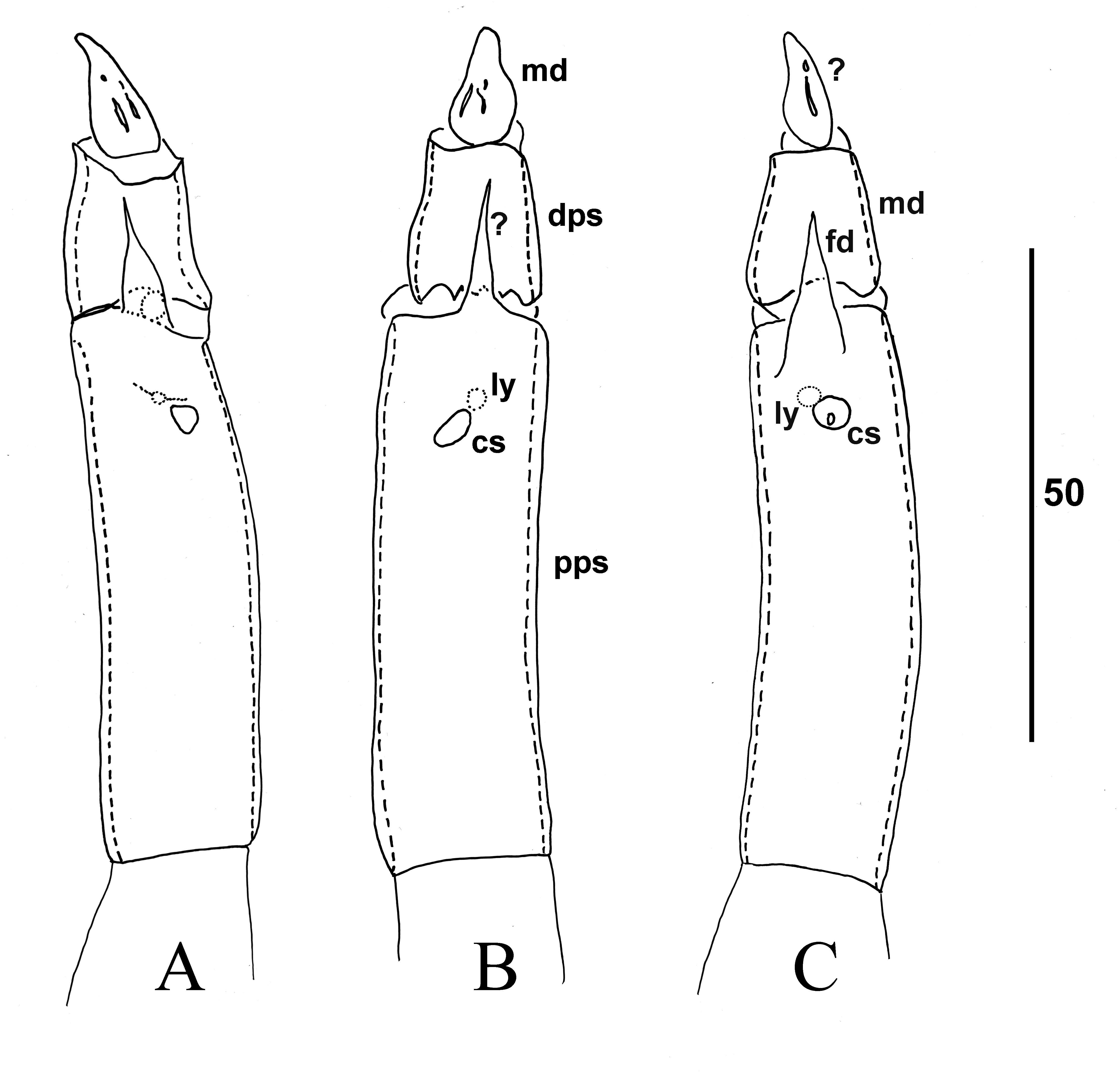
Chelicerae ( Figs. 5 View FIGURE A-C) — Second cheliceral segment seemingly split into two segments (see Remarks); proximal pseudosegment segment 52 – 55, bearing vestigial cheliceral seta and lyrifissure, and distal membranous process; distal pseudosegment 17 – 19; movable digit (12) a hyaline lobe bearing some narrow, straight sclerotised elements; arthrodial corona and fixed digit absent.
Legs ( Figs. 6 View FIGURE A-F) — Excluding ambulacra, lengths of leg I 280 – 295, leg II 370 – 410, leg III 385 – 425, leg IV 410 – 445. Tarsus I (72 – 77) with elongate ambulacrum (20 – 24), without claws; ambulacra II- IV (48 – 55), each with a dorsal ribbed leaf-like pretarsus ( Fig. 6F View FIGURE ); claws I absent, II-IV hyaline. Setation of leg segments I-IV: coxae 2-2-2-2; trochanters 4-4-4-5 (TrI-III with larval complement for Laelapidae ); femora 8-8-8-8 or, as (al -ad / av, pd / pv, pl), I-IV (1-2/1, 2/1-1); genua 8-8-8-8, or I-IV (1-2/1, 2/1-1); tibiae 8-7-7-7, or I (1-2/1, 2/1-1), II-IV (1-1/1, 2/1- 1); tarsi II-IV 17-17-17, with ad1-pd1 seta-like, apical processes 0.4-0.5 × length of pretarsi, ad1 subequal to pd1. Tarsus I with 30 setae: four ribbed blunttipped setae (length 16 – 26) dorsodistally among dorsodistal cluster of nine abruptly tapering setae plus one tiny seta (3), and 16 proximal and ventral strongly tapered setae. All setae on other leg segments unspecialised, smooth, length 13 – 35.
Male ( Figs. 7 View FIGURE ).
Dorsal idiosoma — Dorsal shield (length 650 – 660 × width 550 – 560), similar in ornamentation and setation to female.
Ventral idiosoma ( Fig. 7A View FIGURE ) — Sternoventral shield (420 long × 200 wide at level of st4), fused to exopodal shield, bearing slender sternal setae and 21 opisthogastric setae at posterior margin; of setae st1-5, st5 longest (36), other sternal setae (30 – 32); longitudinal reticulation throughout except near anterolateral margin. Genital operculum in anterior concavity of sternal shield. Anal shield similar to female (length 135 × width 100). Soft cuticle hypertrichous, setal length 30 – 80, longer posteriorly. Poroids on soft cuticle and anal shield absent; lyrifissure iv5 near posterolateral margin of sternoventral shield.
Gnathosoma ( Figs. 7C View FIGURE ) — Similar to female, setae h1 28, h2 14, h3 11, palpcoxal seta absent, palp length 95, corniculi similar to female. Subcapitulum extends medially as two finely spiculate processes reaching level of mid-palp femur; labrum finely spiculate, larger, thicker than in female, reaching level of mid-palp femur. Chelicerae ( Fig. 7B View FIGURE ): fixed digit absent; movable digit represented by hyaline sheath that encloses spermatodactyl (length 73).
Legs — Chaetotaxy as in female, legs subequal in length compared with female. Tarsus I as in female; all other leg setae unspecialised, as in female.
Remarks — The only previous record for M. brevipes is from Italy with the ant T. erraticum ( Berlese 1904) . Our new specimens are from Greece, but from the same host species, and we expect M. brevipes to be present throughout the intervening terrain in the Balkan Peninsula and central Europe where T. erraticum is known to occur.
Species of Myrmozercon are highly host specific, with no species known from more than one species of ant, although in three known cases two species of Myrmozercon are known to utilize the same species of ant (Shawand Seeman 2009). In these cases, the species pairs conform to a Hutchinsonian size ratio (Shawand Seeman 2009). Therefore, the host species and size of the mite is informative. Our specimens match Berlese’s (1904) illustrations in all regards, including the bizarre chelicerae of the female and male. Their size is also similar. Berlese (1902) measured the female as 800 µm × 609 µm and the male at 700 µm × 560 µm. Dr Roberto Nannelli (Berlese Collection, Florence) measured all available intact females, including the holotype (n = 4, on slides 5/39 and 5/40, holotype measurement first followed by range of values based on three other females at the Berlese Collection), and recorded the length as 792 µm (720-768) and width as 600 µm (552 – 576). Berlese’s male was remeasured at 720 µm × 552 µm. Our females (640 – 760 µm × 550 – 640 µm) and males (650 – 660 µm × 550 – 560 µm) tended to be slightly shorter, and Berlese (1904) also illustrated the species with a slightly tapering idiosoma, while ours are ovate. We regard our specimens as the same species, attributing the small differences in size and shape to intraspecific variation.
The chelicerae of M. brevipes are unlike any other member of the genus (or perhaps the Acari) and are difficult to interpret. Two hypotheses are given. In the first, the lateral cheliceral lyrifissure has enlarged to such a size that it separates the second cheliceral segment into two pseudosegments ( Fig. 5B View FIGURE ). The dorsal lyrifissure and vestigial cheliceral seta remain on the proximal pseudosegment, while the distal pseudosegment terminates in a membranous movable digit that includes small rod-like sclerotised elements. The dorsal, membranous flange at the margin of the pseudosegmental division is a novel structure. The lyrifissures and setae are absent or not visible in males. The terminology in the description is based on this hypothesis.
An alternative interpretation is that the movable digit is equivalent to the distal pseudosegment of the first hypothesis, and this bears a novel structure distally ( Fig. 5C View FIGURE ). In support of this is Berlese’s (1904) drawing of cheliceral musculature for M. brevipes , showing the adductor muscle of the movable digit attaching to the first separation in the second cheliceral digit. In this interpretation, the small membranous flange is a much reduced fixed digit. Cheliceral musculature was poorly visible in our specimens, but what was visible supported Berlese’s drawing.
| HNHM |
Hungarian Natural History Museum (Termeszettudomanyi Muzeum) |
| T |
Tavera, Department of Geology and Geophysics |
| QM |
Queensland Museum |
No known copyright restrictions apply. See Agosti, D., Egloff, W., 2009. Taxonomic information exchange and copyright: the Plazi approach. BMC Research Notes 2009, 2:53 for further explanation.
|
Kingdom |
|
|
Phylum |
|
|
Class |
|
|
Order |
|
|
Family |
|
|
Genus |
Myrmozercon brevipes Berlese, 1902
| Kontschán, J. & Seeman, O. D. 2015 |
Myrmozercon brevipes:
| Berlese A. 1904: 313 |
Myrmozercon brevipes
| Berlese A. 1902: 700 |