
Vietnamella sinensis ( Hsu, 1936 )
|
publication ID |
https://doi.org/ 10.11646/zootaxa.4286.3.5 |
|
publication LSID |
lsid:zoobank.org:pub:CDC90512-2065-450B-AA9A-9ADD54B9A362 |
|
DOI |
https://doi.org/10.5281/zenodo.6045237 |
|
persistent identifier |
https://treatment.plazi.org/id/03A98792-FFCC-875A-FF2A-FCFEFA6CF805 |
|
treatment provided by |
Plazi |
|
scientific name |
Vietnamella sinensis ( Hsu, 1936 ) |
| status |
|
Vietnamella sinensis ( Hsu, 1936) View in CoL
Ephemerella sinensis Hsu, 1936: 325 . Holotype: male imago, from Shang Jao (=Shangrao), Jiangxi Province, China.
Ephemerella sinensis: Gui, 1985: 89 .
Ephemerellina sinensis: Allen & Edmunds, 1963: 15 ; Tshernova, 1972: 614; You & Gui, 1995: 138.
Ƒietnamella sinensis: Wang & McCafferty, 1995: 194 ; McCafferty & Wang, 1997: 395.
Ƒietnamella dabieshanensis You & Su, 1987: 176 . Syntypes: male and female adults, nymphs, from Anhui and Fujian Provinces, NEW SYNONYM.
Ƒietnamella dabieshanensis: Edmunds & Murvosh, 1995: 159 ; McCafferty & Wang, 1997: 395; Su & Zhou, 1998: 28.
Cincticostella (Ƒietnamella) dabieshanensis: You & Gui, 1995:140 ; Gui et al., 1999: 341.
Ƒietnamella qingyuanensis Zhou & Su, 1995: 47 . Holotype: nymph; paratypes: nymphs, from Zhejiang Province, NEW SYNONYM .
Ƒietnamella qingyuanensis McCafferty & Wang, 1997: 395 .
Ƒietnamella guadunensis Zhou & Su, 1995: 48 . Holotype: nymph; paratypes: nymphs, from Fujian Province, NEW SYNONYM .
Ƒietnamella qingyuanensis McCafferty & Wang, 1997: 395 .
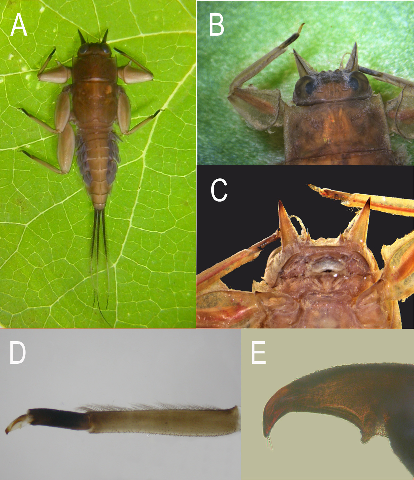
Description. Nymph (in alcohol): body length 13.0–16.0 mm, cerci 6.0– 9.5 mm, terminal filament 8.0–11.0 mm. body greenish brown to dark brown, with dense setae laterally and on free margins of head.
Head ( Figs 1 View FIGURE 1 A–C): two pairs of projections below eyes near slightly concave anterior margin of head capsule; inner pair of projections small, spine-like and sharp, outer pair large, triangular, cone-shaped with length slightly less than width of head capsule; antenna length more than half width of head and positioned between projections; ocelli elevated on small tubercles; vertex of head rough; compound eyes large, dark.
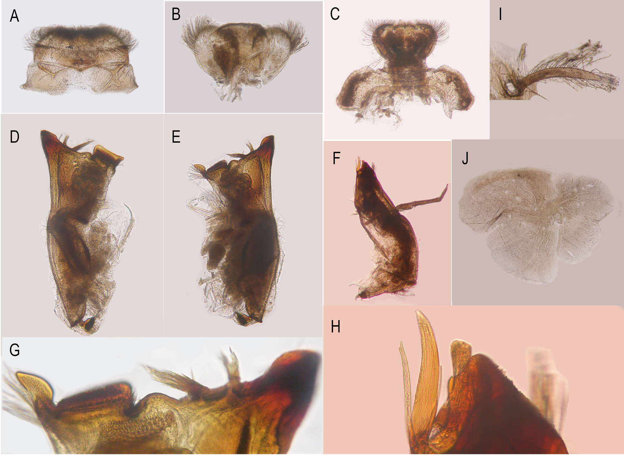
Mouthparts: Labrum ( Figs 2 View FIGURE 2 A, 3A): anterior half of dorsal surface and margins with relatively long setae, ventral surface with shorter setae but those near margins longer and denser, two setal tufts near anteromedian corners. Left mandible ( Figs 2 View FIGURE 2 D, 3D): slender, with very thin setae on lateral surface; outer incisor totally fused, spoon-like, inner incisor transformed into two tufts of spines with common base; prostheca stouter than inner incisor, consisting of numerous spines; molar block-like with rough surface, additional small tuft of spine-like setae on mesal apex. Right mandible ( Figs 2 View FIGURE 2 E, 2G, 3E): similar to left but molar with apparent independent tooth and larger tuft of spine-like setae. Maxilla ( Figs 2 View FIGURE 2 F, 2H, 3F): slender, apex with 3 fused medio-apical teeth or canines, 2 dentisetae and an additional spine-like seta at apex; a small tuft of setae located dorsally ( Fig 2 View FIGURE 2 H); a median seta on inner margin; maxillary palpi 3-segmented, with tiny setae, length ratio from basal to apical 1: 1.6: 1, tip of apical segment sclerotized; cardo with setae. Labium ( Figs 2 View FIGURE 2 C, 3C): glossae and paraglossae almost fused, with dense setae on surface, setae on dorsal surface and margins longer; labial palpi 3-segmented, basal segment broader and longer than the second, apical segment very small; palpi with tiny setae; submentum broad. Hypopharynx ( Figs 2 View FIGURE 2 B, 3B): lingua and superlinguae nearly round, with setae on surface, those near margins longer.
Thorax ( Figs 1 View FIGURE 1 A–E): pronotum with sharp anterolateral projections and small, slightly rounded protuberances mesal to anterolateral projection. Forelegs: coxae with setae on dorsal surface but trochanters without setae; length ratio of femora: tibiae: tarsi = 3: 3: 1; femora strongly expanded except at base, expansion forming distinct transverse ridge dorsally, leading margin of forefemora serrated with projections or teeth progressively smaller distally; inner, outer, basal margins and ridge with a row of setae; tibiae with small spines on surface, inner margin with dense setal row; tarsi with very sparse setae. Midlegs: Length ratio of femora: tibiae: tarsi = 3: 2.5: 1; coxae with obvious dorsal projection and setae; trochanters with setae; femora wider and flatter than tibiae and tarsi, inner, outer and basal margin with setae. Hindlegs: Setal pattern similar to midleg; length ratio of femora: tibiae: tarsi = 4: 3: 1; tarsi of all legs dark brown; all claws similar, with basal projection and several tiny apical setae ( Figs 1 View FIGURE 1 D, 1E).
Abdomen ( Fig 1 View FIGURE 1 A): terga 1–10 with a pair of median ridges or tubercles progressively larger posteriorly; posterolateral angles of terga 1–9 extended into sharp projections, lateral margins of terga with dense setae; sterna with a pair of short dark submedian stripes. Gills on terga 1–7; gills 1 long, finger-like, with setae ( Fig 2 View FIGURE 2 I); gills 2– 6 similar in structure, with dorsal and ventral lamellae, the latter further divided into 2 clusters, each with several smaller lobes; gills 7 smaller, usually covered by gills 6, also with 2 lamellae but ventral lamella divided into 3 lobes only ( Fig 2 View FIGURE 2 J). Caudal filaments dark brown to black with broad median pale band, with dense lateral setae on inner and outer margins ( Fig 1 View FIGURE 1 A).
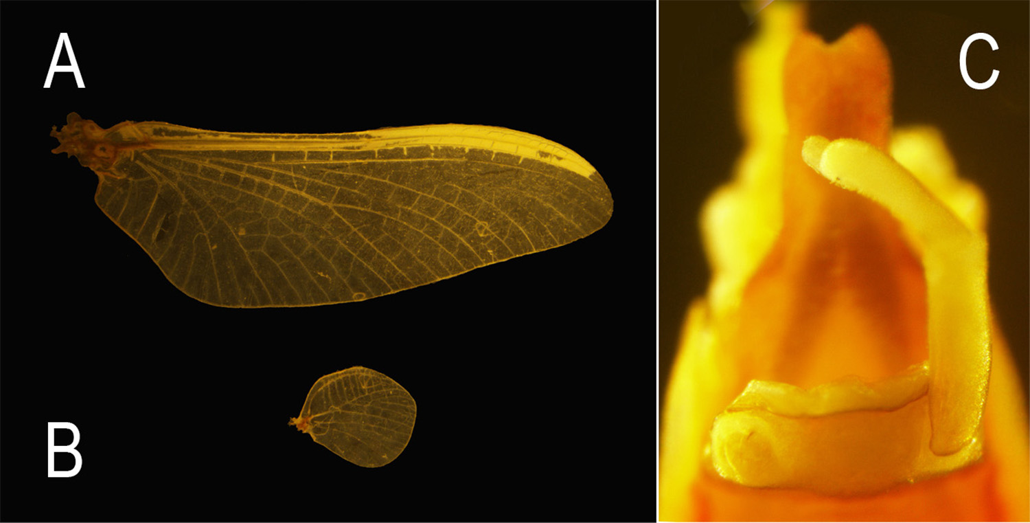
Male imago (in alcohol, Figs 4 View FIGURE 4 , 5 View FIGURE 5 ): stigmatic area of forewing divided by a thin longitudinal vein into upper and lower series of cells. MA forked near middle of wing, MP forked basally, 3–4 long intercalaries between MP1 and MP2; CuA and CuP adjacent at base; independent marginal intercalaries present ( Figs 4 View FIGURE 4 A, 5A). Hind wing nearly round, leading margin slightly concave, with clear crossveins ( Figs 4 View FIGURE 4 B, 5B). Genitalia ( Figs 4 View FIGURE 4 C, 5C): forceps 3-segmented, basal segment a little longer and broader than second, apical segment small, nearly round; penes totally fused with a shallow median cleft; subgenital plate slightly convex.
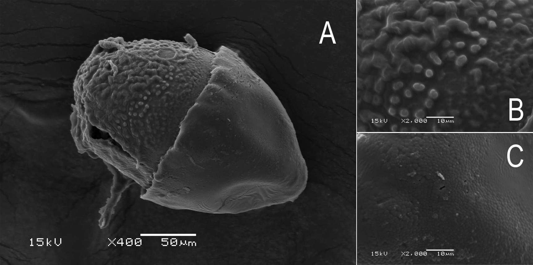
Egg ( Figs 6 View FIGURE 6 A–C, dissected from females collected in 2006): Length 0.223 mm, width 0.15 mm; oval, chorionic surface without distinct structure but with small protuberances. Half of egg covered with helmet-shaped polar cap.

| NEW |
University of Newcastle |
No known copyright restrictions apply. See Agosti, D., Egloff, W., 2009. Taxonomic information exchange and copyright: the Plazi approach. BMC Research Notes 2009, 2:53 for further explanation.
|
Kingdom |
|
|
Phylum |
|
|
Class |
|
|
Order |
|
|
Family |
|
|
Genus |
Vietnamella sinensis ( Hsu, 1936 )
| Hu, Ze, Ma, Zhen-Xing, Luo, Juan-Yan & Zhou, Chang-Fa 2017 |
Ƒietnamella qingyuanensis
| McCafferty 1997: 395 |
Ƒietnamella qingyuanensis
| McCafferty 1997: 395 |
sinensis:
| McCafferty 1997: 395 |
| Wang 1995: 194 |
dabieshanensis:
| Su 1998: 28 |
| McCafferty 1997: 395 |
| Edmunds 1995: 159 |
Cincticostella (Ƒietnamella) dabieshanensis:
| Gui 1999: 341 |
| You 1995: 140 |
Ƒietnamella qingyuanensis
| Zhou 1995: 47 |
Ƒietnamella guadunensis
| Zhou 1995: 48 |
Ƒietnamella dabieshanensis
| You 1987: 176 |
Ephemerella sinensis:
| Gui 1985: 89 |
Ephemerellina sinensis:
| You 1995: 138 |
| Tshernova 1972: 614 |
| Allen 1963: 15 |
Ephemerella sinensis
| Hsu 1936: 325 |