
Cryptodacus Hendel
|
publication ID |
https://doi.org/ 10.5281/zenodo.182184 |
|
DOI |
https://doi.org/10.5281/zenodo.6228036 |
|
persistent identifier |
https://treatment.plazi.org/id/03A99C64-EE50-CD00-5B9C-2696FA10DC9E |
|
treatment provided by |
Plazi |
|
scientific name |
Cryptodacus Hendel |
| status |
|
Key to the species of Cryptodacus Hendel View in CoL View at ENA
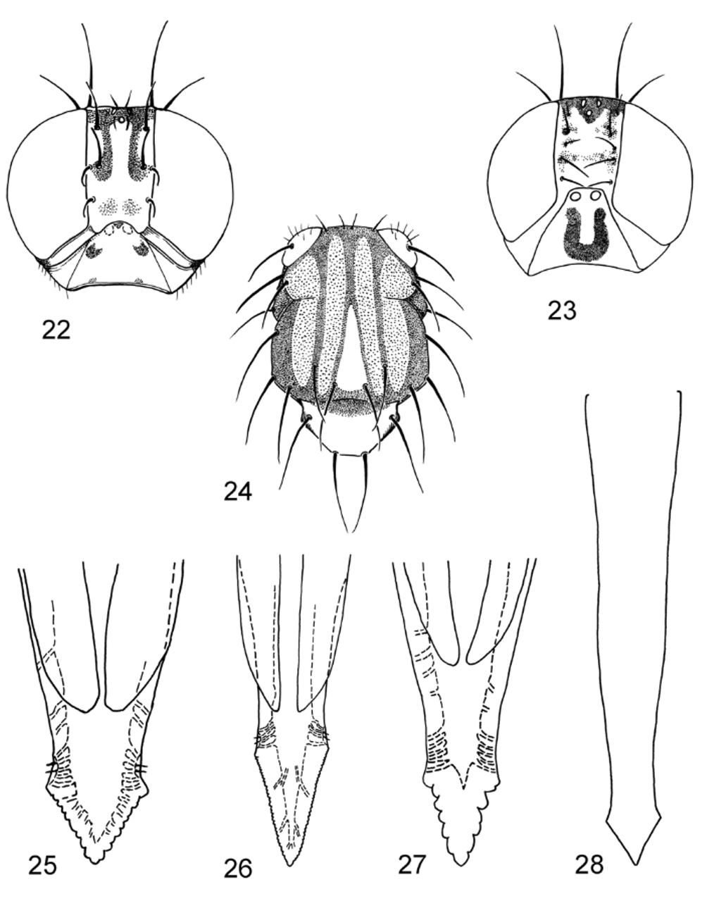
1. Frons yellow except between ocelli. Face entirely yellow. Scutum without white band along transverse suture and without sublateral white vitta or spot at base of intra-alar seta ( Fig. 24 View FIGURES 22 – 28. 22 – 23 ). Discal band not extended across crossvein dm-cu ( Figs. 12–13 View FIGURES 11 – 21 ). Orbital setae separated by less than distance from anterior seta to eye margin. Panama, Bolivia, Peru. ........................................................................ obliquus Hendel
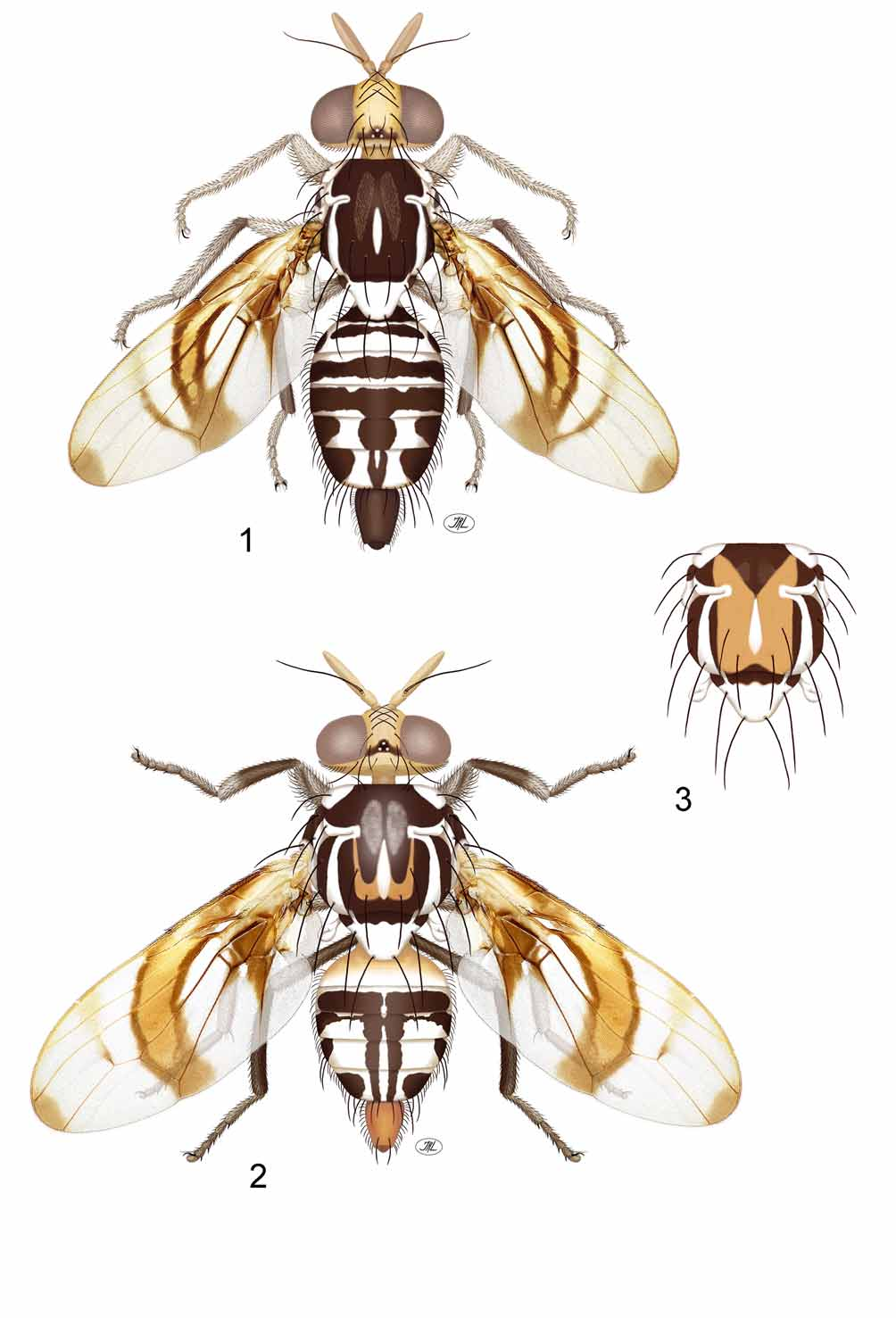
- Frons with brown mark extended lateral to ocelli ( Figs. 1–2 View FIGURES 1 – 3. 1 – 2 , 4 View FIGURES 4 – 7. C , 8 View FIGURES 8 – 10. C , 22–23 View FIGURES 22 – 28. 22 – 23 ). Face with dark brown spot or spots. Scutum with white band along transverse suture and with sublateral white vitta ( Figs. 1–3 View FIGURES 1 – 3. 1 – 2 , 6 View FIGURES 4 – 7. C , 8 View FIGURES 8 – 10. C ) or
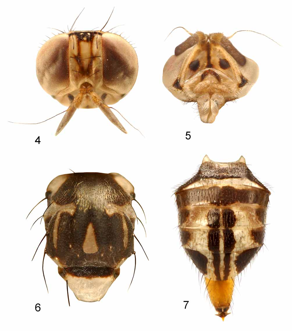
at least white spot at base of intra-alar seta. Discal band extended across at least posterior end of dm-cu ( Figs. 11, 14–21 View FIGURES 11 – 21 ). Orbital setae separated by more than distance from anterior seta to eye margin ( Figs. 1– 2 View FIGURES 1 – 3. 1 – 2 , 4 View FIGURES 4 – 7. C , 8 View FIGURES 8 – 10. C , 22–23 View FIGURES 22 – 28. 22 – 23 ). ............................................................................................................................................. 2 2. Occiput and postgena entirely yellow or orange. Female tergites 5 and 6 with sublateral white areas bordering medial brown vitta or vittae ( Figs 1–2 View FIGURES 1 – 3. 1 – 2 , 7 View FIGURES 4 – 7. C ). Crossvein dm-cu entirely covered by discal or subapical band ( Figs. 11, 17, 19 View FIGURES 11 – 21 ). Mexico, Central America. 3
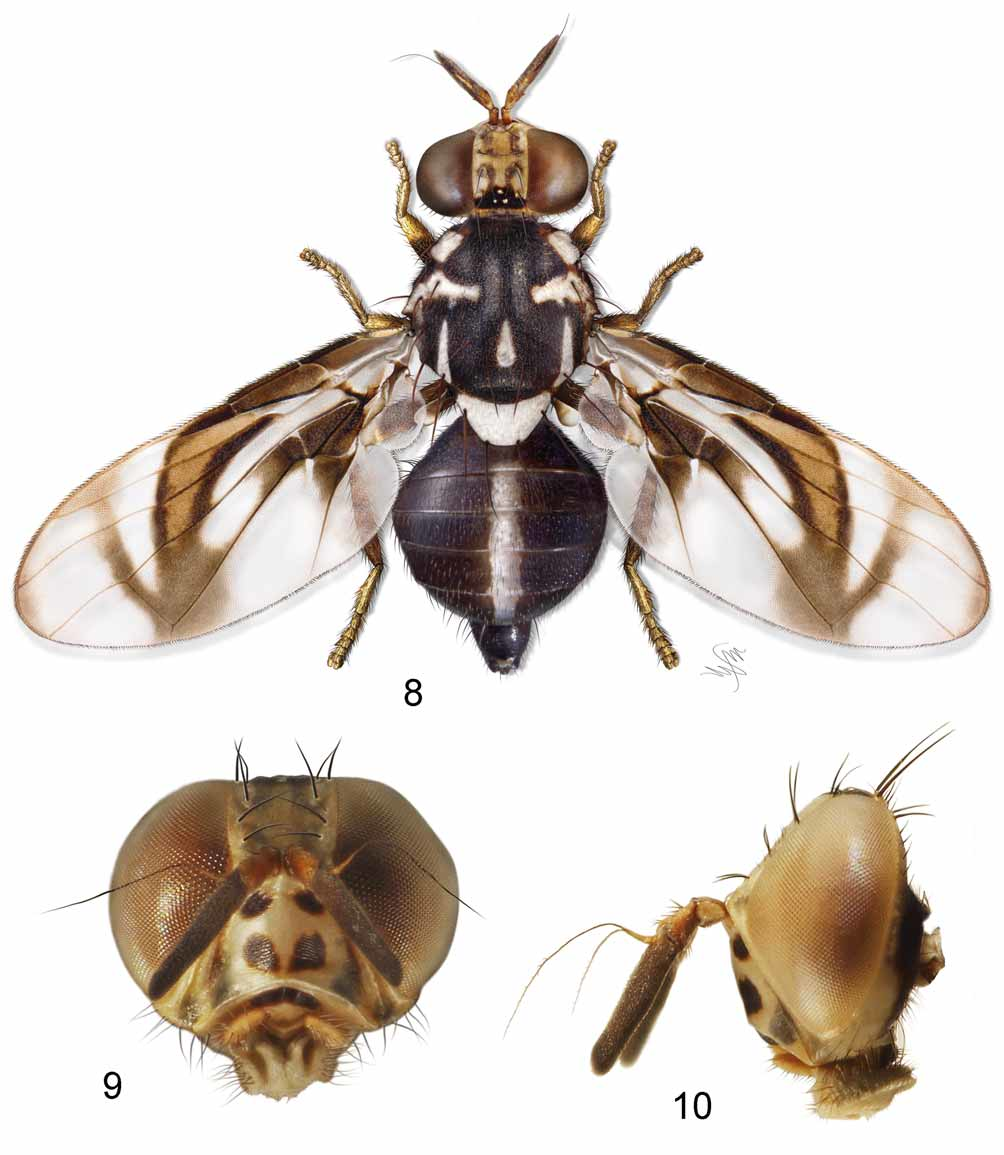
- Occiput and postgena dark brown except anterior margin. Female tergites 5 and 6 without sublateral white areas ( Fig. 8 View FIGURES 8 – 10. C ). Crossvein dm-cu not bordered by brown medially ( Figs. 14–16 View FIGURES 11 – 21 ) except in tigreroi and univittatus ( Fig. 18, 20–21 View FIGURES 11 – 21 ). Costa Rica, South America............................................................................5
3. Crossvein r-m less than 0.75 distance from bm-cu to dm-cu ( Fig. 11 View FIGURES 11 – 21 ). Discal band extended across only posterior end of dm-cu. Face with single, medial dark brown spot. Gena without dark spot. Female tergites 5–6 with undivided medial dark brown vitta ( Fig. 1 View FIGURES 1 – 3. 1 – 2 ). Oviscape dark brown. Guatemala....................... .............................................................................................................................................. lopezi Norrbom
- Crossvein r-m more than 0.80 distance from bm-cu to dm-cu ( Figs. 17, 19 View FIGURES 11 – 21 ). Discal band extended across all of dm-cu. Face with 2–4 brown markings including pair of sublateral spots. Gena with dark brown spot. Female tergites 5–6 with 2 narrowly separated submedial dark brown vittae ( Figs. 2 View FIGURES 1 – 3. 1 – 2 , 7 View FIGURES 4 – 7. C ). Oviscape orange...........................................................................................................................................................4
4. Face, in addition to pair of large dark brown sublateral spots, with unpaired narrow medial brown vitta or 2 spots aligned along midline ( Fig. 5 View FIGURES 4 – 7. C ). Clypeus dark brown. 2 frontal setae. Wing ( Fig. 19 View FIGURES 11 – 21 ) with orange band from apical part of cell r1 with distal margin in cell r4+5 closer to level of apex of vein R2+3 than to level of crossvein dm-cu; apex of cell r1 entirely orange. Syntergite 1+2 mostly dark brown except for white band on apical margin ( Fig. 7 View FIGURES 4 – 7. C ). Panama. ......................... trinotatus Norrbom & Korytkowski, n. sp.
- Face with only dark brown sublateral spots, lacking brown vitta or spots along midline. Clypeus yellow to orange. 3 frontal setae. Wing ( Fig. 17 View FIGURES 11 – 21 ) with orange band from apical part of cell r1 with distal margin in cell r4+5 equidistant between levels of apex of vein R2+3 and crossvein dm-cu or closer to dm-cu; apex of cell r1 entirely hyaline. Syntergite 1+2 mostly yellow except for white band on apical margin ( Fig. 2 View FIGURES 1 – 3. 1 – 2 ). Mexico, Guatemala, Costa Rica. tau (Foote)
5. Cell dm with only apical corner hyaline ( Fig.14 View FIGURES 11 – 21 ). Abdominal tergites 3 and 4 each with apical white band. Scutum with white spot at base of intra-alar seta, but dark between it and white mark on transverse suture. Face with 2 or 4 brown spots, each side with large dorsal and small ventral spot ( Fig. 22 View FIGURES 22 – 28. 22 – 23 ) or one elongate spot. Frons with distinct dark brown anterior extensions from band across ocelli ( Fig. 22 View FIGURES 22 – 28. 22 – 23 ). Colombia, Ecuador, northern Brazil. ................................................................................................... ornatus Norrbom
- Cell dm with basal and apical hyaline areas ( Figs. 15–16, 18, 20–21 View FIGURES 11 – 21 ). Abdominal tergites 3 and 4 without apical white bands. Scutum with narrow sublateral white vitta from intra-alar seta to or almost to white mark on transverse suture ( Fig. 8 View FIGURES 8 – 10. C ), except in quirozi. Face with 3–4 brown spots, sometimes fused into singe U-shaped mark ( Fig. 23 View FIGURES 22 – 28. 22 – 23 ). Frons sometimes with irregular brownish or grayish areas ( Fig. 8 View FIGURES 8 – 10. C ), but without distinct dark brown anterior extensions from dark band across ocelli...........................................6
6. Postgena brown except anterior margin. Scutum with sublateral white vitta extended anteriorly at least to level of postsutural supra-alar seta ( Fig. 8 View FIGURES 8 – 10. C ). Postpronotal lobe with at least small white spot (possibly variable in P. p a r k e r i).........................................................................................................................................7
- Postgena entirely yellow. Scutum with white spot posterior to intra-alar seta, not extended anteriorly to level of postsutural supra-alar seta. Postpronotal lobe entirely brown. Mexico. .................quirozi Norrbom
7. Subapical band ( Figs. 18, 20–21 View FIGURES 11 – 21 ) connected to discal band posteriorly, covering all of crossvein dm-cu. Antennal first flagellomere more than 6 times as long as wide. Abdominal tergites 3 and 4 with uninterrupted medial white vitta ( Fig. 8 View FIGURES 8 – 10. C ).................................................................................................................8
- Subapical band ( Fig. 15 View FIGURES 11 – 21 ) not connected to discal band, middle part of dm-cu not bordered by brown. Antennal first flagellomere less than 5.5 times as long as wide. Abdominal tergites 3 and 4 with or without
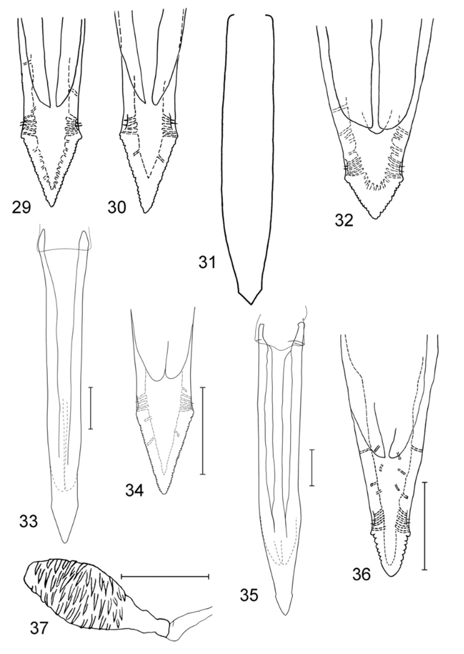
uninterrupted medial white vitta...................................................................................................................9 8. Aculeus tip ( Figs. 31–32 View FIGURES 29 – 37 ) stout, serrate part slightly wider than long. Subapical band anterior to vein R4+5 and apical band dark brown and well delimited ( Fig. 18 View FIGURES 11 – 21 ). Syntergite 1+2 with white band on posterior half except on lateral margin. Gena with brown spot. Ecuador. ................................................ tigreroi Norrbom
- Aculeus tip ( Figs. 35–36 View FIGURES 29 – 37 ) slender, serrate part longer than wide. Subapical band anterior to vein R4+5 and apical band orange and poorly defined ( Figs. 20–21 View FIGURES 11 – 21 ). Syntergite 1+2 mostly dark brown, white only narrowly on posterior margin medially ( Fig. 8 View FIGURES 8 – 10. C ). Gena without brown spot. Panama. ........................................ .................................................................................................. univittatus Norrbom & Korytkowski, n. sp.
9. Face with 2 ventral and 2 dorsal brown spots. Abdomen with medial white vitta interrupted on tergites 3 and 4. Apical band ( Fig. 15 View FIGURES 11 – 21 ) faintly covering all of cells r1 and r2+3 apical to subapical band. Costa Rica.... ............................................................................................................................................ parkeri Norrbom
- Face with 1 ventral and 2 dorsal brown spots, sometimes fused into large U-shaped mark ( Fig. 23 View FIGURES 22 – 28. 22 – 23 ). Abdominal tergites 3 and 4 with uninterrupted medial white vitta. Apical band faint and narrow in cells r1 and r2+3. Southern Brazil. .............................................................................................................. silvai Lima

No known copyright restrictions apply. See Agosti, D., Egloff, W., 2009. Taxonomic information exchange and copyright: the Plazi approach. BMC Research Notes 2009, 2:53 for further explanation.