
Sennertionyx manicati ( Giard, 1900 )
|
publication ID |
https://doi.org/ 10.11646/zootaxa.4857.1.9 |
|
publication LSID |
lsid:zoobank.org:pub:75043663-E121-4CFB-BE93-8BCAD5F3BD92 |
|
DOI |
https://doi.org/10.5281/zenodo.4421436 |
|
persistent identifier |
https://treatment.plazi.org/id/03AA0770-FFDE-FFF6-1BA7-BD6A25F4006B |
|
treatment provided by |
Plazi |
|
scientific name |
Sennertionyx manicati ( Giard, 1900 ) |
| status |
|
Description of Sennertionyx manicati ( Giard, 1900)
( Figures 1–24 View FIGURE 1 View FIGURE 2 View FIGURE 3 View FIGURE 4 View FIGURE 5 View FIGURE 6 View FIGURE 7 View FIGURE 8 View FIGURE 9 View FIGURE 10 View FIGURE 11 View FIGURE 12 View FIGURE 13 View FIGURE 14 View FIGURE 15 View FIGURE 16 View FIGURE 17 View FIGURE 18 View FIGURE 19 View FIGURE 20 View FIGURE 21 View FIGURE 22 View FIGURE 23 View FIGURE 24 ; Table 1–3)
ADULT FEMALE (n=5; Figs 1–3 View FIGURE 1 View FIGURE 2 View FIGURE 3 , 7 View FIGURE 7 ). Idiosoma oval, white to light brown; cuticle smooth, without obvious striation.
Dorsum ( Fig. 1 View FIGURE 1 ). Prodorsal shield vase-shaped, extended anterolaterally, joining supracoxal sclerites, faintly and evenly punctate. Supracoxal sclerites terminate in a flattened and fringed Grandjean’s organ (11–18 teeth), orifice of supracoxal gland prominent, about as long as width of supracoxal sclerite; supracoxal setae scx spiniform, tapering from base to tip. All dorsal idiosomal setae smooth, sci longer than other prodorsal setae, vi about twice as long as ve; sce anterolateral to sci, ratios: sci: sce= 3.2–4.5, sci–sci: sci–sce= 3.1–4.7. Sejugal furrow a distinctive suture. Hysterosoma bearing 12 pairs of setae, c 1, c 2, c p, c 3, d 1, d 2, e 1, e 2, and f 2 similar in length; h 2 prominently longer than other setae; h 3 slightly longer than h 1; opisthonotal gland opening gla right in middle between d 2 and e 2.
Venter ( Figs 2 View FIGURE 2 and 7C View FIGURE 7 ). Coxal apodemes I joined at midline, forming a prosternal apodeme directed posteromedially; coxal plate I posteriorly extending beyond apex of prosternal apodeme and expanded laterally; ventral setae 1a inserted laterad of coxal plate I; coxal apodemes II directed posteromedially, posterior margin of coxal plates clearly concave; apodemes III and IV freely directed anteromedially. Genital opening in shape of inverted V, situated between coxae III–IV; ventral setae 3a at level of anterior rim of genital opening, genital setae g lateral to genital opening; setae 4a lateral to posterior parts of genital opening. Anal opening clearly posterior to and about as long as genital opening, surrounded by 3 pairs of adanal setae (ad 1–3) and 3 pairs of pseudanal setae (ps 1–3); ad 1–3 about half to 2/3 of ps 1–3. Copulatory opening posterior to anus, at level of ps 1; spermathecal duct a narrow cylindrical tube ( Figs 2 View FIGURE 2 and 7C View FIGURE 7 ); sclerotised base of spermathecal sac bowler-hat-shaped, a pair of bell-like sclerites of oviducts situated at end of base of spermathecal sac.
Gnathosoma ( Fig. 7 View FIGURE 7 A–B). Chelicera robustly chelate, fixed digit bearing one subterminal tooth, two paraxial and two antiaxial teeth medially, and one trapezoidal proximal tooth; movable digit bearing two large teeth medially; cheliceral setae cha falcate, nearly one fourth of movable digit; chb filiform, about one third as long as movable digit; subcapitular setae m filiform. Palpal supracoxal setae elcp attenuate, dorso-lateral, subequal to subcapitular setae; dorsal and lateral palptibial setae filiform, dorsal palptarsal setae filiform, terminal palptarsal solenidion ω tiny, rod-like ( Fig. 7D View FIGURE 7 ).
Legs ( Fig. 3 View FIGURE 3 ) I and IV longer than legs II and III; setae on all segments smooth; tarsal empodial claws strong; condylophores symmetrical and stout.
Leg I ( Fig. 3A View FIGURE 3 ). Trochanter with a simple seta, nearly reach apical rim of trochanter; femoral seta vF filiform, extending beyond apical rim of genu; genual solenidia σ’ and σ” subequal in length, nearly reach middle of tibia; setae cG conical and subequal to mG, mG slightly thinner than cG; tibial solenidion φ whip-like, extending far beyond tip of claw, setae gT and hT conical, gT slightly longer; tarsus I about twice as long as basal width, solenidion ω 1 nearly parallel sided basally, then gradually narrowing before forming a small head, ε adjacent to ω 1, solenidia ω 2 situated closely to ω 3, on apical half of tarsus, ω 2 slightly longer than ω 3, setae wa, ba and la stouter than ra, d filiform, e and f setiform; apicoventral setae s, u and v stout and conical; p and q filiform, adjacent to u and v, respectively, and slightly longer than u and v; empodium reduced, empodial claw a strong hook.
Leg II ( Fig. 3B View FIGURE 3 ). Trochanter with a simple seta as in trochanter I; femoral seta vF filiform; genu with a solenidion (σ) and two conical setae (cG, mG); tibial solenidion φ whip-like, extending far beyond tip of claw, setae gT conical and hT slender; tarsus nearly twice as long as basal width; ω gradually narrowing before forming a small head; setae wa, ba and la stouter than ra, d filiform, e and f setiform; apicoventral setae s, u and v stout and conical, setae p and q setiform, adjacent to u and v, respectively, and slightly longer than u and v; empodium reduced, empodial claw a strong hook.
Leg III ( Fig. 3C View FIGURE 3 ). Trochanter with a simple seta as in trochanter I; femur nude; genu III with a solenidion (σ) and a seta (nG), σ about one half of nG; tibia with a solenidion (φ) and a seta (kT), φ rod-like, slightly longer than kT; tarsus about twice as long as basal width, w and r conical, d filiform, e and f setiform; apicoventral setae s, u and v stout and conical, setae p and q setiform, adjacent to u and v, respectively, and slightly longer than u and v; empodium reduced, empodial claw a strong hook.
Leg IV ( Fig. 3D View FIGURE 3 ). Trochanter nude; femur with a simple seta; genu nude; tibia with a solenidion (φ) and a seta (kT), φ rod-like, shorter than kT; tarsus about 2.5 times as long as wide, w and r conical, d filiform, e and f setiform; apicoventral setae s, u and v stout and conical, setae p and q setiform, adjacent to u and v, respectively, and slightly longer than u and v; empodium reduced, empodial claw a strong hook.
ADULT MALE (n=7; Figs 4–8 View FIGURE 4 View FIGURE 5 View FIGURE 6 View FIGURE 7 View FIGURE 8 , 24 View FIGURE 24 ). With two morphs (homeomorphic and heteromorphic) present. Idiosomal shape, colour and cuticle structure similar to those in female.
Dorsum ( Figs 4–5 View FIGURE 4 View FIGURE 5 , 24 View FIGURE 24 ). Prodorsal shield similar to that of female. Grandjean’s organ anteriorly fringed, ending in 10–16 teeth; supracoxal setae scx smooth, tapering from base to tip; sce distinctly longer than sci. Ratios: sci: sce= 3.1–4.2 sci–sci: sci–sce= 3.0– 4.3 in homeomorphic male; sci: sce= 3.5–4.6 sci–sci: sci–sce= 2.5–2.8 in heteromorphic. Sejugal furrow a simple suture. Opisthonotal gland opening gla in middle between d 2 and e 2. Opisthosomal setae h 2 prominently longer than other idiosomal setae. Setae ve, c 1, c 2, c p, c 3, d 1, d 2, e 1, e 2 and f 2 subequal.
Venter ( Fig. 6 View FIGURE 6 , 7 View FIGURE 7 E–F). Shapes and structures of coxal apodemes and plates similar to those in adult female. Ventral setae 1a inserted posterolateral to coxal plate I, 3a and 3b mediad of coxae III; genital opening shaped as an inverted V, situated between coxae IV, genital setae g lateral to genital opening, anterior to genital papillae, 4a posterolateral to genital opening. Lateral arms supporting aedeagus turning inwards. Aedeagus (53–56) gradually tapered from base to middle, nearly parallel sided subterminally, then gradually narrowing before forming an obvious head ( Fig. 24 View FIGURE 24 ); canal of aedeagus smooth, without lumps. Distance between anterior rim of anal opening and posterior margin of aedeagus short, about a half of genital opening. Anal suckers each bears a small anal disc. Pseudanal setae ps 1 slightly longer than ps 2, ps 2 about twice as long as ps 3.
Gnathosoma ( Fig. 7D View FIGURE 7 ). Chelicera robustly chelate as in female, fixed digit and movable digit with same numbers of teeth; cheliceral setae cha drop point blade shaped, chb seta-like; subcapitular setae m filiform. Palpal supracoxal setae elcp attenuate, dorso-laterally; palptibial and palptarsal setae filiform, palptarsal solenidion peg-like.
Legs ( Fig. 8 View FIGURE 8 ) I and IV longer than legs II and III; setae on all segments smooth; tarsal empodial claws strong; condylophores symmetrical and stout.
Leg I ( Fig. 8A View FIGURE 8 ). Trochanter with a simple seta, nearly reaches apical rim of trochanter; femoral seta vF filiform, extending beyond apical rim of genu; genual solenidia σ’ and σ” subequal, σ’ slightly shorter than σ”, nearly reach middle of tibia; setae cG and mG stout, subequal; tibial solenidion φ whip-like, extending far beyond tip of claw, setae gT and hT stout; tarsus I about twice as long as basal width, solenidion ω 1 nearly parallel sided basally, then gradually narrowing before forming a small head, ε adjacent to ω 1, solenidion ω 2 posteriad of d, ω 3 enlarged and situated at same level of d; setae s, u and v spine-like, setae p and q slender, adjacent to u and v, respectively; empodium reduced, claw a strong hook.
Leg II ( Fig. 8B View FIGURE 8 ). Trochanter with a simple seta as in trochanter I; femoral seta vF filiform; genu with a solenidion (σ) and two conical setae (cG, mG); tibia II with φ whip-like, extending far beyond tip of claw; tarsus less than twice as long as basal width; ω gradually narrowing before forming a small head; setae s, u and v spine-like, p and q slender, adjacent to u and v, respectively; empodium reduced, claw a strong hook.
Leg III ( Figs 8C, 8E View FIGURE 8 ). Both sides or one side of leg III enlarged in heteromorphic male ( Fig. 8C View FIGURE 8 ). Trochanter with a simple seta as in trochanter I; femur nude; genu III with a solenidion and a seta, σ nearly two thirds as long as nG; tibia with a solenidion and a seta, φ rod-like, about as long as kT in homeomorphic male ( Fig. 8E View FIGURE 8 ) and longer than kT in heteromorphic male ( Fig. 8C View FIGURE 8 ); tarsus about twice as long as basal width in homeomorphic male ( Fig. 8E View FIGURE 8 ), or shortened and claw enlarged in heteromorphic male( Fig. 8C View FIGURE 8 ); setae s, u and v spine-like, p and q slender, adjacent to u and v, respectively; empodium reduced, claw a strong hook.
Leg IV ( Fig. 8D View FIGURE 8 ). Trochanter nude; femur with a seta; genu nude; tibia with a solenidion and a seta, φ rod-like, longer than kT; tarsus about 2.5× as long as wide, d sucker, e sucker, f filiform; setae s, u and v spine-like, p and q slender, adjacent to u and v, respectively; empodium reduced, claw a strong hook.
TRITONYMPHS (n=3; Figs 9–12 View FIGURE 9 View FIGURE 10 View FIGURE 11 View FIGURE 12 ). Idiosoma oval, white to light brown; cuticle plain, without obvious striation.
Dorsum ( Fig. 9 View FIGURE 9 ). Prodorsal shield vase-shaped, anterior parts extended laterally, narrowly joining supracoxal sclerites, faintly and evenly punctate; Supracoxal sclerite elongate, orifice of supracoxal gland prominent, about as long as width of supracoxal sclerite; Grandjean’s organ anteriorly fringed, ending in 8–15 teeth; supracoxal setae scx smooth, tapering from base to tip. All dorsal idiosomal setae smooth, sci longer than other prodorsal setae, vi about twice as long as ve, c 1, c 2, d 1, e 1, e 2 and h 1 subequal; sci obviously longer than sce, ratios: sci: sce= 3.5–3.6, sci–sci: sci–sce= 3.2–4.2.
Venter ( Fig. 10 View FIGURE 10 ). Coxal apodemes I joined at midline, forming a prosternal apodeme directed posteromedially; coxal plate I posteriorly extending beyond apex of prosternal apodeme and expanded laterally; coxal apodemes II directed posteromedially; apodemes III and IV freely directed anteromedially. Ventral setae 1a inserted laterad of coxal plate I, 3a at level of anterior rim of genital opening, genital setae at level of genital papillae, 4a lateral to posterior parts of genital opening. Genital opening a longitudinal slit, situated centrally between coxae III–IV. Anal opening far posterior to genital opening, about as long as genital opening, surrounded by 3 pairs of pseudanal setae (ps 1–3), ad 1–3 absent.
Gnathosoma ( Fig. 11 View FIGURE 11 ). Chelicera robustly chelate, movable digit, cheliceral setae cha lanceolate, chb setiform; subcapitulum bearing a pair of subcapitular setae m. Palpal supracoxal setae elcp dorso-laterally; dorsal palptibial setae filiform, lateral palptibial setae filiform, dorsal palptarsal setae filiform, terminal palptarsal solenidion tiny.
Legs ( Fig. 12 View FIGURE 12 ) I and IV longer than legs II and III; setae on all segments smooth; tarsal empodial claws strong; condylophores symmetrical and stout.
Leg I ( Fig. 12A View FIGURE 12 ). Trochanter with a simple seta, nearly reach apical rim of trochanter; femoral seta vF filiform, extending beyond apical rim of genu; genual solenidia σ’ and σ” subequal in length, nearly reach middle of tibia; setae cG and mG stout, subequal; tibial solenidion φ whip-like, extending far beyond tip of claw, setae gT and hT stout; tarsus I about twice as long as basal width, solenidion ω 1 nearly parallel sided basally, then gradually narrowing before forming a small head, ε adjacent to ω 1, solenidia ω 2 and ω 3 about at same level at apical half of tarsus, similar in length; setae s, u and v spine-like, setae p and q slender, adjacent to u and v, respectively; empodium reduced, claw a strong hook.
Leg II ( Fig. 12B View FIGURE 12 ). Trochanter with a simple seta; femoral seta vF filiform; genu with σ slightly shorter than cG and mG; tibia with φ whip-like, extending far beyond tip of claw, setae gT and hT stout; tarsus about twice as long as its basal width; ω gradually narrowing before forming a small head, setae s, u and v spine-like, setae p and q slender, empodium reduced, claw a strong hook.
Leg III ( Fig. 12C View FIGURE 12 ). Trochanter with a simple seta; femur nude; genu III with a solenidion and a seta, σ about half as long as nG; tibia with φ rod-like, kT spine-like; tarsus about twice as long as basal width, setae s, u and v spinelike, setae p and q slender, empodium reduced, claw a strong hook.
Leg IV ( Fig. 12D View FIGURE 12 ). Trochanter nude; femur with a simple seta; genu nude; tibia with a solenidion and a seta, φ rod-like, about as long as kT; tarsus about 2.5 times as long as wide, setae s, u and v spine-like, setae p and q slender, empodium reduced, claw a strong hook.
DEUTONYMPH (n=5; Figs 13–16 View FIGURE 13 View FIGURE 14 View FIGURE 15 View FIGURE 16 ). Idiosoma flat, nearly oval in dorsoventral view, brown in colour.
Dorsum ( Fig. 13 View FIGURE 13 ). Lateral prodorsum covered with oblique striae, central area anterior to sejugal furrow bearing horizontal and oblique striae. Setae vi mediodistal, a small area around bases of vi punctate; ve posterolateral to vi; sci anteromedial to sce; scx spine-like. Hysterosoma mainly covered with longitudinal striae, bearing 12 pairs of setae, h 2 longer than others. Opisthonotal gland openings gla close to e 2. Anterior cupules ia posterior to c 2, im posteromedial to e 2.
Venter ( Figs 14 View FIGURE 14 , 15B View FIGURE 15 ). Coxal apodemes I joined at midline, forming a prosternal apodeme directing posteromedially, reaching middle level of apodemes II; coxal setae 1a filiform, situated on cuticle between prosternal apodeme and anterior apodemes II; posterior apodemes I fused with anterior apodemes II; posterior apodemes II extending posteromedially, nearly reaching anterior apodemes III; anterior apodemes III extending medially, then curved posteromedial, merging with anterior apodemes IV, each bearing a micro seta (3a); 3b as vestigial alveoli, situated on cuticle surrounded by apodemes; anterior apodemes IV extending medially, then curved posteromedial, ending freely, 4a as vestigial alveoli. Genital setae filiform, anterolateral to genital opening; genital papillae apically attenuate. Suctorial plate large, median suckers (ad 1–2) larger than other suckers, ps 1 and ps 2 conoidal; ps 2 anterolateral to ad 1–2; cupules ih lateral to ps 2.
Gnathosoma ( Fig. 15A View FIGURE 15 ). Subcapitular remnant developed; palpal remnants vestigial; bearing a filiform dorsal seta, palpal solenidion much longer than dorsal setae.
Legs ( Fig. 16 View FIGURE 16 ) I and II longer and thicker than leg III; leg IV smallest; tarsi I – IV each with a twisted claw.
Leg I ( Fig. 16A View FIGURE 16 ). Trochanter and femur each with a filiform seta; genu with a stout solenidion (σ) and two setae, cG spiniform and mG thinner; tibia with solenidion φ stout, extending to base of tarsal solenidion ω 3, gT and hT setiform; tarsus with eight setae, three solenidia and a famulus; setae wa, la, d, e and s foliate, ba, ra and f setiform; solenidia ω 1 parallel sided, extending to base of claw, ω 2 and ω 3 rod-like, ω 3 anterior to ω 2; famulus ε peg-like.
Leg II ( Fig. 16B View FIGURE 16 ). Trochanter and femur each with a filiform seta; genu with a stout solenidion (σ) and two setae, cG spiniform and mG thinner; tibia with solenidion φ stout, extending nearly to base of tarsal seta d, setae gT and hT setiform; tarsus with eight setae and a solenidion; setae wa, la, d, e and s foliate, ba, ra and f setiform; solenidia ω parallel sided, extending to base of claw.
Leg III ( Fig. 16C View FIGURE 16 ). Trochanter with a filiform seta; femur nude; genu with a small solenidion (σ) and a setiform seta (nG); tibia with φ extending to base of tarsal claw, setae kT spiniform; tarsus with eight setae, r and e foliate, w, d, f, s, u and v setiform, d whip-like.
Leg IV ( Fig. 16D View FIGURE 16 ). Trochanter nude, femur with a filiform seta; genu nude; tibia with φ extending beyond base of tarsal setae d, setae kT spiniform; tarsus with eight setae, d, e and f whip-like and longer then leg IV, w, r and s spiniform, u and v filiform.
PROTONYMPH (n=5; Figs 17–20 View FIGURE 17 View FIGURE 18 View FIGURE 19 View FIGURE 20 ). Idiosoma oval, white; cuticle plain, without obvious striation.
Dorsum ( Fig. 17 View FIGURE 17 ). Prodorsal shield vase-shaped, anterior parts extending laterally, narrowly joining supracoxal sclerites; Grandjean’s organ anteriorly fringed, ending in several teeth; supracoxal setae scx smooth, attenuate; dorsal idiosomal setae smooth, sci longer than other prodorsal setae, vi nearly three times as long as ve; ratios: sci: sce= 3.3–4.3, sci–sci: sci–sce= 3.0–3.5. Hysterosomal setae h 1 prominently longer than other setae, c 1, c 2, c p, c 3, d 1, d 2, e 1, e 2, f 2 and h 1 subequal.
Venter ( Fig. 18 View FIGURE 18 ). Coxal apodemes I joined at midline, forming a prosternal apodeme directed posteromedially; setae 1a inserted laterad of coxal apodemes I; coxal apodemes II directed posteromedially; apodemes III and IV freely directed anteromedially; setae 3a and 4a absent. Genital opening a longitudinal slit, situated between coxae IV. Anal opening close to genital opening, about 1.2 times as long as genital opening, surrounded by 3 pairs of pseudanal setae (ps 1–3); ad 1–3 absent.
Gnathosoma ( Fig. 19 View FIGURE 19 ). Chelicerae, palpi and subcapitulum same as those in tritonymph.
Legs ( Fig. 20 View FIGURE 20 ). Legs I and II longer and thicker than legs III and IV.
Leg I ( Fig. 20A View FIGURE 20 ). Trochanter nude; femoral seta vF extending beyond apical rim of genu; genual solenidion σ’ slightly shorter than σ”; setae cG and mG subequal and spiniform; tibial solenidion φ whip-like, extending far beyond tip of tarsal claw, setae gT and hT spiniform; tarsus nearly twice as wide as long, ω 1 mostly parallel sided, famulus ε adjacent to ω 1, ω 2 at terminal half of tarsus, ω 3 absent; seta wa a stout spine, ba and la spiniform but thinner than wa; ra, e and f filiform, d whip-like; ventro-terminal setae s, u and v spiniform, p and q setiform; claw strong.
Leg II ( Fig. 20B View FIGURE 20 ). Trochanter nude; femoral seta vF extending beyond apical rim of genu; genual solenidion σ nearly reach base of gT, setae cG and mG subequal and spiniform; tibial solenidion φ whip-like, extending far beyond tip of claw, setae gT and hT spiniform; tarsus nearly twice as wide as long, ω mostly parallel sided, setae wa a stout spine; ba and la slender than wa; ra, e and f filiform, d whip-like; setae s, u and v spiniform, p and q setiform; claw strong.
Leg III ( Fig. 20C View FIGURE 20 ). Trochanter and femur nude; genu bearing a peg-like solenidion (σ) and a spiniform seta (nG); tibia bearing a rod-like solenidion (φ) and a spiniform seta (kT); tarsus twice as wide as long, seta w a stout spine; r slender and longer than w; e and f filiform, d whip-like; setae s, u and v spiniform, p and q setiform; claw strong.
Leg IV ( Fig. 20D View FIGURE 20 ). Trochanter, femur, genu and tibia nude; tarsus more than twice as wide as long, seta w and r spiniform and subequal; e and f absent, d whip-like; setae s absent, u and v spiniform, p and q setiform; claw strong.
LARVA (n=5; Fig. 21–22 View FIGURE 21 View FIGURE 22 ). Idiosoma elongate oval, white, cuticle plain.
Dorsum ( Fig. 21A View FIGURE 21 ). Prodorsal shield very faint; posterior margin convex. Grandjean’s organ anteriorly fringed, ending in a few teeth; supracoxal setae scx smooth, setiform; sci obviously longer than sce; ratios: sci: sce= 2.1–2.5, sci–sci: sci–sce= 2.3–2.6. Opisthosomal setae h 2 prominently longer than other idiosomal setae.
Venter ( Fig. 21B, C View FIGURE 21 ). Coxal apodemes I joined at midline, forming a prosternal apodeme; Claparède organ ( Fig. 21C View FIGURE 21 ) situated between coxae I and II, nearly four times as wide of terminal part (12) as long (40); genital opening and setae (g) absent. Ventral setae 3a absent. Anal opening postero-terminally, pseudanal setae and adanal setae absent.
Gnathosoma ( Fig. 21D View FIGURE 21 ). Chelicerae as in protonymph; subcapitular setae (m) subequal to palpal supracoxal setae (elcp); palptibial and palptarsal setae filiform, solenidion (ω) about half as long as tarsal setae.
Legs ( Fig. 22 View FIGURE 22 ): leg I thicker and longer than legs II and III; all setae smooth.
Leg I ( Fig. 22A View FIGURE 22 ). Trochanter nude; femoral seta vF extending beyond base of mG; genual solenidion σ’ slightly longer than σ”; cG and mG spiniform, cG slightly longer than mG; tibial solenidion φ whip-like, extending far beyond tip of tarsal claw, setae gT and hT spiniform, gT longer than hT; tarsus nearly twice as wide as long, ω 1 mostly parallel sided, famulus ε adjacent to ω 1, ω 2 and ω 3 absent; seta wa a stout spine, ba and la thinner than wa; ra, e and f filiform, d whip-like; ventro-terminal spine s, u and v spiniform, p and q setiform; claw strong.
Leg II ( Fig. 22B View FIGURE 22 ). Trochanter nude; femoral seta vF extending beyond base of mG; genual solenidion σ nearly reach to middle of tibia, setae cG and mG spiniform, cG longer than mG; tibial solenidion φ whip-like, extending far beyond tip of claw, hT spiniform, gT slender and longer than hT; tarsus less than twice as wide as long, ω mostly parallel sided, setae wa a stout spine; ba and la spiniform but slender than wa; ra, e and f filiform, d whip-like; ventro-terminal spine s, u and v spiniform, p and q setiform; claw smaller than claw of tarsus I.
Leg III ( Fig. 22C View FIGURE 22 ). Trochanter and femur nude; genu bearing a peg-like solenidion (σ) and a spiniform seta (nG); tibia bearing a rod-like solenidion (φ) and a spiniform seta (kT); tarsus twice as wide as long, seta w a spine; r setiform, e and f filiform, d whip-like; ventro-terminal spine s, u and v spiniform, p and q setiform; claw larger than claw of tarsus I.
EGG (n=5; Fig. 23 View FIGURE 23 ). Oval, without any obvious ornamentation.
Ontogenetic changes of idiosomal and leg chaetotaxy are summarised in Table 2 and Table 3 View TABLE 3 , respectively. A key to developmental stages is provided below.
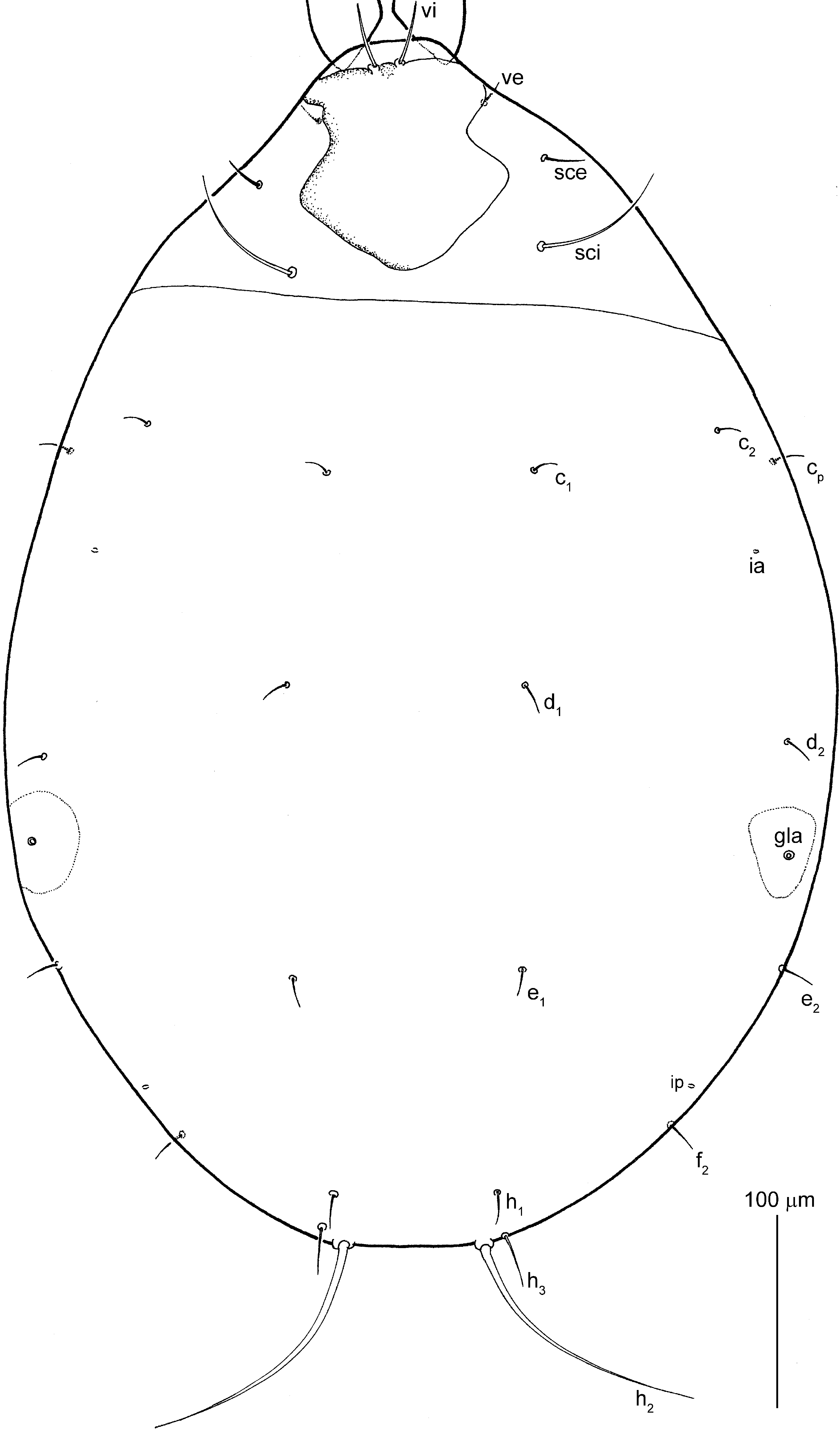
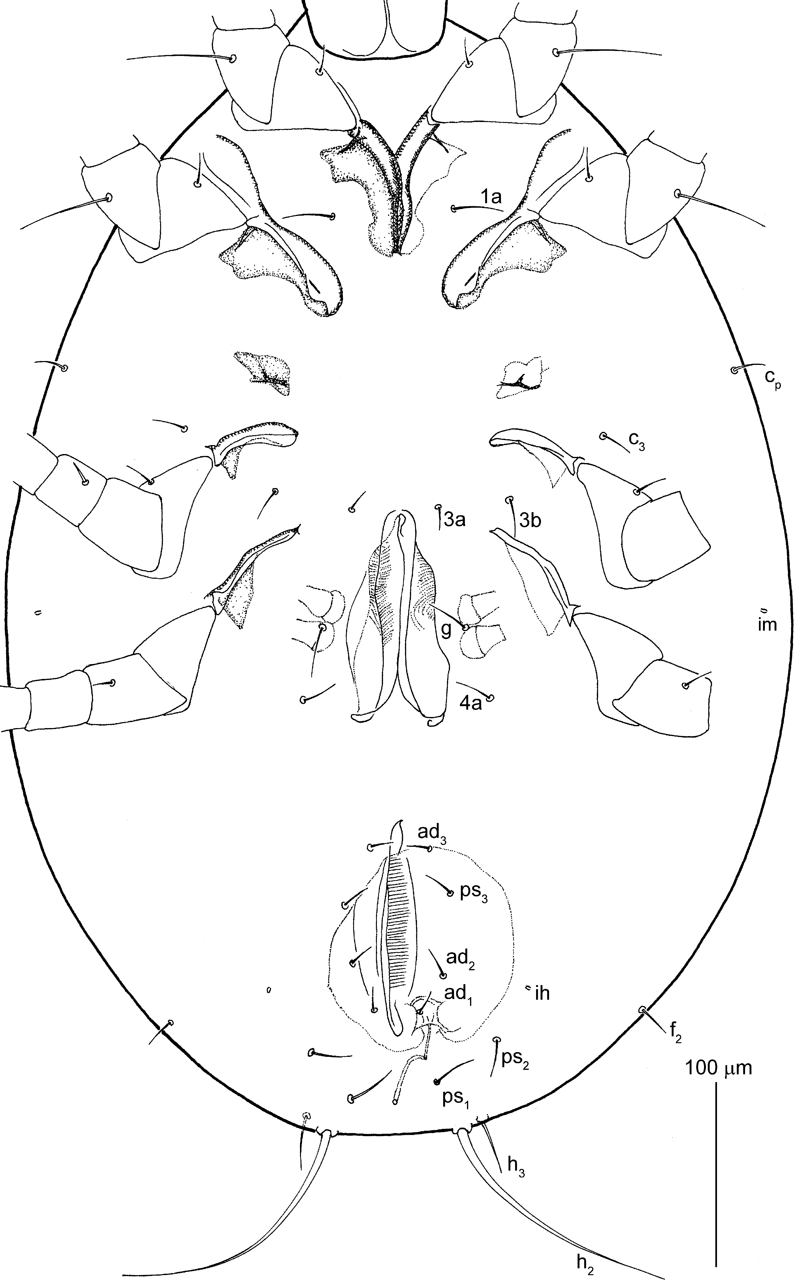
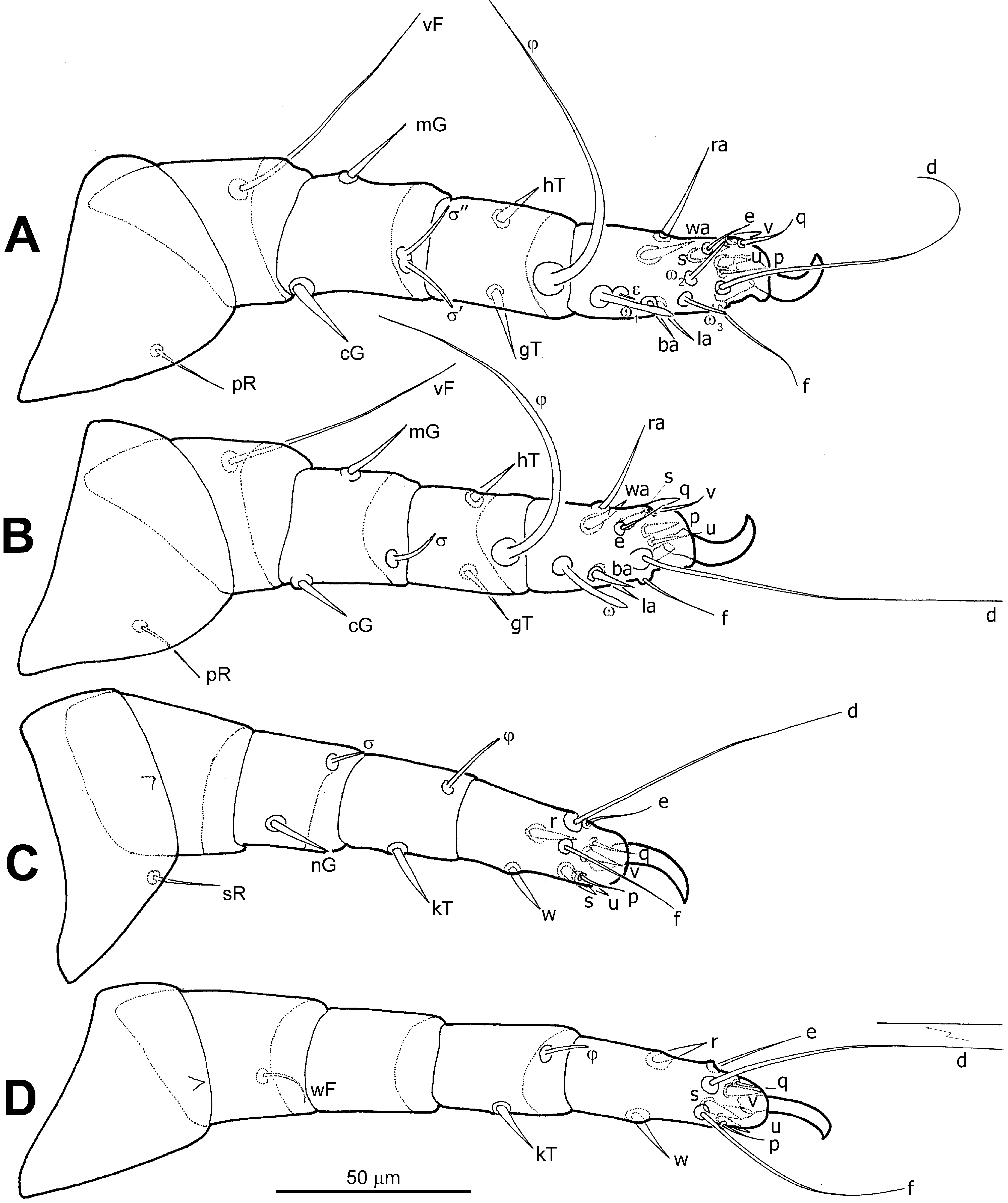
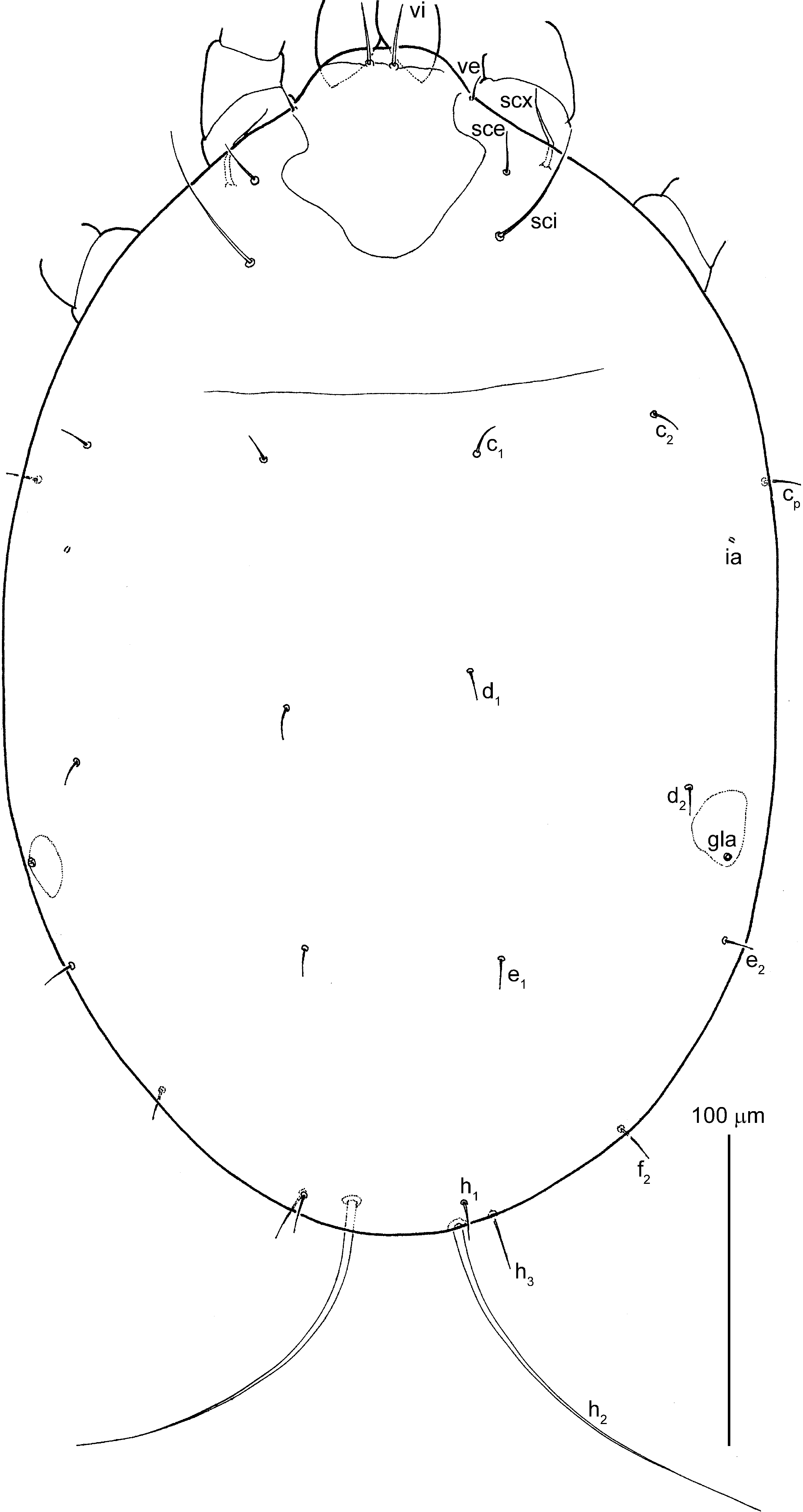
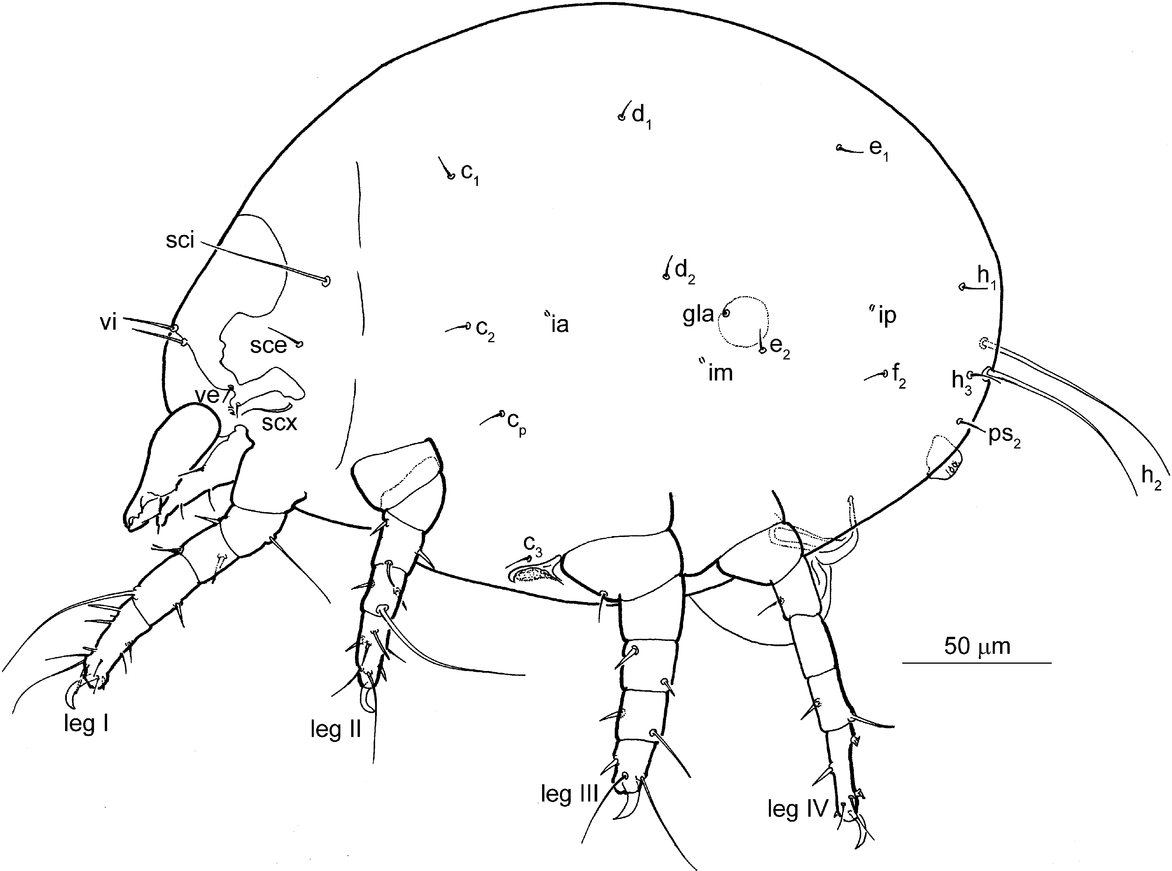
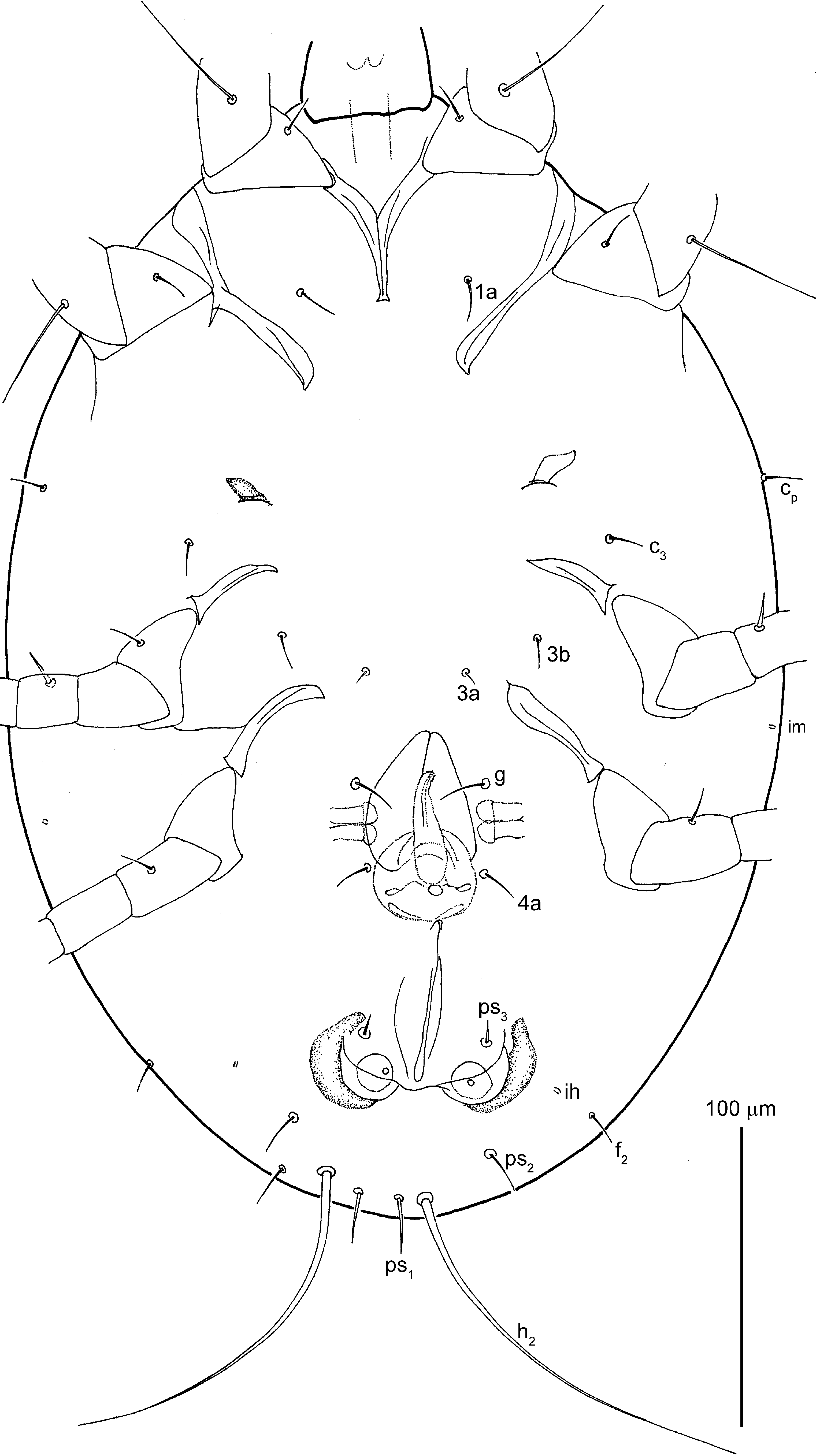

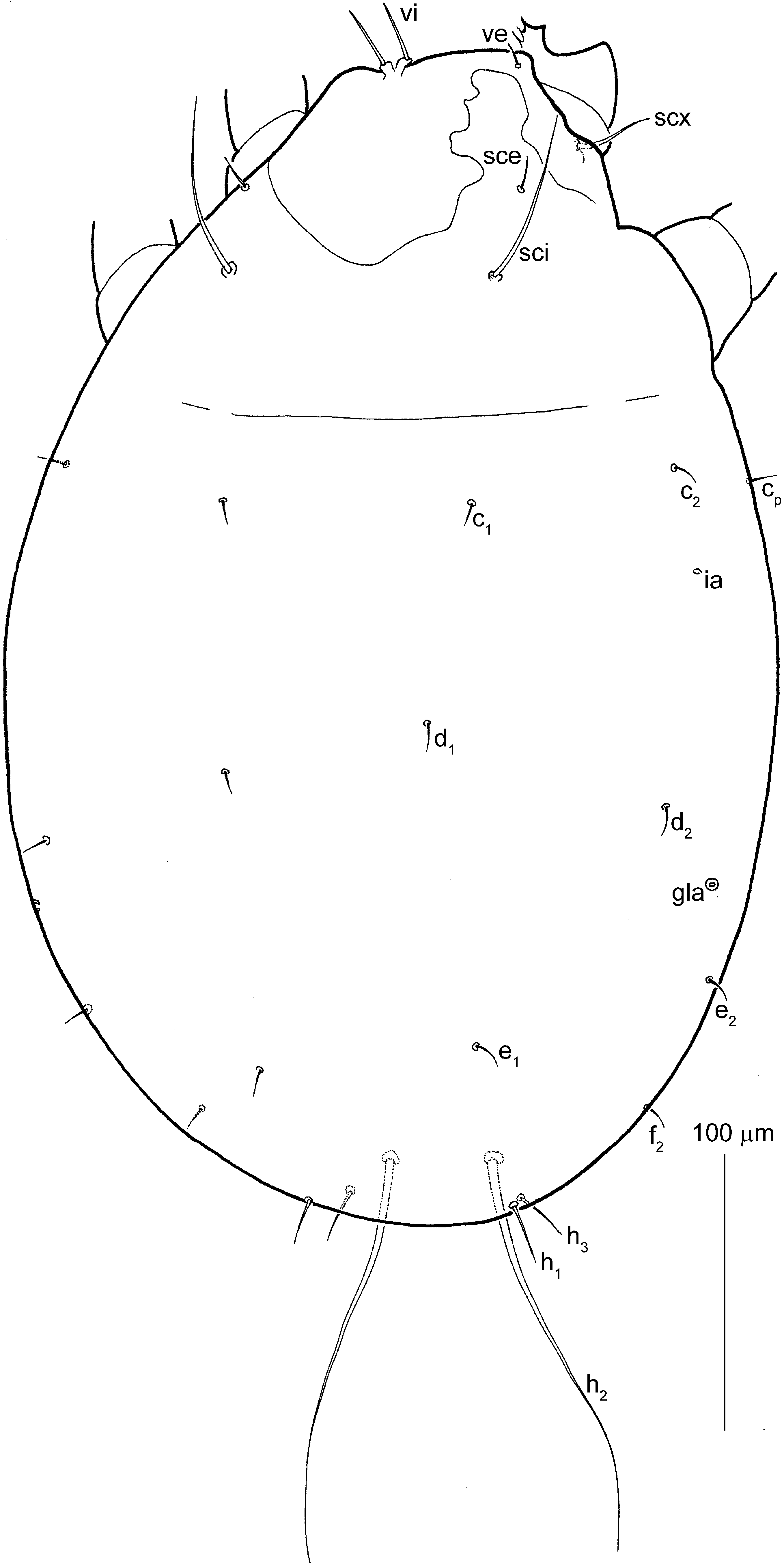
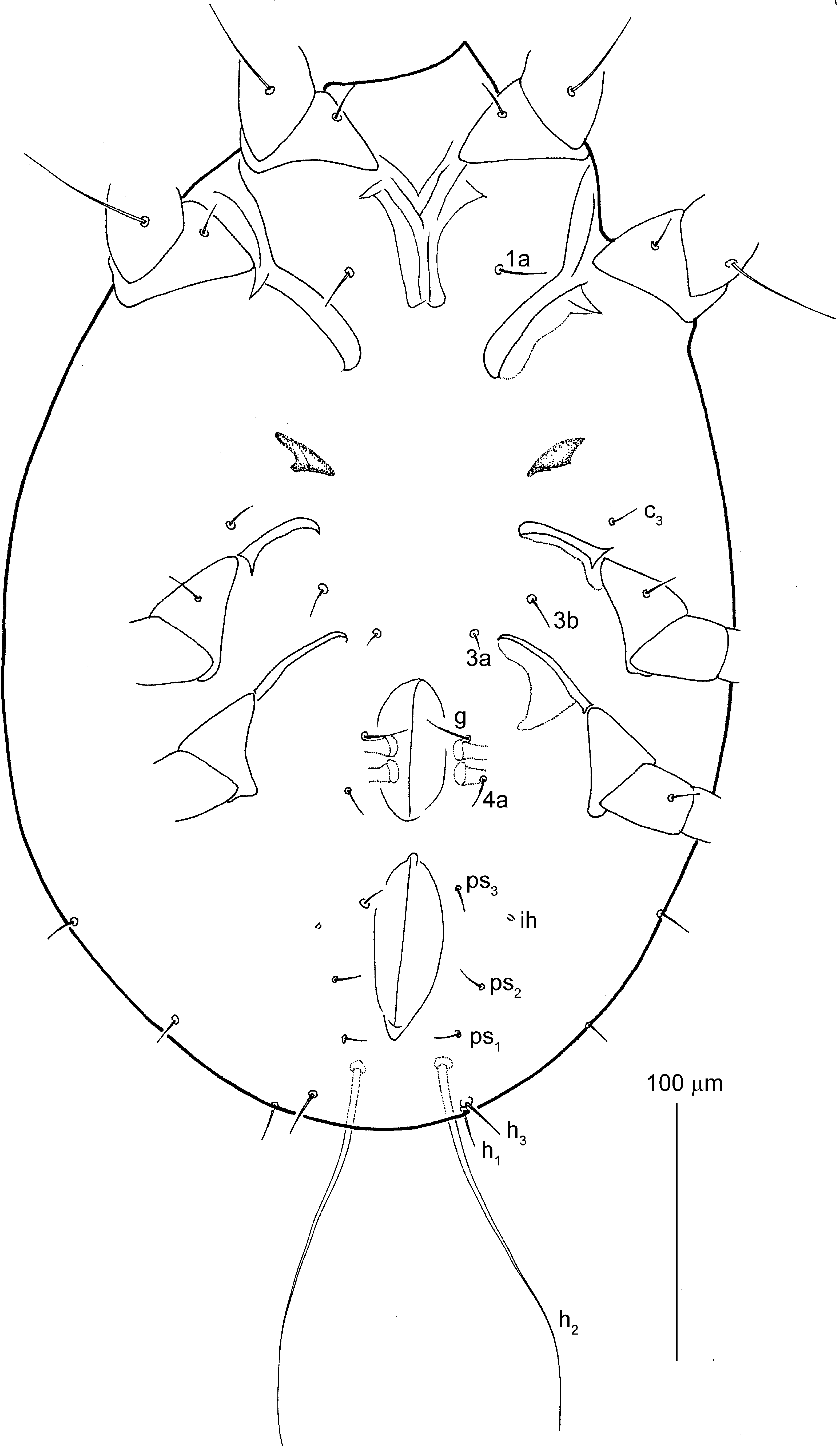
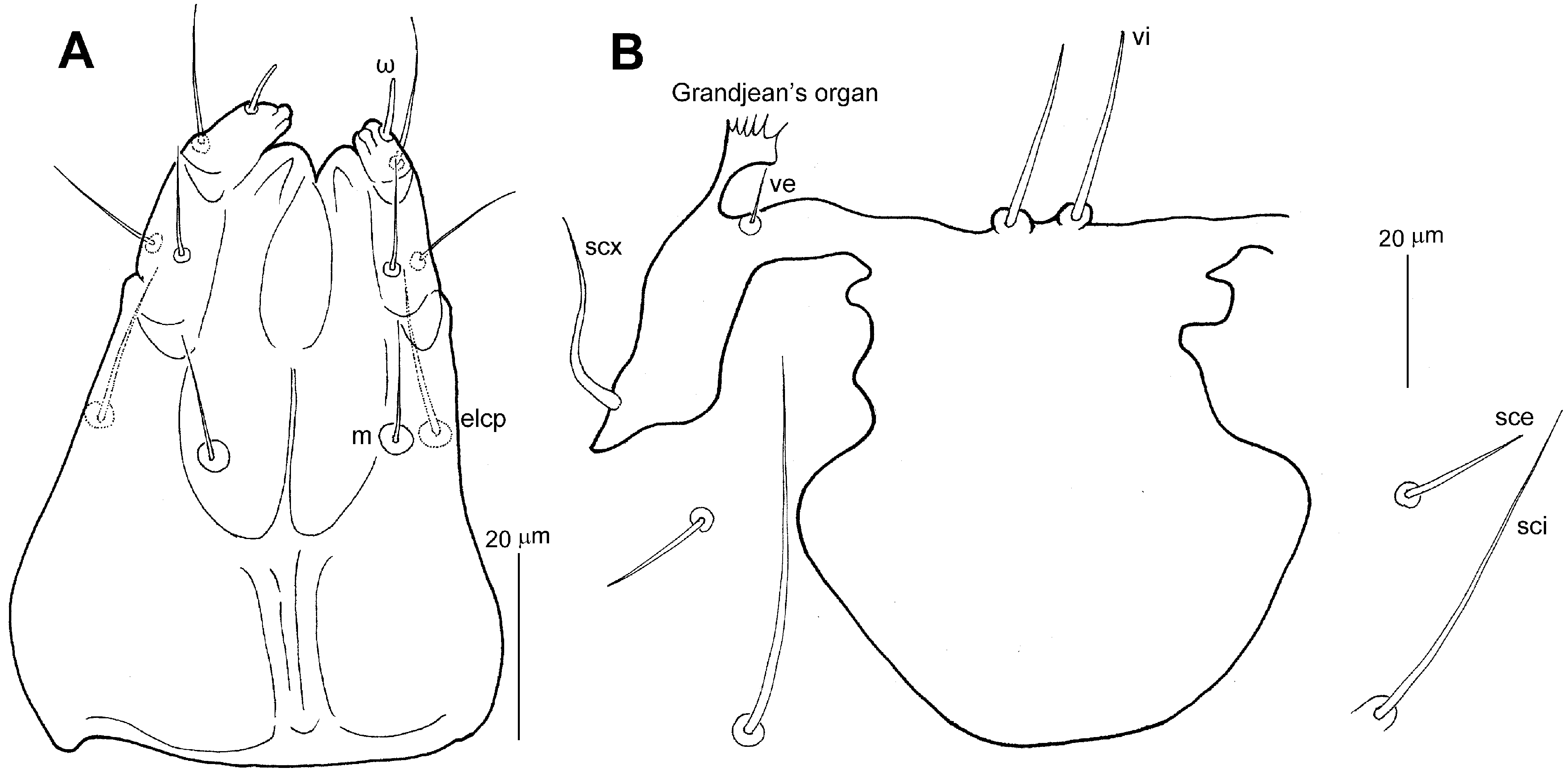
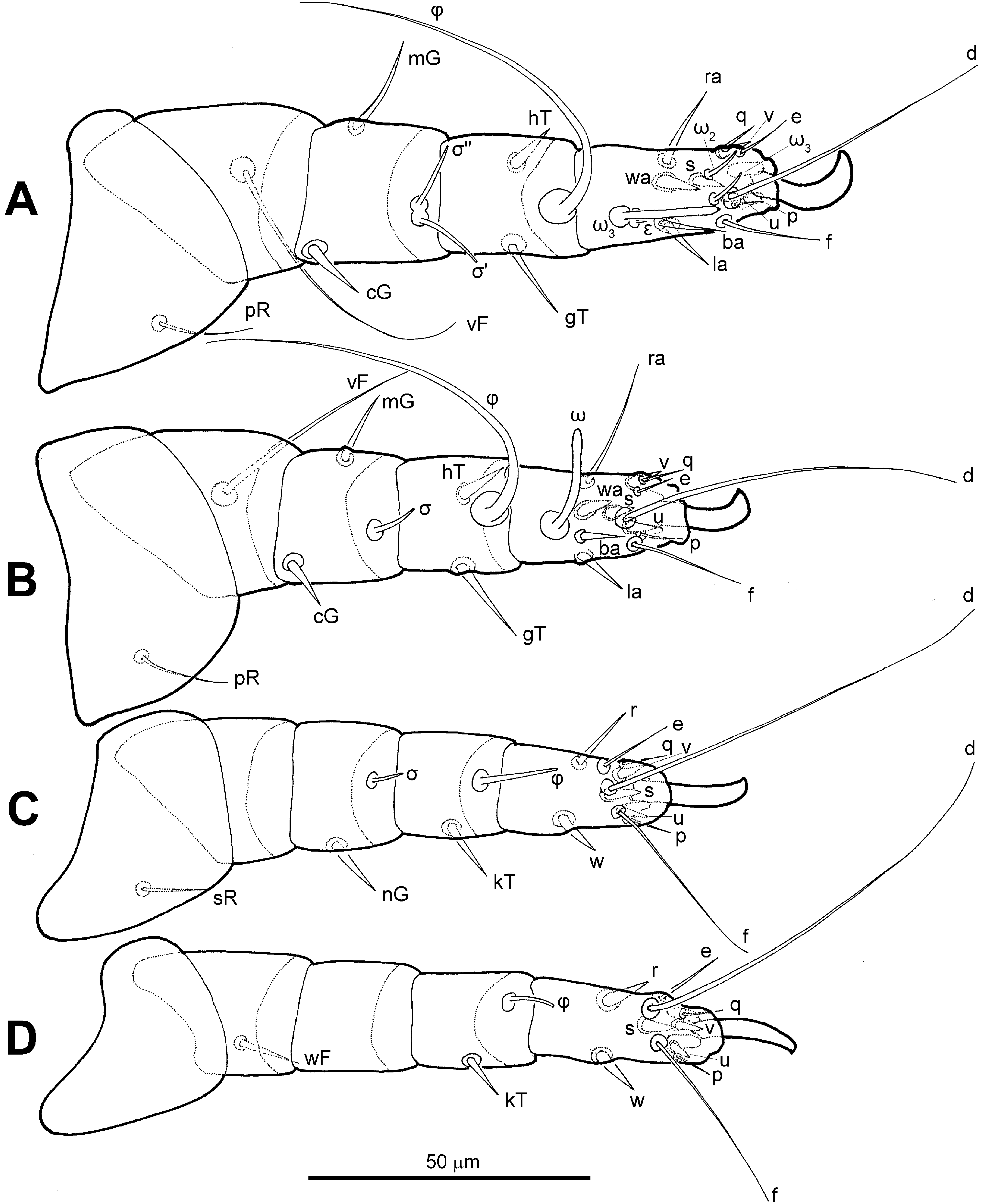
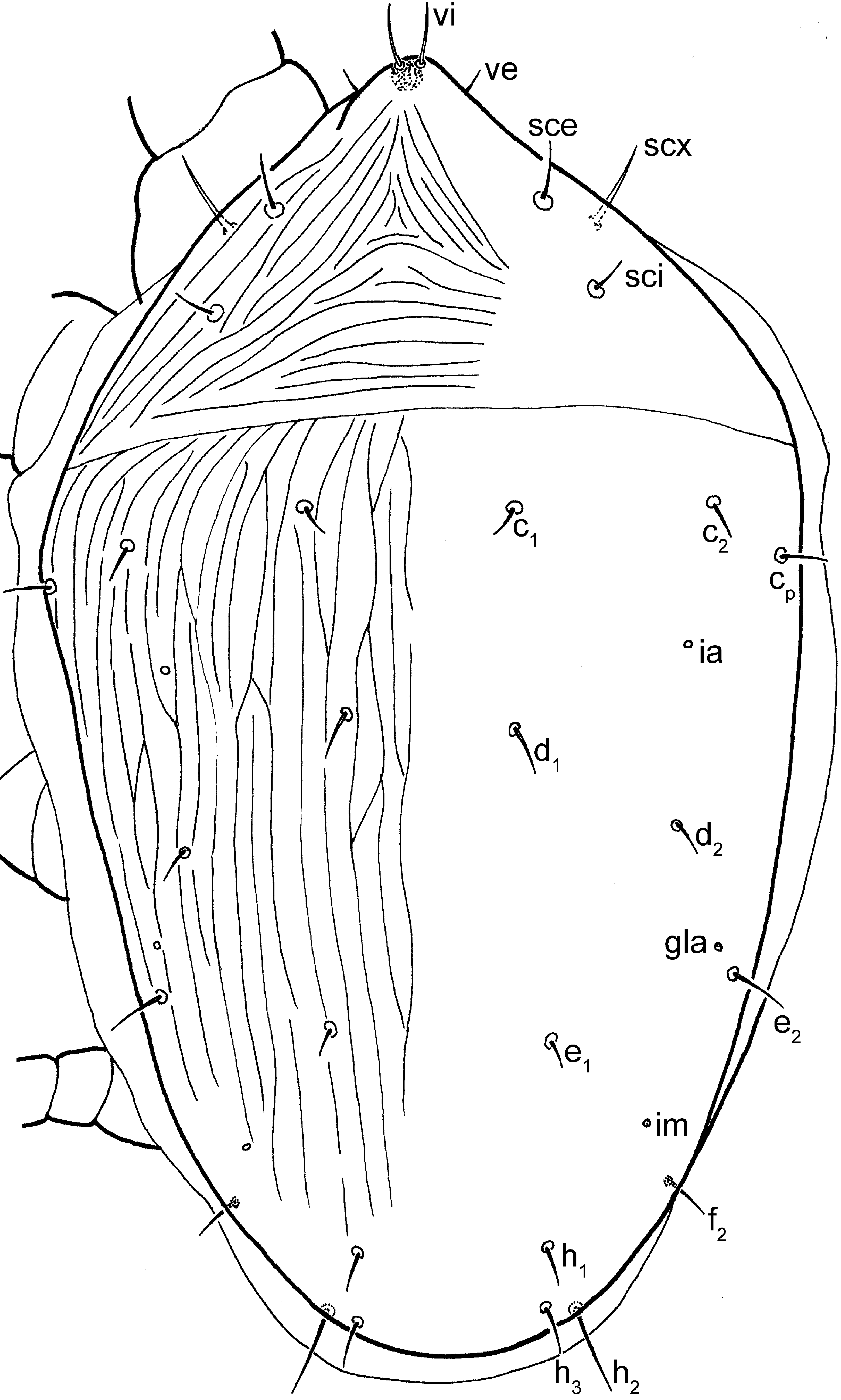
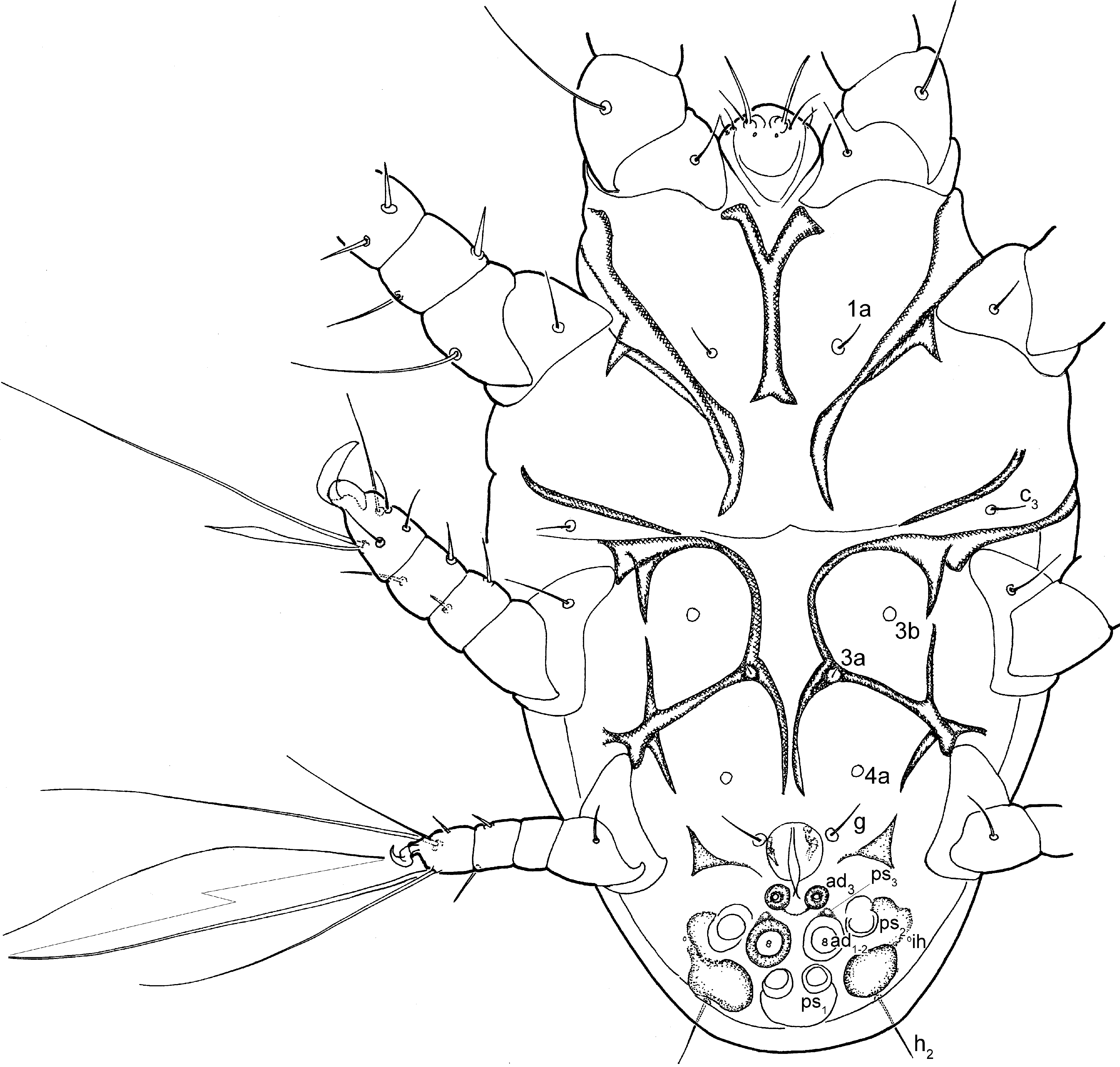
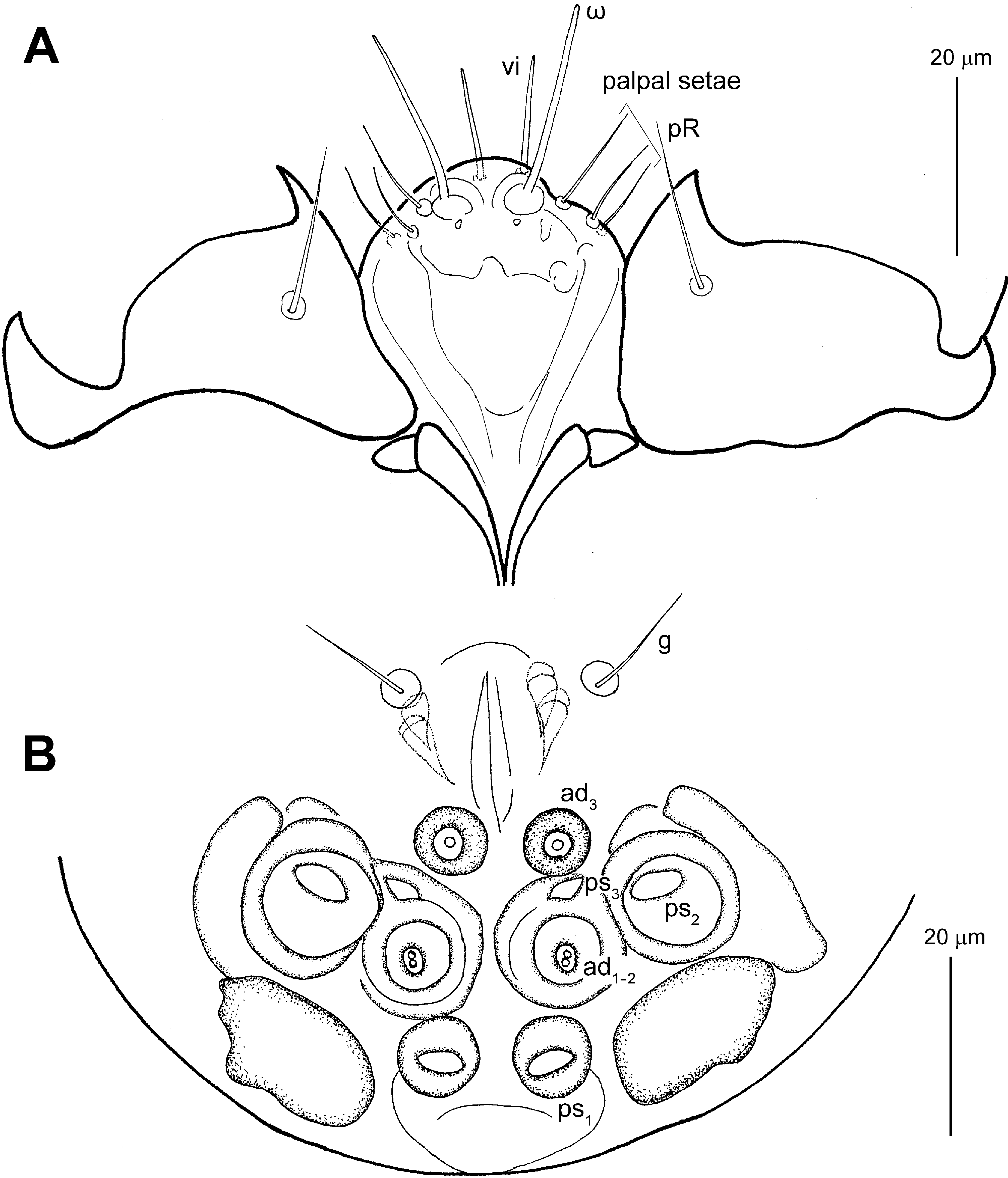
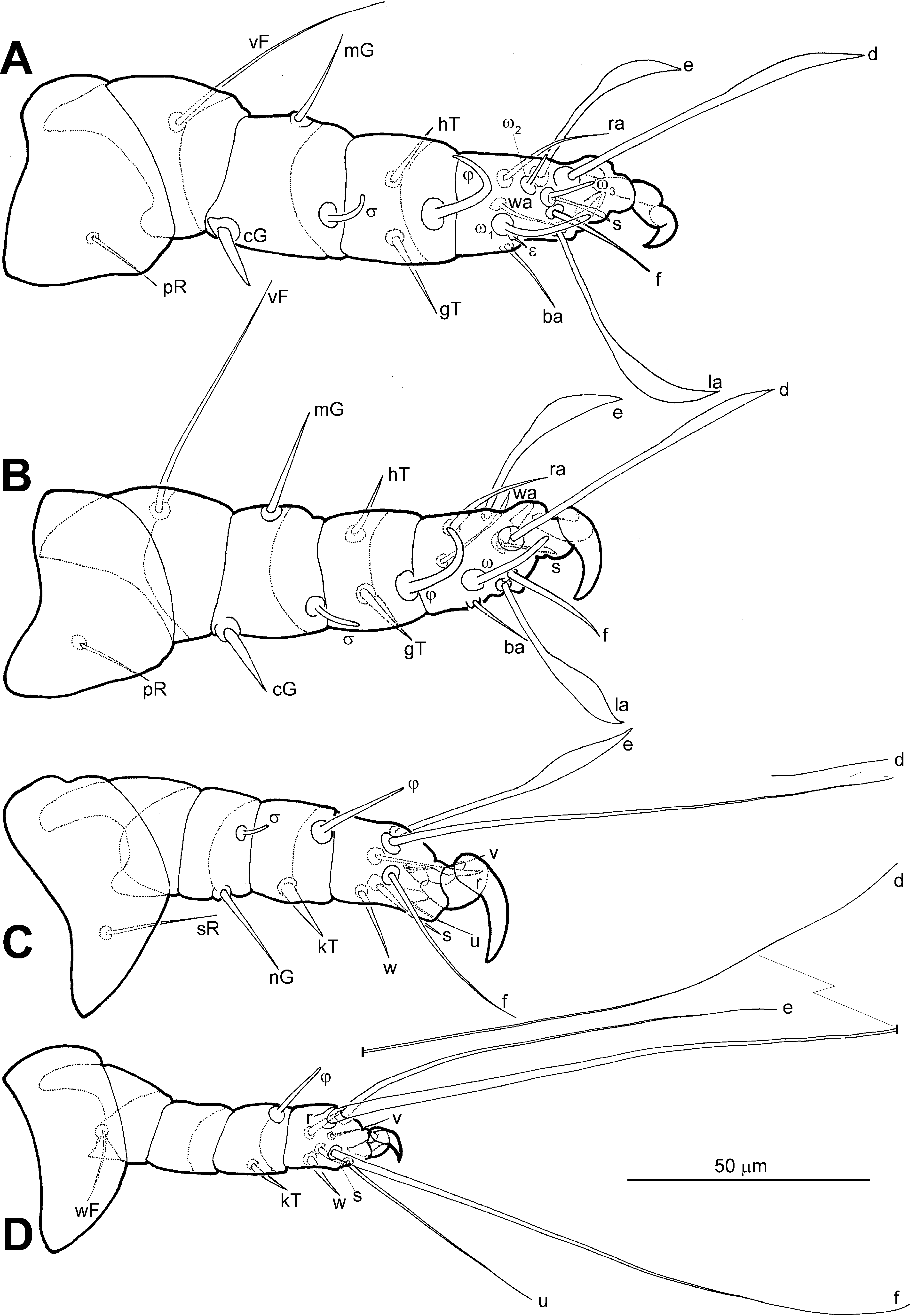
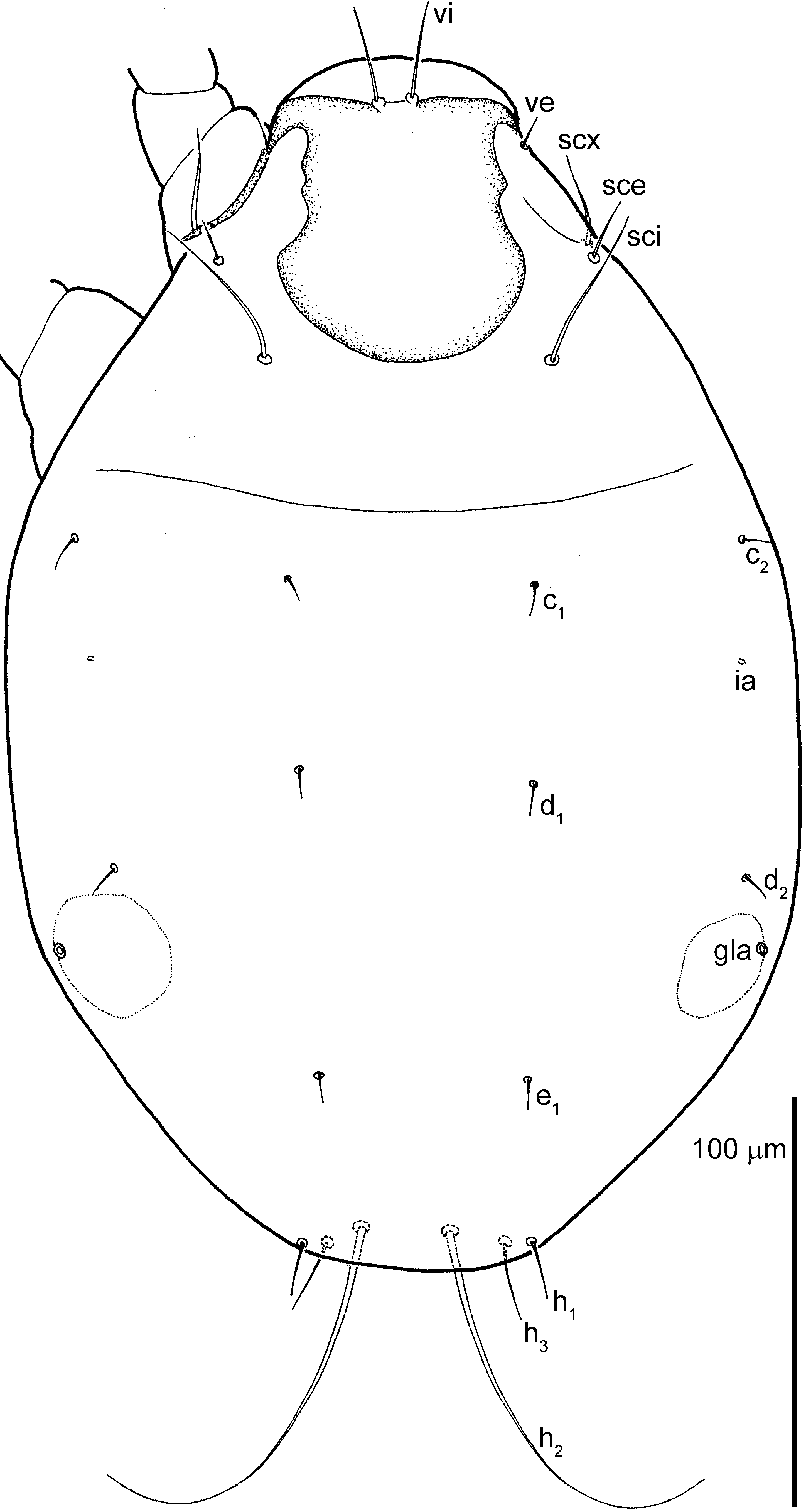
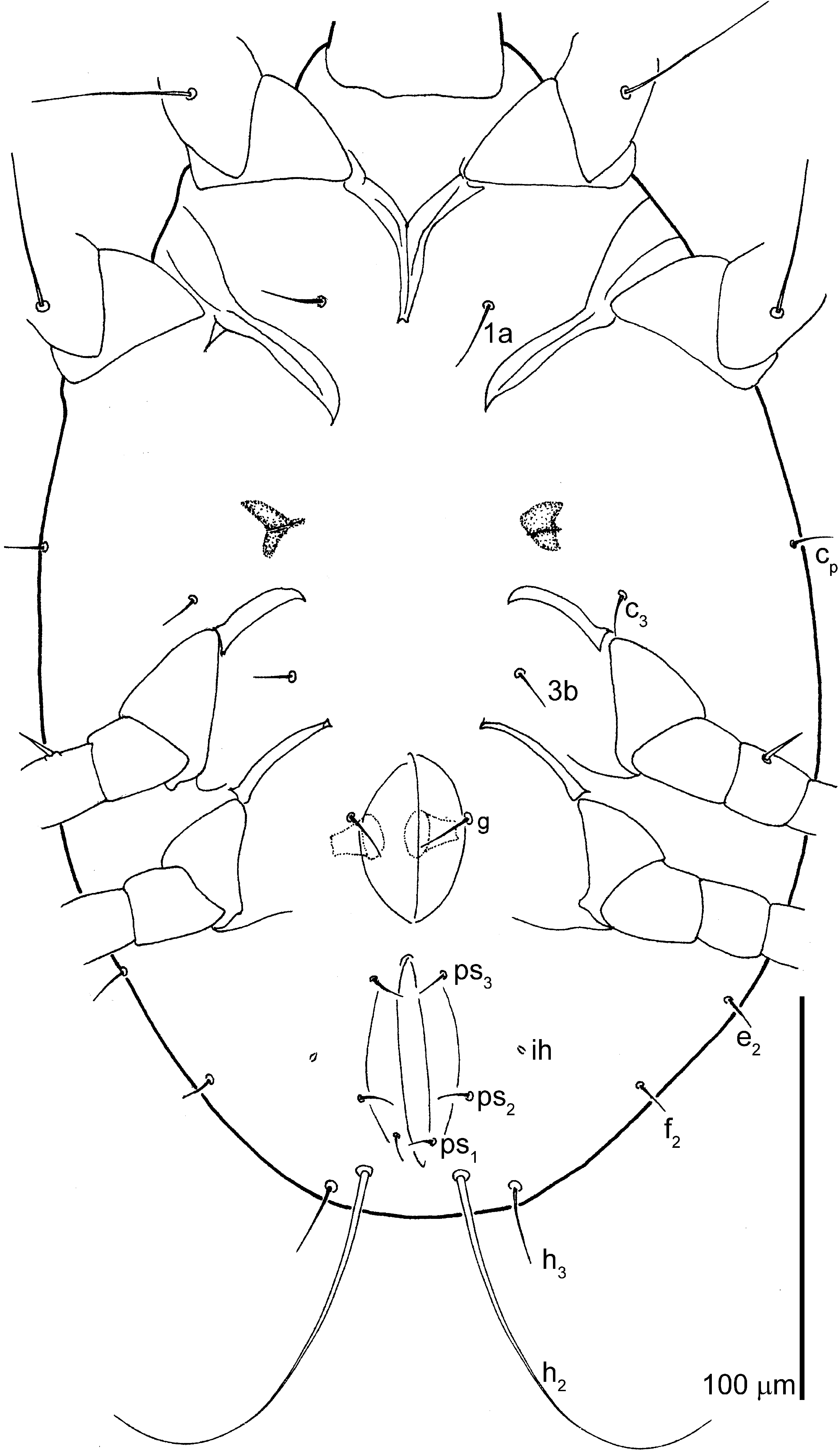
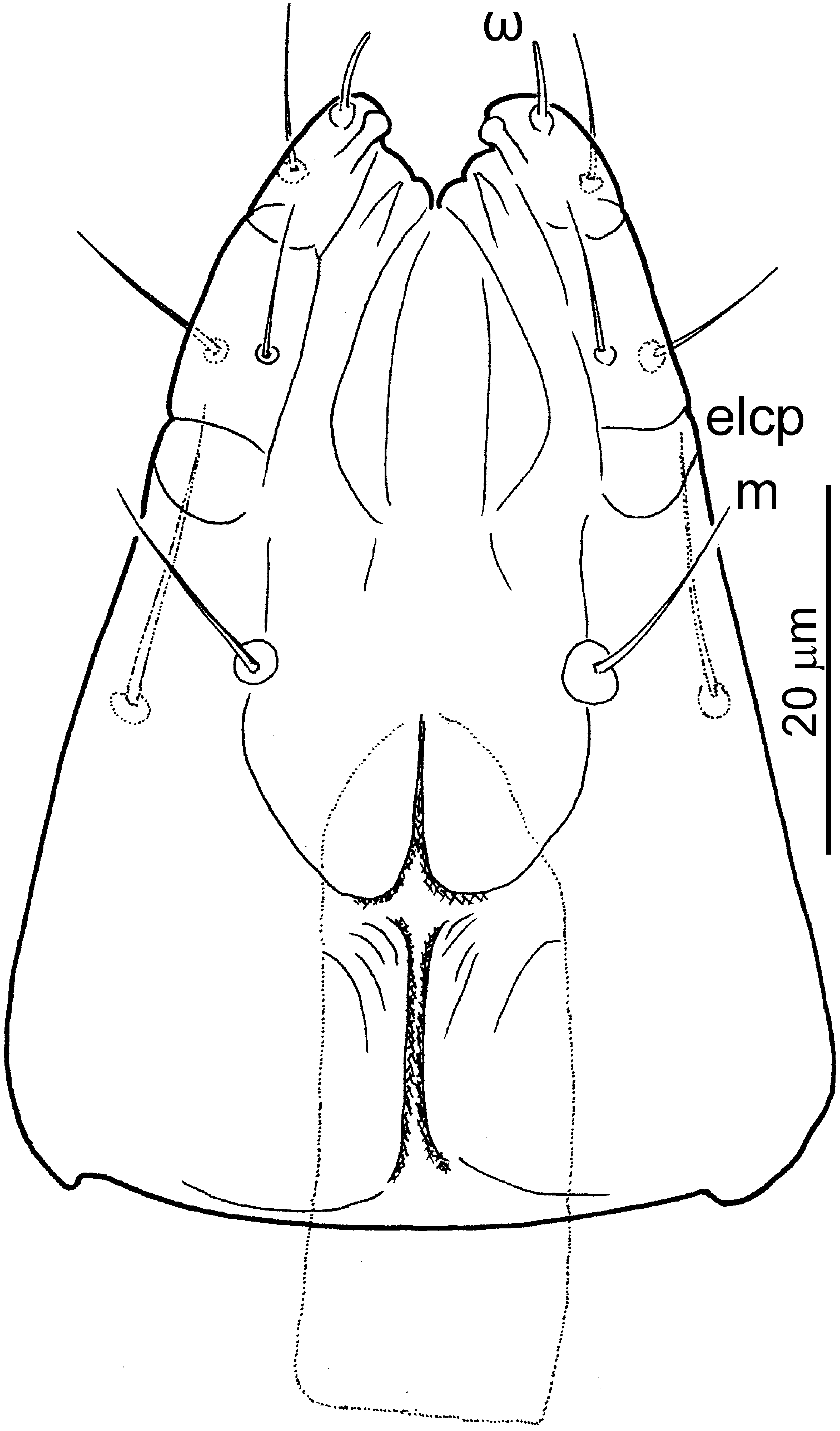
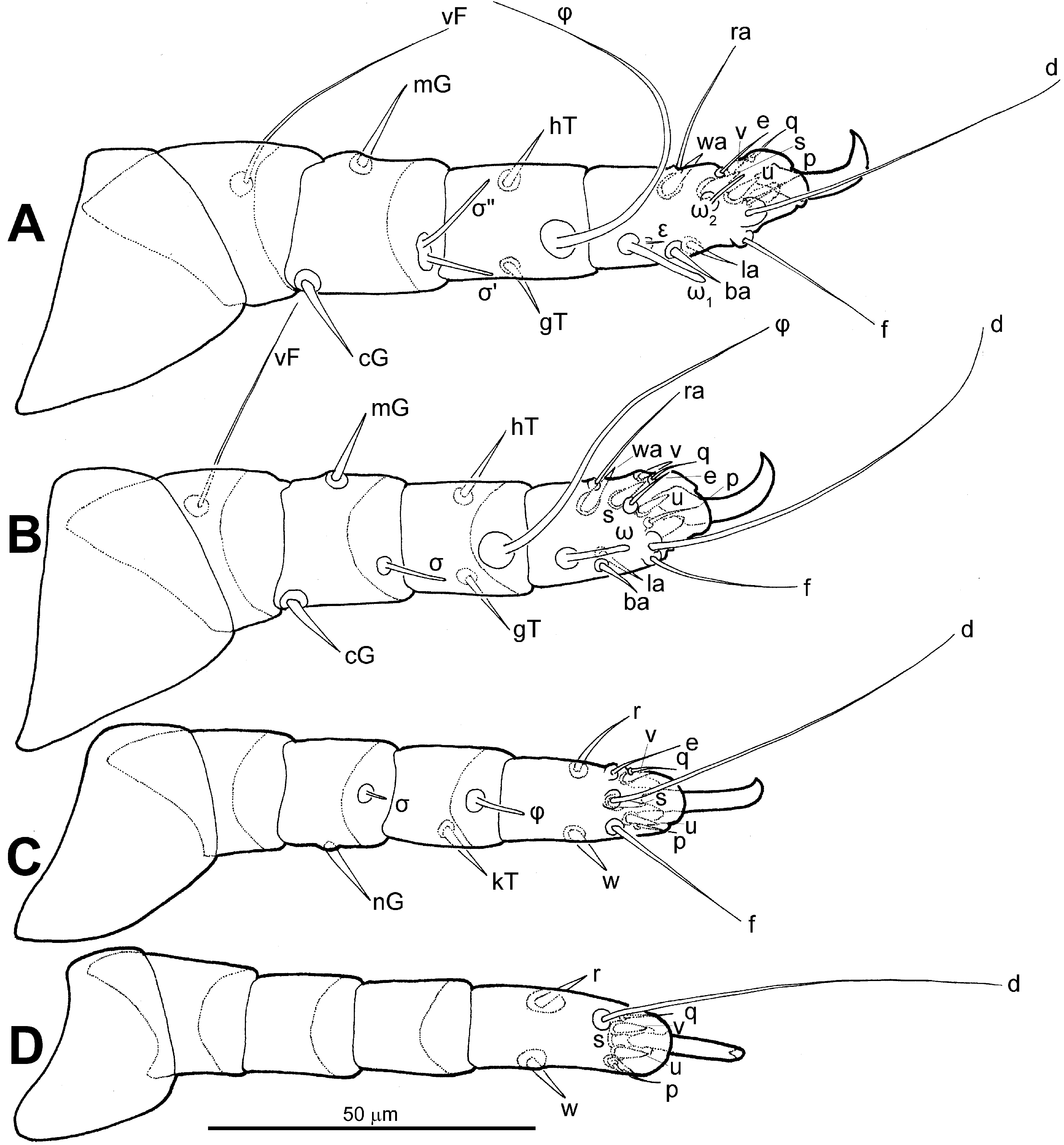
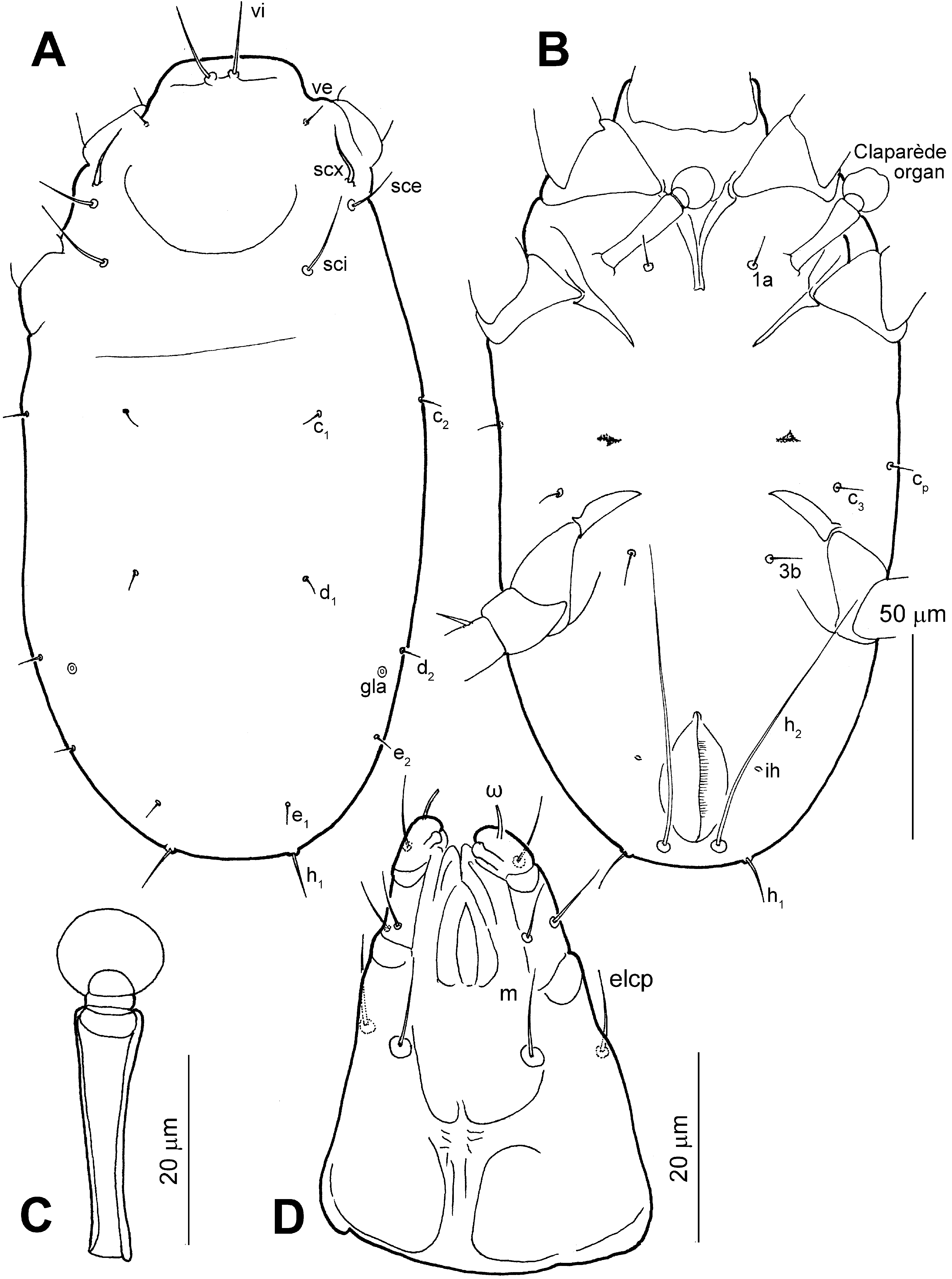
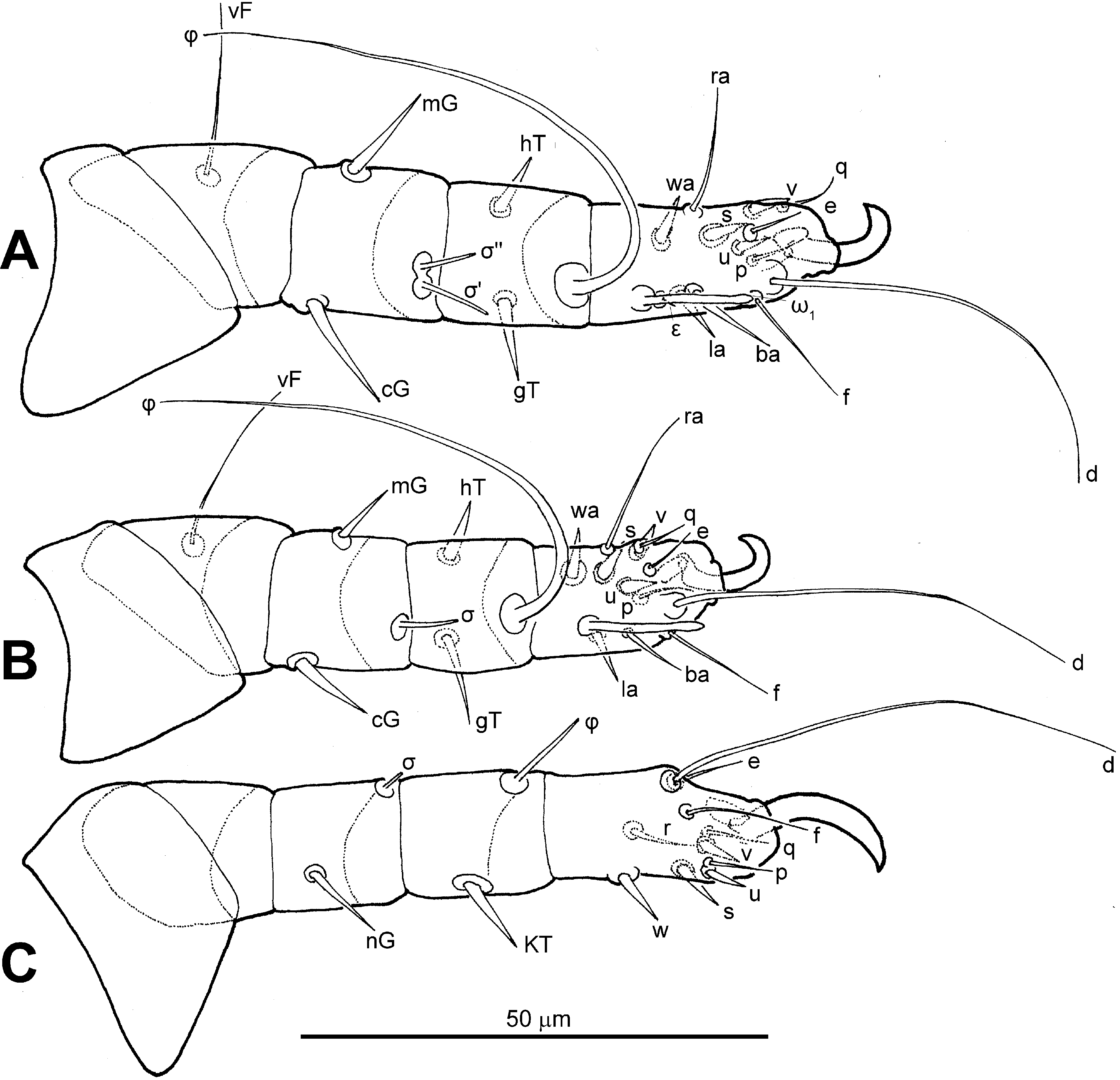
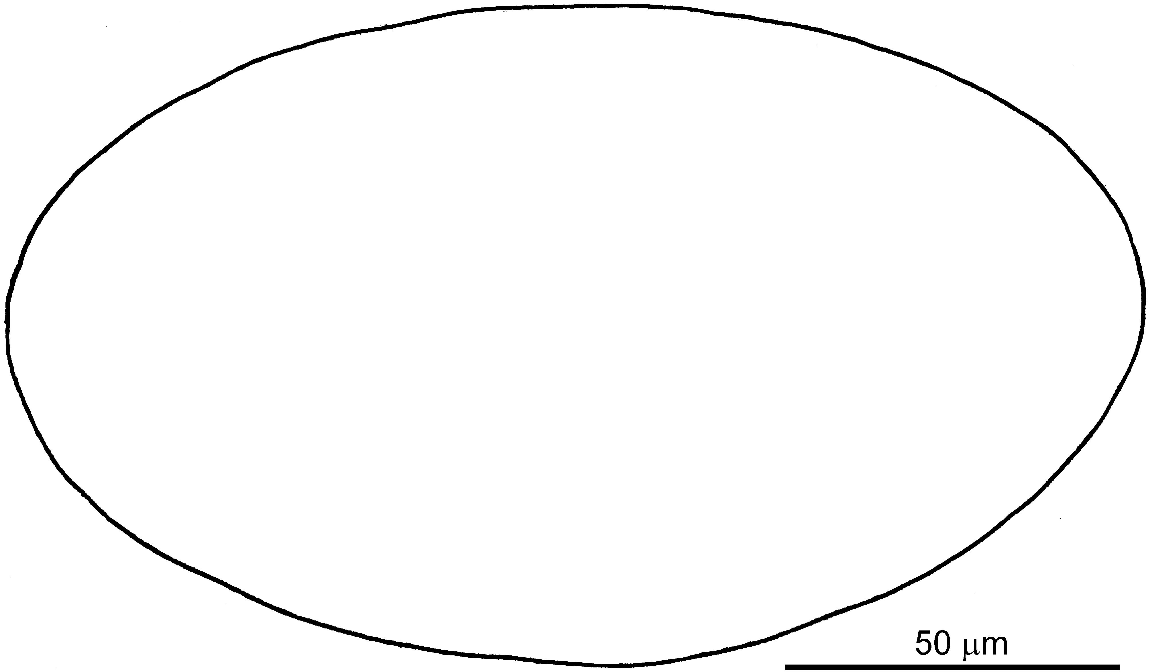

TABLE 3. Development of leg chaetotaxy in Sennertionyx manicati (Setae are indicated where they first appear, unless stated otherwise).
| Leg I | Trochanters | Femora | Genua | Tibiae | Tarsi |
|---|---|---|---|---|---|
| Larva | - | vF | cG, mG, σ’, σ’’ | gT, hT, φ | ba, ra, wa, la, d, f, e, s, u, v, p, q, ω 1, ε |
| Protonymph | - | - | - | - | ω 2 |
| Deutonymph | pR | - | - (σ’’ lost) | - | ω 3 (u, v, p & q lost) |
| Tritonymph | - | - | - (σ’’ added) | - | (u, v, p & q added) |
| Adult | - | - | - | - | - |
| Leg II | |||||
| Larva | - | vF | cG, mG, σ | gT, hT, φ | ba, ra, wa, la, d, f, e, s, u, v, p, q, ω |
| Protonymph | - | - | - | - | - |
| Deutonymph | pR | - | - | - | - (u, v, p & q lost) |
| Tritonymph | - | - | - | - | (u, v, p & q added) |
| Adult | - | - | - | - | - |
| Leg III | |||||
| Larva | - | - | nG, σ | kT, φ | r, w, d, f, e, s, u, v, p, q |
| Protonymph | - | - | - | - | - |
| Deutonymph | sR | - | - | - | - (u and v lost) |
| Tritonymph | - | - | - | - | (u and v added) |
| Adult | - | - | - | - | - |
| Leg IV | |||||
| Larva | - | - | - | - | - |
| Protonymph | - | - | - | - | r, w, d, u, v, p, q |
| Deutonymph | - | wF | - | kT, φ | f, e, s, (u and v lost) |
| Tritonymph | - | - | - | - | (u and v added) |
| Adult | - | - | - | - | - |
No known copyright restrictions apply. See Agosti, D., Egloff, W., 2009. Taxonomic information exchange and copyright: the Plazi approach. BMC Research Notes 2009, 2:53 for further explanation.