
Trachytherus ramirezi, Shockey, Bruce J., Billet, Guillaume & Salas-Gismondi, Rodolfo, 2016
|
publication ID |
https://doi.org/ 10.11646/zootaxa.4111.5.3 |
|
publication LSID |
lsid:zoobank.org:pub:A6BEC731-0447-4BF3-BBB3-B50E4A64135B |
|
DOI |
https://doi.org/10.5281/zenodo.5681050 |
|
persistent identifier |
https://treatment.plazi.org/id/03AA8786-633D-FF8F-FF49-4774FE6DFBED |
|
treatment provided by |
Plazi |
|
scientific name |
Trachytherus ramirezi |
| status |
sp. nov. |
Trachytherus ramirezi sp. nov.
Figs. 2 View FIGURE 2 , 3, 4 View FIGURE 4 , & 5
Synonyms. Trachytherus sp. ( Shockey et al. 2006: p. 206 & fig. 2); Trachytherus sp. “mid-sized species” or “medium size species” ( Shockey et al. 2009: pp. 18, 20, fig. 1C & 8B, table 5).
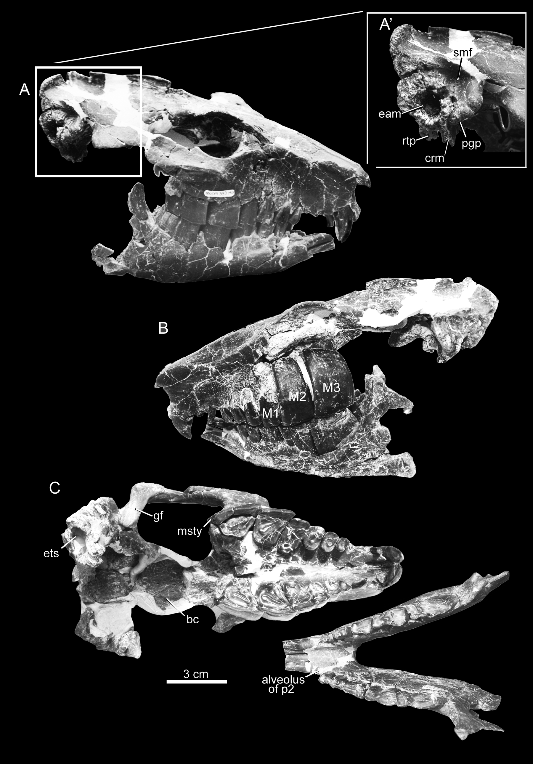
Holotype. MUSM 350; nearly complete cranium with jaws preserving all of the teeth except right I3 –C, left I2–3; the missing p 2 may have been lost during the life of the animal; see Discussion). The holotype was discovered by RSG near the summit of Pan de Azúcar, in the Upper Moquegua formation in August, 2002.
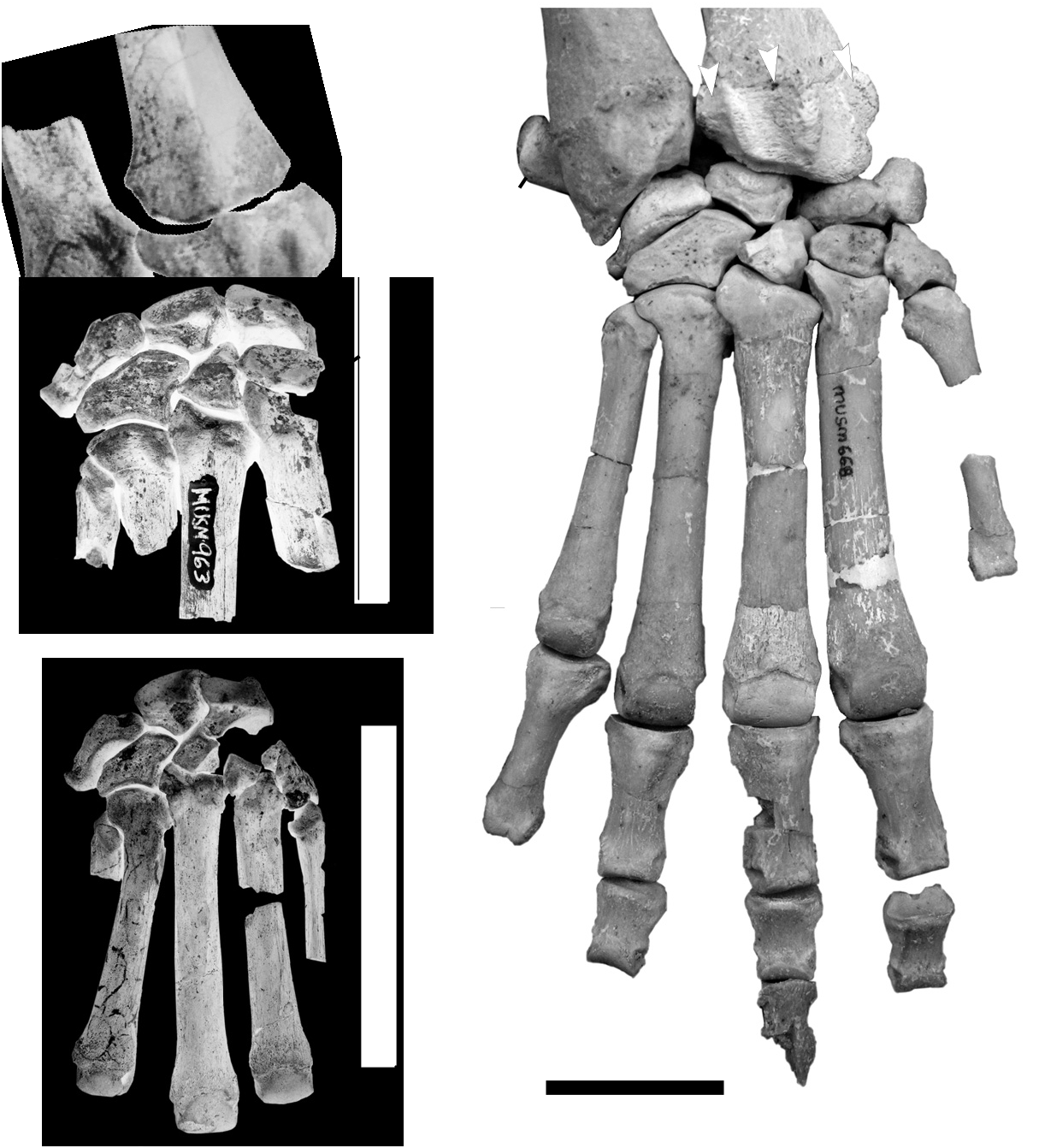
Referred specimens. MUSM 963, partial right manus, including distal radius and ulna, cuneiform, lunar, unciform, and magnum, along with proximal Mc II–V; MUSM 961, right astragalus; MUSM 962, right astragalus; MUSM 966, right astragalus.
Occurrence. Trachytherus ramirezi is only known from near the summit of Pan de Azúcar (elevation 1,663 m), located at S 17° 13.100”, W 71° 0.665’, nine km WSW of the city of Moquegua, Peru, in the Departamento de Moquegua. The holotype ( MUSM 350) and all the referred specimens were collected within the upper Moquegua Formation, from about 10 to 20 meters below the summit of Pan de Azúcar, likely (but not certainly) above the level of the Sugarloaf Ash (the specimens were buried in weathered sediments that obscured their precise stratigraphic context).
Etymology. The specific name, ramirezi , is a tribute to our late colleague and friend, Gregorio Ramírez Andrade, who helped excavate the holotype and provided essential help in the field at Moquegua and in the lab at MUSM during his all too brief tenure there.
Diagnosis. Relatively small non-mesotheriine (“trachytheriine”) mesotheriid with incisive foramina being parallel, rather than having a triangular or “heart-shaped” form, typical of mesotheriids; enlarged molars (M1–3 occupy> 2/3 of the mesiodistal length of the cheektooth row in mature adult); M1–2 with transverse dimensions nearly twice that of the preceding premolars; upper canine retained and larger than I3; unique pattern of central fossettes co-occurrence on upper cheek teeth, that is, well-isolated central fossettes co-occuring on P3–4 and M2– 3.
A B 3 cm
m1 Trachytherus ramirezi differs from T. subandinus by upper molars with much greater transverse dimensions, relatively small premolars, relatively smoother ectolophs of cheekteeth, M1 that forms roots and is lower crowned; and closed fossettes co-occurring on P3–4 and M2–3, whereas the lingual apertures of M2–3 remain open in T. subandinus when P3–4 have closed and almost disappearing fossettes.
Differs from T. spegazzinianus and T. alloxus , by presence of upper canine (usually absent in mature specimens of T. alloxus ); greater difference in size and form between upper premolars and molars; M1 hypsodont (not hypselodont as generally occurs in T. alloxus ); closed fossettes co-occurring on P3–4 and M2–3 (see description for a detailed comparison on this aspect); and presence of well-developed suprameatal fossa.
Description. Trachytherus ramirezi is a relatively small mesotheriid, smaller than T. alloxus , as evidenced by the palatal length of 100 mm of the holotype of T. ramirezi , whereas sampled specimens of T. alloxus from Salla (n=8) had a mean of 121 mm and ranged from 111–126 mm ( Shockey et al. 2006; see also Billet and Martin, 2011). Astragali referred to T. ramirezi also are distinctly smaller and fall outside the range of those referred to T. alloxus ( Fig. 5).
Upper Dentitions. The holotype has gliriform incisors, hypsodont and rooted premolars, M1 hypsodont and rooted, and M2–3 euhypsodont and apparently hypselodont. The dental formula is 3/2, 1/0, 3/3, 3/3. As generally occurs in other known mesotheriids, the teeth are covered with cementum. The upper series is essentially closed, save for the small sizes of I2 –C, which, due to their diminutive form, leave small gaps between them ( Fig. 3 A).
As is typical for Trachytherus , and mesotheriids in general, the I1 is broad and strongly curved, though that of T. ramirezi is neither as broad or as curved as those of T. spegazzinianus or T. alloxus . Enamel covers the I1 labial surface, whereas it is lacking from the lingual side. The lingual surface of I1 occludes with the tip of the crown of the procumbent lower incisors (i1–2), resulting in uneven wear on I1 ( Fig. 2 View FIGURE 2 A & B). The more durable enamel on the labial surface also likely contributed to the uneven wear. I2 is much smaller than I1, being little more than a peg-like cylinder, subcircular in cross section, and with a crown height <1/2 that of the I1. I2 occludes with the lingual surface of the procumbent i2 ( Fig. 2 View FIGURE 2 A). Both I3 s are broken, but judging from their roots in the alveoli they were the smallest teeth in the dental series.
Unlike most mature specimens of T. alloxus ( Billet et al. 2008) , an upper canine is present and is larger than I3. It is slightly spatulate and curved, having a concave lingual surface and convex labial surface. The canine is slightly procumbent, with the tip of the crown lying further from the P2 than the base of the tooth.
The holotype does not have a P1 (or retained dP1). The P2 is much smaller than the remaining premolars. Like the other upper premolars, the transverse dimension is greater than the distal-mesial dimension. The ectoloph of P2 forms a gentle curve in occlusal view, lacking any distinctive convolutions aside from a subtle, blunt paracone ridge. The lingual border is slightly more curved than is the ectoloph. The tooth is worn such that there is no fossette. P3 and P4 are similar to P2, but they are progressively larger and each retains a small oval fossette that has an obliquely oriented long axis.
There is a marked size discontinuity between the premolars and the much larger molars, exceeding that observed in any ontogenetic/wear stage of T. spegazzinianus , T. alloxus , or T. subandinum . M1 is heavily worn, leaving no trace of a fossette, and its transverse dimension is greater than the mesiodistal, as in heavily worn M1 of T. alloxus ( Billet et al., 2008: Appendix 3). M1 is conspicuously lower crowned than M2–3, and unlike the M2–3 it has closed roots ( Fig. 2 View FIGURE 2 B). Enamel is absent from the lingual and posterior borders of the tooth. The parastyle of M2 slightly overlaps the M1. The M2 is less worn and much higher crowned than the M1 and, unlike M1, is openrooted. It retains an oblique central fossette, which presumably resulted with the basal union of protoloph with a metaloph/posterior cingulum as occurs in T. spegazzinanus and T. alloxus ( Billet et al., 2008) . Like the M1, the lingual and posterior borders lack enamel. M3 differs from M1–2 by having a distinct metastyle that contributes to the greater mesiodistal dimension of the tooth, resulting in this dimension greater than the transverse (note that the M3 of T. alloxus , contrarily to M1–2, also has a greater mesiodistal diameter than the transverse one, at all stages; Billet et al. 2008). It is fully erupted, as evidenced by wear of the posterior occlusal surface. This posterior border has no enamel, but enamel is present on the lingual side as well as the mesial and labial borders of the tooth.
Central fossettes on the P3–4 and M2–3 of the type are contemporaneous. This is in stark contrast with T. spegazzinianus , which loses the central fossettes of P3–4 well before the central fossettes of M2–3 are formed (FMNH P13281, UNPSJB PV 112). The pattern exhibited by T. subandinus is also close to that of T. spegazzinianus in contrast to the form of T. ramirezi . Indeed, the central fossettes of P3–4 are very small (thus close to disappearance; see Billet et al., 2008) in the unique specimen of T. subandinus while M2–3 are far removed from isolating a central fossette (a stage possibly never attained for M 2–3 in this species; Villarroel et al., 1994 p 29); thus the central fossette probably disappears from P3–4 before being isolated on M 2–3 in T. subandinus . The situation in T. alloxus is rather intermediate as it barely loses the central fossettes of P3–4 before the central fossettes are isolated on both M2 and M3 (see diagnosis above and Billet et al. 2008: Appendix 2: stages 12-13-14; MNHN-Bol-V 0 0 5011 and 003478).
Mandible and Lower Dentitions. The mandible is fairly complete, except for the ascending rami, which were badly crushed ( Fig. 2 View FIGURE 2 B). One of the mandibular condyles was preserved and its long axis (transverse dimension) is 21 mm. The jaw is smaller and more gracile than that of T. alloxus . Likely indicative of its late ontogenetic age, the molars of the holotype are quite large relative to the premolars, occupying about 70% of the cheektooth length ( Fig. 3 B; Table 2).
The incisors (i1–2) are columnar, procumbent, and relatively and absolutely thinner than those of T. alloxus . The wear surface of i1 is flat and perpendicular to the long axis of the tooth, whereas i2 shows significant wear on its lingual surface from occlusion with I2.
There is no sign of either a canine or first premolar (neither dp1 or p1). The p2 is lost from both sides, but the shallow alveoli remaining on each side indicate that a single-rooted tooth was present and smaller than the remaining premolars. These posterior premolars (p3–4) are high crowned, but very small relative to the molars. Both p3s show significant wear; the trigonid/talonid fossettid had worn away on the left p3, but a minute part remains on the lingual side of the right p3. This tooth is nearly rectangular in occlusal outline, with just a barely discernable sulcus demarcating the trigonid and talonid boundary. The mesiodistal dimension of the tooth is greatest at the occlusal level and tapers ventrally nearly to a point at the root such that the length of the occlusal surface decreases with wear (the opposite is true of m3). The p4 has a more distinctive trigonid/talonid fossettid, which lies close to the lingual border of the tooth. The p4 trigonid is slightly larger than the talonid.
The m1 has an asymmetric figure-eight shape in occlusal view, with the talonid being longer and wider than the trigonid. The vertical groove of the labial sulcus is better defined than those of the premolars. The m1 has significant wear, lacking any hint of a trigonid/talonid fossettid. The m2 is larger than m1. Consistent with its later eruption, it shows less wear, as evidenced by the presence of the trigonid/talonid fossettid. The m3 is the largest molar and has a similar form as m2, except that the talonid is longer and extends distally to a point, whereas m1–2 have blunt posterior ends. The base of the m3 talonid extends beyond the terminal border of the occlusal surface, indicating that the total length of the tooth would increase with continued wear.
Skull. The area containing the incisive foramina does not present the typical triangular (heart-shaped) outline observed in mesotheriids by Billet et al. (2008); in T. ramirezi , the distal end of these foramina is not pointed and the lateral border of each foramen is more parallel to its medial border than it is in T. alloxus and other mesotheriids.
The maxillae overlap the nasals, as occurs in other mesotheres and a few other typotheres (e.g., Protypotherium AMNH 9226; Hegetotherium AMNH 9501). The nasal/frontal sutures are at the level of the anterior margin of the orbits and the nasals extend anteriorly such that they overlap the nasal aperture. The frontals have well developed triangular postorbital processes that define the dorsoposterior region of the orbit. These do not reach the zygomatic arch, leaving a gap of over 1 cm.
The long axis of the glenoid fossa is transversely oriented, but slightly oblique, with its lateral margin projecting slightly anterior to the medial edge. Its long axis has a dimension of 24.4 mm. The post glenoid process is robust and abuts a distinctive descending crest of the auditory meatus, the “crista meatus” of Patterson (1934) and subsequent workers ( Fig. 2 View FIGURE 2 A’). The contact of the postglenoid process and crista meatus is unlike the condition seen in Pseudotypotherium ( Patterson, 1934:fig. 8), Mesotherium (e.g., AMNH 14965), or some specimens of T. alloxus (e.g., UF 91933), in which there is a significant gap between the crista meatus and postglenoid process. The crista meatus appears to be shorter than those of other mesotheres, but breakage obscures its complete form. A suprameatal fossa defines the dorsal boundary between the postglenoid process and the dorsal region of the external auditory meatus. An associated foramen is superficial to the deep part of the fossa, and lies in the same position as the suprameatal foramen of the leontiniid notoungulates, Ancylocoelus ( Gabbert, 2004: fig. 14.2) and the mesotheriid T. alloxus ( Billet et al., 2008: 163) . The retrotympanic (or posttympanic) process of the squamosal is small, much shorter than the crista meatus. The posterior portion of the external auditory meatus lies at the posterolateral border of the skull. Breakage at the dorso-posterior region of the skull reveals a hollow epitympanic sinus (visible in Fig. 2 View FIGURE 2 C).
grooves for
D A
cf. Trachytherus 40 spegazzinianus 35 T. alloxus L total
30
Indet. taxon 25 of Moquegua 20
10 12 14 16 18 20 22
Wtrochlear
W
B
fib f
cot f
troch
troch ppa (tib f) L total tib
stop
fg nav f fib f
ect f ppa sus
1 cm f
nav f
T. ramirezi T. alloxus cf. T. spegazzinianus
The paroccipital process is broken and the occipital condyles are missing. The basisphenoid and sphenoid were recovered, but we are unable to connect these to the rest of the skull due to damage. This damage and missing pieces also makes it impossible to observe the ventral foramina.
The braincase is not large, its broadest dimension being distinctively less than the width reached by the postorbital process of the frontals. A sagittal crest is present, its modest size exaggerated by the smallness of the braincase it overlies.
Postcranial Elements. A variety of postcranial elements of Trachytherus were found alongside the holotype skull as well as an m3 of a different individual of T. ramirezi . They apparently do not represent the same individual as the type since they appear to be from ontogenetically young individuals, whereas the dentitions of the holotype are heavily worn, suggestive of a more advanced ontogenetic age. Of these elements, we refer the partial manus of MUSM 963 ( Fig. 4 View FIGURE 4 A), as well as some tarsals found near the holotype, to T. ramirezi . A smaller mesothere manus (MUSM 968; Fig 4 View FIGURE 4 B) may also pertain to T. ramirezi , but we do not refer it to this taxon since it is so small that it may represent a different species.
The manus of Trachytherus ramirezi (MUSM 963; Fig 4 View FIGURE 4 A) is similar to that of cf. T. spegazzinianus from Cerro Mono of Moquegua ( Fig. 4 View FIGURE 4 C; Shockey et al. 2009: fig. 1g [map] for locality) and T. alloxus from Salla ( Fig. 4 View FIGURE 4 D). We note the unfused epiphyses of the distal radius of the specimen referred to T. ramirezi ( Fig. 4 View FIGURE 4 A) that indicates its relatively young ontogenetic age. The form of the carpals of T. ramirezi is nearly identical to those of T. spegazzinianus , T. alloxus , and the small indeterminate species ( Figs. 4 View FIGURE 4 ), save for being intermediate in size; i.e., larger than the diminutive MUSM 965 and smaller than both those of cf. T. spegazzinianus and T. alloxus ( Fig. 4 View FIGURE 4 ). Where known, mesotheriid hands are pentadactyl, however, since neither the Mc I nor trapezoid of T. ramirezi were recovered, we cannot assert that it also was pentadactyl. (Note, even the geologically younger, Pleistocene Mesotherium had five digits on its manus [ Serres 1867; Ameghino 1891].)
Astragali referred to T. ramirezi (MUSM 961, 962, and 966) are like those of known mesotheriids, having assymetric trochlea (lateral trochlear ridge much larger than the medial), a separate groove for the flexor hallucis longus (suggestive of a pentadactyl manus), distinct constricted neck (though not greatly elongated) and a well developed medial process ( Fig. 5; see also Shockey et al. 2007, 2009). The referral of these specimens specifically to T. ramirezi was based primarily upon size; i.e., they include the larger astragali from Cerro Pan de Azúcar, which are distinctly smaller than the one associated with the partial skeleton of cf. T. spegazzinianus (MUSM 668) from Cerro Mono ( Table 2; Fig. 5). Due to our uncertainty of the alpha taxonomy of mesotheriid postcranial specimens that are discontinuously smaller than those referred to T. ramirezi , we did not refer the diminutive astragalus (MUSM 967) or a partial manus (MUSM 965) of Pan de Azúcar to that, or any, taxon. These remain as an indeterminate small taxon.
No known copyright restrictions apply. See Agosti, D., Egloff, W., 2009. Taxonomic information exchange and copyright: the Plazi approach. BMC Research Notes 2009, 2:53 for further explanation.