
Stillabothrium lunae, Ruhnke & Pommelle & Aguilar & Hudson & Reyda Key Words Abstract, 2022
|
publication ID |
https://doi.org/ 10.1645/21-94 |
|
publication LSID |
lsid:zoobank.org:pub:D4CBA609-D7EE-45D5-9E86-064ADFD8B866 |
|
DOI |
https://doi.org/10.5281/zenodo.7753681 |
|
persistent identifier |
https://treatment.plazi.org/id/AF7E0704-B792-4AFD-BC27-317D118CBE31 |
|
taxon LSID |
lsid:zoobank.org:act:AF7E0704-B792-4AFD-BC27-317D118CBE31 |
|
treatment provided by |
Felipe |
|
scientific name |
Stillabothrium lunae |
| status |
sp. nov. |
Stillabothrium lunae View in CoL n. sp. Ruhnke, Aguilar, and Reyda (Informal synonym: Rhinebothriinae New genus 3 n. sp. 5 of Healy et al. [2009], Caira et al. [2014], Ruhnke et al. [2015], Marques and Caira [2016])
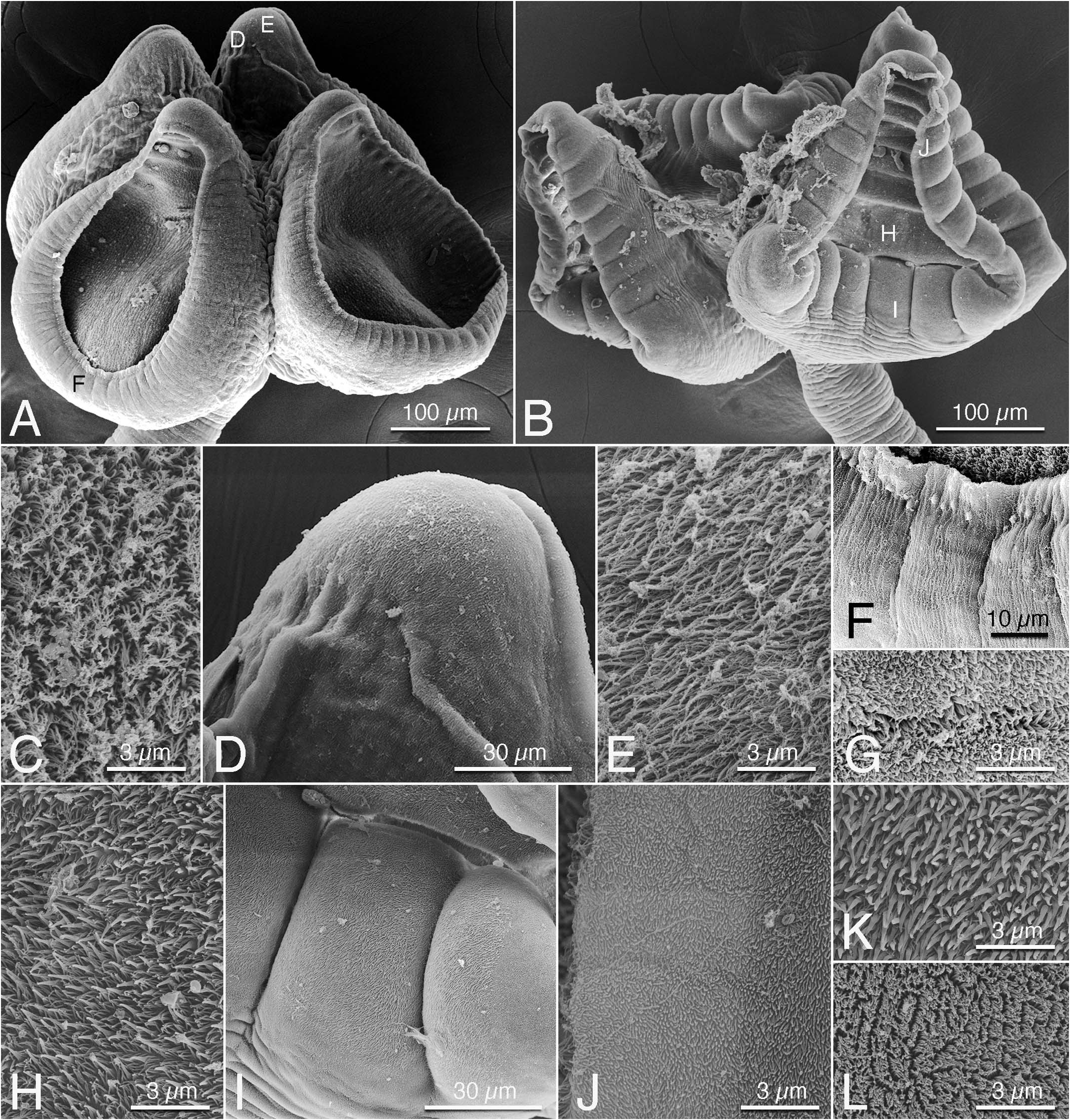
( Figs. 4B, 4H–4L View Figure 4 , 5 View Figure 5 )
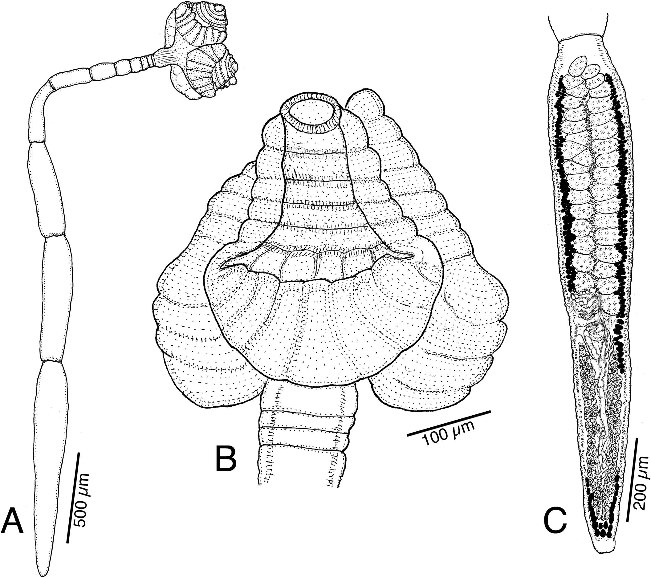
Description (based on whole mounts of 13 complete mature worms and 2 scoleces examined with SEM): Worms ( Fig. 5A View Figure 5 ) euapolytic, acraspedote 2.58–4.74 mm (3.72 ± 6.15; n ¼ 13) long, greatest width 450–600 (515 ± 43; n ¼ 13) at level of scolex; 8–14 (11 ± 1.4; n ¼ 13) proglottids per worm. Cephalic peduncle absent; darkly staining germinative zone present.
Scolex ( Figs. 4B View Figure 4 , 5B View Figure 5 ) consisting of scolex proper bearing 4 stalked bothridia. Stalks 40–75 (54 ± 10; n ¼ 12) long by 55–100 (78 ± 16; n ¼ 12) wide, attached slightly posterior to middle of bothridia. Bothridia ( Figs. 4B View Figure 4 , 5B View Figure 5 ) finely to shallowly deltoid, facially loculated, 300–440 (364 ± 40; n ¼ 11) long by 240–365 (314 ± 33; n ¼ 13) wide; bothridial margins with thin rim of tissue ( Fig. 4B View Figure 4 ). Anterior region of bothridia ( Figs. 4B View Figure 4 , 5B View Figure 5 ) with 7–8 horizontally oriented loculi (i.e., loculi that are wider than long). Posterior-most loculi in anterior bothridial region somewhat muscular (see Fig 5B View Figure 5 ). Anterior loculus 35–60 (43 ± 8; n ¼ 10) long by 50–90 (63 ± 13; n ¼ 11) wide. Posterior region of bothridia ( Fig. 4B View Figure 4 ) with 6 (6 ± 0; n ¼ 10) nonmedial longitudinal septa dividing region into 7 loculi longer than wide; longitudinal septa in posterior region of bothridium not overlapping transverse septa in anterior region of bothridium. Marginal loculi absent.
Loculi and septa ( Fig. 4H View Figure 4 ) of distal bothridial surfaces bearing capilliform filitriches and coniform spinitriches. Bothridial rim bearing capilliform filitriches and several cilia ( Fig. 4J View Figure 4 ). Proximal bothridial surface away from rim bearing acicular filitriches ( Fig. 4J View Figure 4 ). Posterior portion of proximal bothridial surface ( Figs. 4I, 4K View Figure 4 ) bearing patch of coniform spinitriches near, but not extending to, bothridial rim. Bothridial stalks and strobila ( Fig. 4L View Figure 4 ) bearing capilliform filitriches only.
Strobila ( Fig. 5A View Figure 5 ) with 2–7 (4.0 ± 1.4; n ¼ 13) proglottids wider than long followed by 3–9 (6.0 ± 1.7; n ¼ 13) proglottids longer than wide. Strobila widest at terminal proglottid; terminal proglottid 720–1,875 (1,299 ± 293; n ¼ 13) long by 110–225 (160 ± 34; n ¼ 13) wide; genital pore located 37–56% (46 ± 5.5; n ¼ 13) of proglottid length from proglottid posterior margin. Immature proglottids 6–10 (8 ± 1.4; n ¼ 13) in number. Mature proglottids 1–3 (2 ± 0.7; n ¼ 13) in number, including 0–1 (0.2 ± 0.4; n ¼ 13) proglottids with sperm-filled vas deferens and atrophied testes.
Testes in mature proglottid 23–38 (31 ± 4.2; n ¼ 11) in total number, 1 layer deep, arranged in 2 columns ( Fig. 5C View Figure 5 ), 27–50 (37 ± 6; n ¼ 13) long by 25–63 (47 ± 11; n ¼ 12) wide; columns extending from anterior margin of proglottid to anterior margin of cirrus sac. Vas deferens coiled, entering anterior margin of cirrus sac, extending from level of ovarian isthmus anteriorly to overlap several posterior-most testes ( Fig. 5C View Figure 5 ). Cirrus sac thinwalled, round to oval, extending past midline of proglottid. Cirrus sac in terminal mature proglottid 62–101 (84 ± 15; n ¼ 7) long by 43–85 (65 ± 14; n ¼ 6) wide. Cirrus spinitriches present.
Vagina ( Fig. 5C View Figure 5 ) thick-walled, sinuous, overlapping medial portion of cirrus sac, extending past midline of proglottid from ootype region to level somewhat anterior to anterior margin of cirrus sac, recurved anterior to cirrus sac, then extending laterally to open to genital atrium anterior to cirrus sac; vaginal sphincter absent. Seminal receptacle present. Ovary near posterior end of proglottid, H-shaped in frontal view, ovarian lobes symmetrical; poral and aporal ovarian lobes in terminal mature proglottids 200–580 (386 ± 164; n ¼ 5) and 205–580 (387 ± 164; n ¼ 5) long, respectively. Poral and aporal ovarian lobes in vas deferensmature proglottids 300–575 (487 ± 100; n ¼ 6) and 320–590 (515 ± 98; n ¼ 6) long, respectively. Maximum width of ovary in mature proglottids 70–190 (106 ± 25; n ¼ 12). Ovarian isthmus near or posterior to midpoint of ovary; poral lobe of ovary stopping 30–90 (52 ± 21; n ¼ 11) short of genital pore, overlapping posterior portion of cirrus sac. Mehlis’ gland posterior to ovarian isthmus, 28–60 (45 ± 11; n ¼ 5) long by 21–35 (26 ± 3; n ¼ 5) wide. Vitellarium follicular; vitelline follicles arranged in 1 dorsal and 1 ventral column on each side of proglottid; columns extending from near anterior to posterior margin of proglottid, interrupted by terminal genitalia, and mostly interrupted by ovary ( Fig. 5C View Figure 5 ). Uterus ventral, sacciform, extending from near isthmus of ovary to near anterior-most testes.
Taxonomic summary
Type host: Himantura leoparda Manjaji-Matsumoto and Last, Leopard whipray. ( Myliobatiformes : Dasyatidae ).
Type locality: Arafura Sea east of Wessel Islands (11°17 ′ 44 ′′ S, 136°59 ′ 48 ′′ E), Northern Territory, Australia (hosts NT-32, NT-37, NT-117) GoogleMaps .
(C) Terminal proglottid of voucher (LRP 10797).
Site of infection: Spiral intestine.
Type material: Holotype QM No. G 239543 . Paratypes LRP Nos. 10803–10807 (including whole mounts, sections, and scoleces prepared for SEM) ; USNM Nos. 1661760–1661763 ; QM Nos. G 239544–G239546; Hologenophore LRP No. 10808 .
Etymology: This species is named after Luna Pearl Pierer, daughter of the parasitologist Dr. Valerie McKenzie.
ZooBank registration: urn:lsid:zoobank.org:act:AF7E0704-B792-4AFD-BC27-317D118CBE31 .
Remarks
Stillabothrium lunae n. sp. can be distinguished from each of the 11 valid species of Stillabothrium in that it possesses bothridia with an anterior region comprising 7–8 loculi that are wider than long (i.e., oriented horizontally) and lacks marginal loculi. Stillabothrium jeanfortiae and S. cadenati are similar to S. lunae in overall bothridial morphology, consisting of muscular septa that do not overlap and a lack of marginal loculi, but are each distinguished from S. lunae in their possession of 3 and 4 respective horizontally oriented loculi, rather than 7–8, in the anterior region of bothridium. Stillabothrium lunae is further distinguished from S. cadenati in that the latter species possesses only 4 horizontally oriented loculi in the anterior region of bothridium and a strongly recurved vagina, unlike S. lunae , and fewer testes (7–13 vs. 23–38). Each of the following 3 species of Stillabothrium possess marginal loculi in their bothridia, distinguishing them from S. lunae : Stillabothrium ashleyae Willsey and Reyda, 2016 , Stillabothrium charlottae Dedrick and Reyda, 2018 , and Stillabothrium davidcynthiaorum Daigler and Reyda, 2016 . Stillabothrium lunae is distinguished from the following 3 species of Stillabothrium in which the septa in the anterior and posterior regions of bothridia markedly overlap one another: Stillabothrium amuletum (Butler, 1987) Healy and Reyda, 2016 , Stillabothrium campbelli Delgado, Dedrick and Reyda, 2016 , and Stillabothrium hyphantoseptum Herzog, Bergman and Reyda, 2016 . Stillabothrium lunae is distinguished from Stillabothrium allisonae Dedrick and Reyda, 2018 , in that the anterior portion of bothridia of the latter species consists of an anterior loculus followed by a row of 4 circular (i.e., not horizontally oriented) loculi. Finally, S. lunae can be distinguished from S. biacetabulatum in that the latter species possesses marginal loculi in addition to facial loculi, whereas S. lunae possesses only facial loculi.
The tapeworm specimen included in the phylogenetic analyses of Healy et al. (2009), Marques and Caira (2016), and Ruhnke et al. (2015) as Rhinebothriinae New genus 3 n. sp. 5 (GenBank FJ177114) is here verified as Stillabothrium lunae n. sp.
| QM |
Queensland Museum |
| USNM |
Smithsonian Institution, National Museum of Natural History |
No known copyright restrictions apply. See Agosti, D., Egloff, W., 2009. Taxonomic information exchange and copyright: the Plazi approach. BMC Research Notes 2009, 2:53 for further explanation.
|
Kingdom |
|
|
Phylum |
|
|
Class |
|
|
Order |
|
|
Family |
|
|
Genus |