
Monographis phuquocensis, Huynh, 2018
|
publication ID |
https://doi.org/10.11646/zootaxa.4402.2.3 |
|
publication LSID |
lsid:zoobank.org:pub:CEA5700E-9F94-42A8-9067-BD8F4D301B9D |
|
persistent identifier |
https://treatment.plazi.org/id/03AB2952-FFE4-5E5D-0FBD-FB8AFD8F0B51 |
|
treatment provided by |
Plazi |
|
scientific name |
Monographis phuquocensis |
| status |
sp. nov. |
Monographis phuquocensis sp. n.
( Fig. 8 View FIGURE 8 )
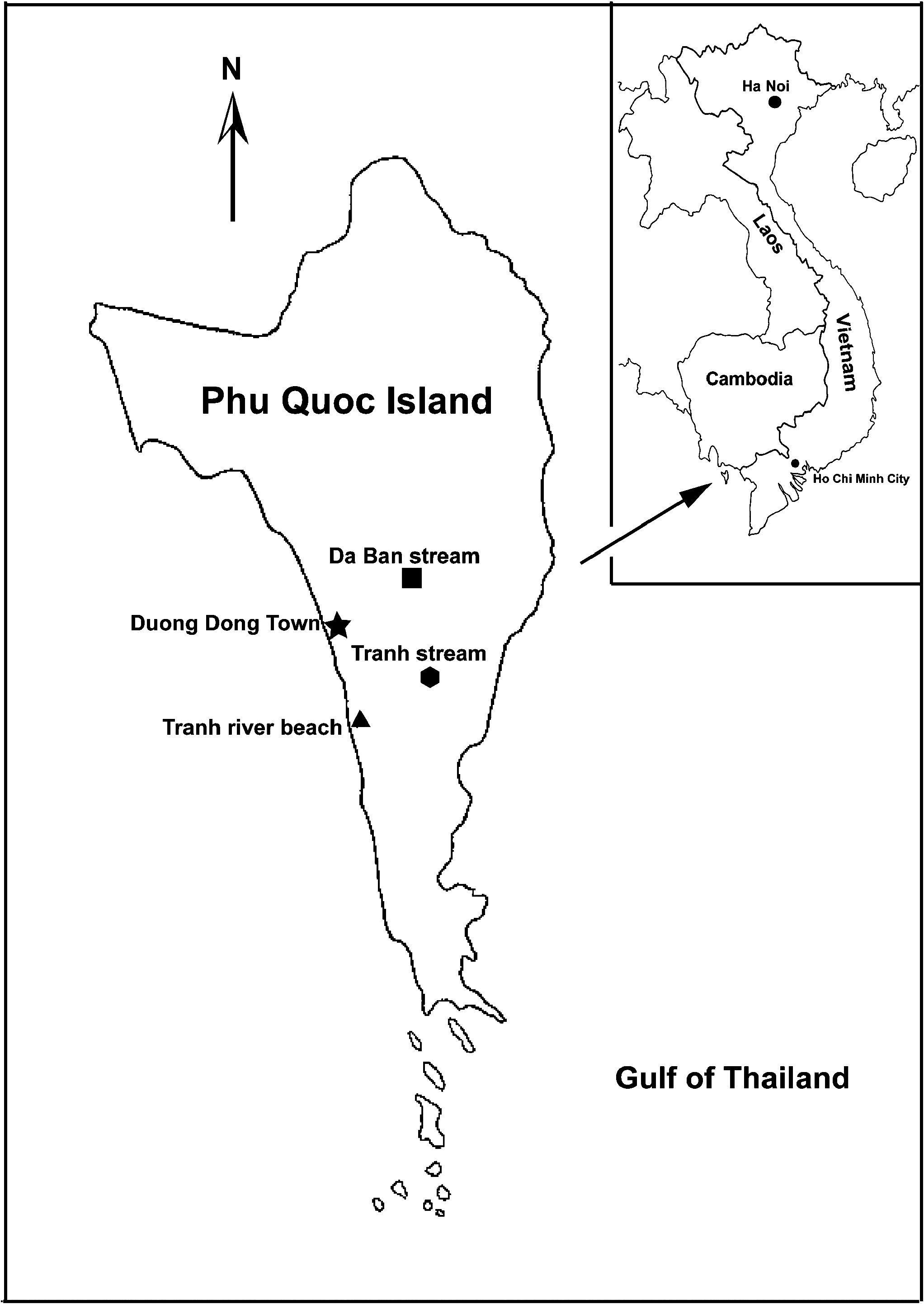
Holotype. Male, Monographis phuquocensis sp. n. was collected by the first author from leaf litter near Tranh Stream (Suoi Tranh) , 10°10'58.96"N, 104° 0'54.72"E; elevation 86 m; 7 km southeast of the main town - Duong Dong town on Phu Quoc Island on 2nd July 2015. Queensland Museum accession number: QMS 108531 (deposited in Queensland Museum, Brisbane, Australia). GoogleMaps
Paratypes. Twelve specimens were collected: 3 adult males, 5 adult females (adult stage- stadium VIII, 13 pairs of legs), and 4 sub-adults (stadium VII, 12 pairs of legs). GoogleMaps At the second location: Da Ban Stream (Suoi Da Ban ), 10°14'42.36"N, 104°01'48.82"E, elevation 43 m; 10 km northeast of Duong Dong town on Phu Quoc Island on 3rd July 2015; 5 specimens were collected: 2 males and 3 females. Both locations are in the Ham Ninh Mountains on Phu Quoc Island ( Fig. 1 View FIGURE 1 ). Queensland Museum accession numbers for 17 paratypes: QMS 108532–108548 (deposited in Queensland Museum, Brisbane, Australia) GoogleMaps .
Etymology. The specific name phuquocensis, noun in apposition, refers to Phu Quoc Island in southwest Vietnam, where this species was first found.
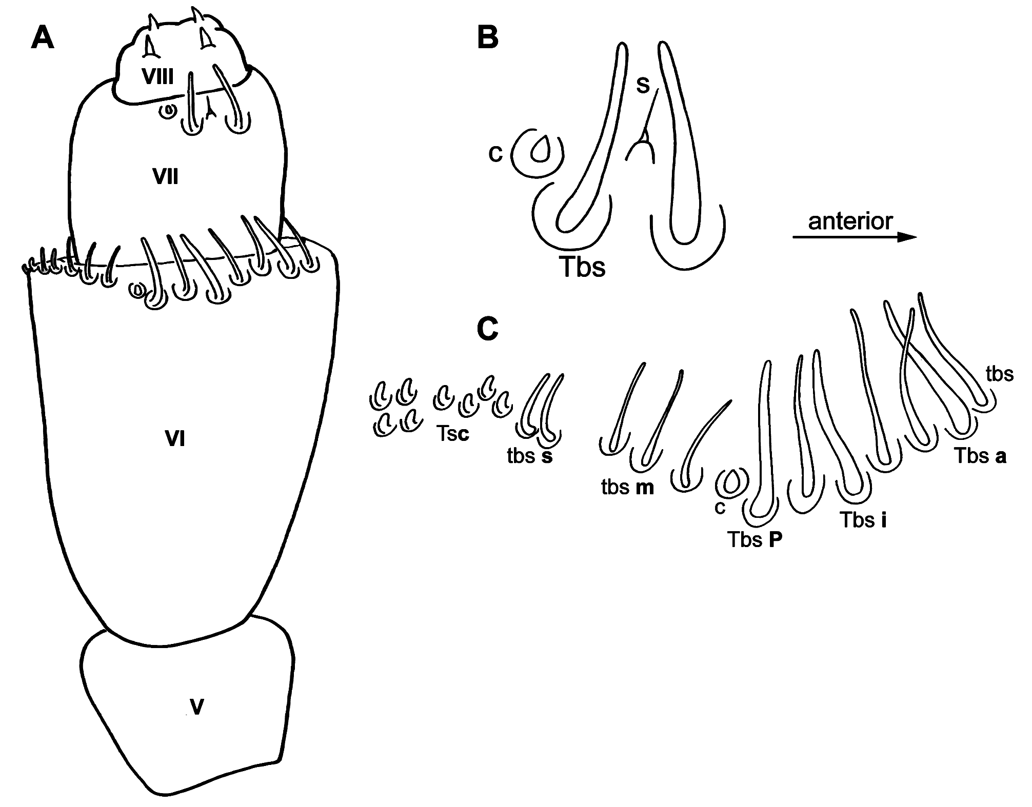
Diagnosis. Colour and trichome patterns of Monographis phuquocensis sp. n. similar in appearance to other described Monographis species including M. queenslandica and M. dongnaiensis ( Huynh and Veenstra, 2013, 2015). Characteristics and arrangement of caudal bundles are quite similar among Monographis species ( Huynh and Veenstra 2015) and this is classified as the caudal bundle type II arrangement (Condé and Nguyen Duy- Jacquemin, 2008)Pattern of sensilla on antennal article VII common to all species of Monographis ( Huynh and Veenstra 2013, 2015). M. phuquocensis sp. n. different from other Monographis species in antennal article VI having 15–20 sensilla of various sizes, forming a crescent-shape with the anterior sensilla longer than posterior ones. These sensilla form 4 distinct groups: 3 thick bacilliform sensilla commonly present in a group of 2–4 thin bacilliform sensilla located anteriorly (Group 1), 2–3 medium length thin bacilliform sensilla (Group 2), 1–2 short thin bacilliform sensilla (Group 3), and a group of 6–8 thick short conical sensilla located posteriorly (Group 4). Surface of labrum asetose and smooth, with a row of setae present near anterior edge of lamellae. Telotarsus bearing a posterior process shorter, half the length of claw. Anterior lateral process and lamella process present; setiform process half the length of claw.
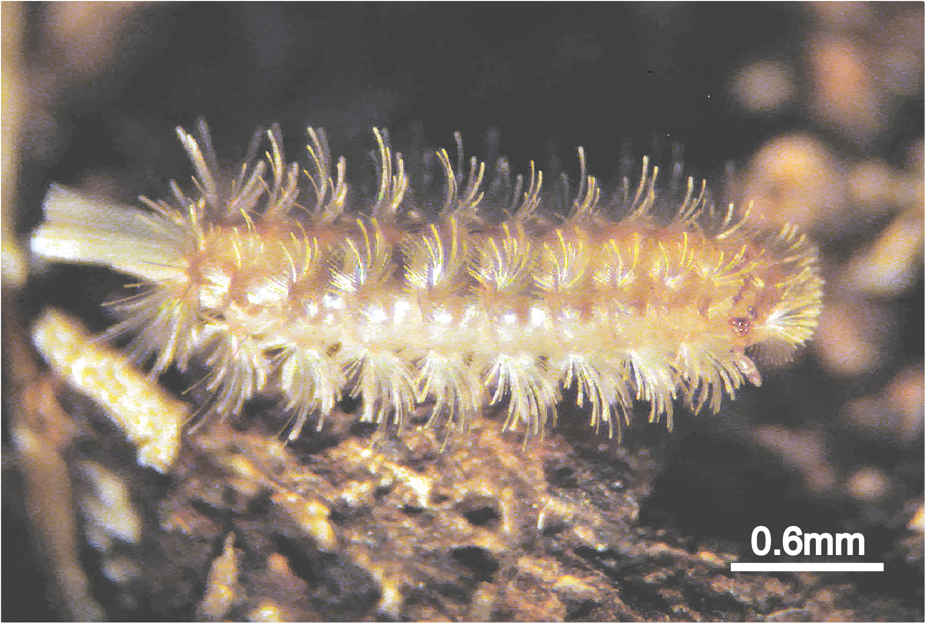
Description. Measurements: Holotype male body length 2.0 mm; ( paratypes) males ranging from 1.4–2.0 mm ( n = 5) and females ( n = 8) ranging from 2.2–2.5 mm. Caudal bundle in male slightly narrower in width and shorter ( 0.5mm) compared to female, the latter wider and longer ( 0.6mm).
Colouration: Body yellowish-brown in colour with 2 darker lateral bands, caudal bundle black-silvery colour ( Fig. 8 View FIGURE 8 ).
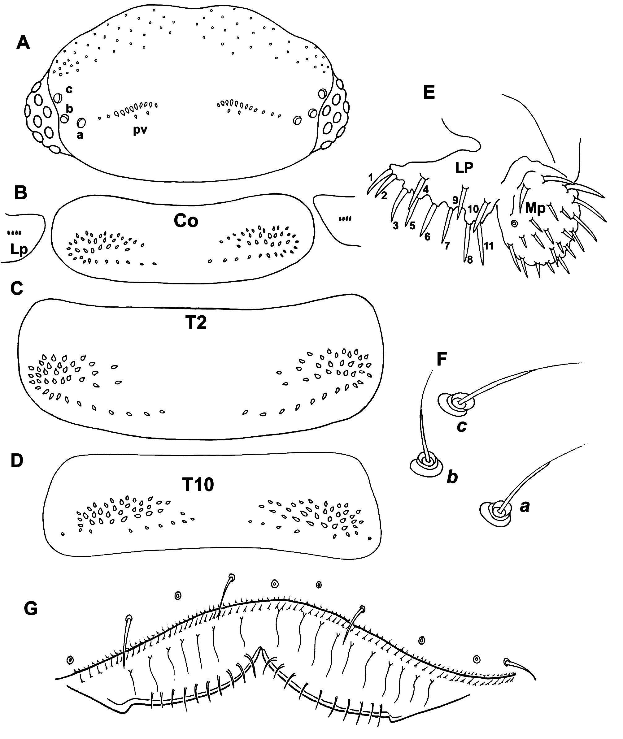
Head: Eight ommatidia on each side: 4 dorsal, 4 lateral (1 anterior, 2 medial and 1 posterior). Vertex with 2 posterior trichome groups comprised of 2 rows, with a large gap present between these groups. Anterior, oblique with a variable number of large trichome sockets in centre, gradually reducing in size toward both ends; posterior row with slightly smaller trichome sockets. A narrow space present between anterior and posterior rows. Holotype male with 11 + 12 trichome sockets in anterior rows and 2 + 2 trichome sockets in posterior rows ( Fig. 9A View FIGURE 9 ). Paratypes with variation common in this species regardless of sex, ranging from 11–15 (anterior rows) and 2–3 (posterior rows). Trichobothria (trichobothrium a, posterior position, trichobothrium b, lateral position and trichobothrium c, anterior position) typically thin sensory hairs with narrow cylindrical funicles. All these trichobothria ( a, b and c) equal in socket size and form an isosceles triangle with an equal distance between ab and bc ( Fig. 9F View FIGURE 9 ).
Antennae: Eight articles and 4 sensory cones present, characteristics typical of Polyxenidae . Antennal article VI ( Figs 10A View FIGURE 10 and 11A View FIGURE 11 ) with 18–20 sensilla. Holotype with 20 sensilla forming a crescent-shape with anterior sensilla longer than posterior ones. These sensilla forming 4 groups: Group 1 located anteriorly with 3 thick bacilliform sensilla (Tbs a, i and p) among 4 thin bacilliform sensilla (tbs), this group followed by a conical sensillum (c); Group 2 with 3 medium length thin bacilliform sensilla (tbs m); Group 3 with 2 short, thin bacilliform sensilla (tbs s); a small gap present between Groups 2 and 3; last group has 8 thick short conical sensilla (Ts c) located posteriorly ( Figure 10C View FIGURE 10 ). Different arrangements of sensilla apparent in scanning electron microscope images of other specimens ( Figs 11A, B and C View FIGURE 11 ). Antennal article VII with 2 thick bacilliform sensilla (Tbs) with a setiform sensillum (s) between them and a conical sensillum (c) located posteriorly ( Figs 10B View FIGURE 10 and 11A View FIGURE 11 ).
Clypeo-labrum: Holotype with 10 setae present on posterior margin, these setae shorter than half the width of labrum. Setae on paratypes ranging from 8–11 in both sexes. Labral surface smooth and asetose. Anterior margin of labrum with a lateral lamella and single broad lamella on each side of median cleft. A row of minute backwardpointing setae present along posterior margin of labrum, another row of 10 slender setae present on each side of anterior margin of lamellae. Number of these anterior slender setae varying among paratypes, ranging from 8–12 on each side. ( Figs 9G View FIGURE 9 and 11D View FIGURE 11 ).
Lateral palp of gnathochilarium: 1.25 times as long as medial palp. 11 conical sensilla on lateral palp and 22 on medial palp observed on both holotype and paratypes ( Fig. 9E View FIGURE 9 ).
Trunk: Body with 10 segments, 9 pleural projections, excluding telson and caudal bundle; 13 pairs of legs. Collum with lateral protuberances bearing 4–5 trichomes on each side. All other tergites, from 2 to 10, each with a pair of pleural projections located antero-laterally. Tergal trichome socket arrangements typically forming 2 broad oval shapes, slightly enlarged laterally, with posterior row curved slightly upward toward centre with a large gap between these trichome socket rows. Collum with 38 (L) and 38 (R) trichome sockets and 4 (L) and 4 (R) trichome sockets on lateral protuberance of the holotype ( Fig. 9B View FIGURE 9 ). Number of trichome sockets varied in paratypes ranging from 36–54 and number of lateral protuberances trichome sockets ranging from 4–6. Tergite 2 with a similar structural pattern with posterior row slightly longer and 39 (L) and 36 (R) trichome sockets ( Fig. 9C View FIGURE 9 ). For tergites 3–9, patterns being similar with characteristic large gaps. Tergite 10 with trichome sockets being smaller and denser, a narrow space present between lateral rosette trichome sockets and posterior row ( Fig. 9D View FIGURE 9 ).
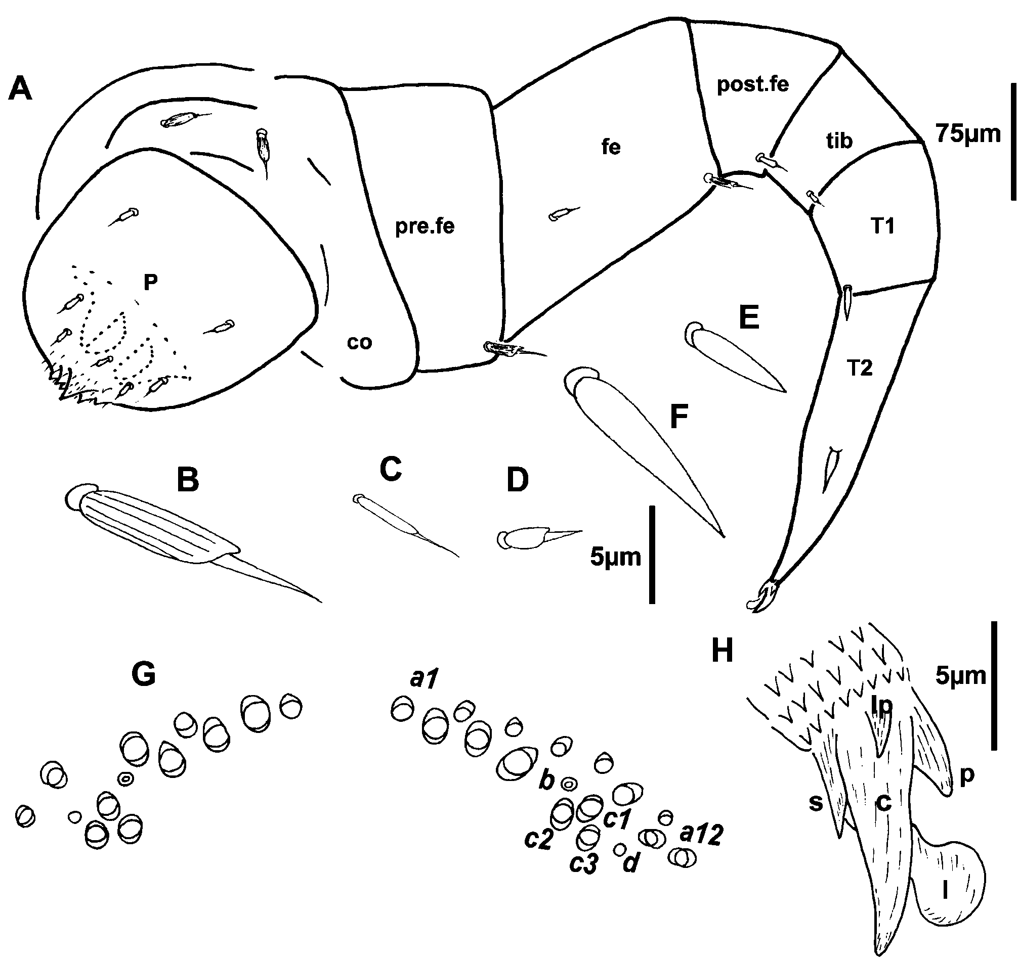
Legs: Leg segments are named following Manton (1956). Legs 1 and 2 are without trochanter, leg 1 also lacks tarsus 1. Chaetotaxy: coxa 1 and 2: 2 setae, coxae 3–13: 2–3 setae; prefemur, post-femur, tibia with 1 seta, except femur with 1–4 setae, tarsus 1 and tarsus 2 with a spine ( Figs 12A View FIGURE 12 and 13A View FIGURE 13 ). Coxa and prefemur, femur with a biarticulated seta with a ridged funicle ( Fig. 12B View FIGURE 12 ). 1–3 smaller biarticulated setae with the ridged funicles present on posterior position of femur and some also on penis ( Figs 12A and 12C View FIGURE 12 ). At distal edge of post-femur and tibia, a setiform seta present ( Fig. 12D View FIGURE 12 ). A spine on tarsus 2 sharply pointed and twice as long as the spine ( Fig. 12F View FIGURE 12 ) in the anterosternal position of tarsus 1 ( Fig. 12E View FIGURE 12 ). Posterior edge of last sternite with 2–4 setae similar to those present on coxa, number of these setae varying; 2 present on holotype and 2–4 on paratypes.
Telotarsus-Claw: A slender structure with an anterior lateral process, a posterior process less than half the length of claw. Anterior setiform process half of the length of claw; a lamella process present ( Figs 12H View FIGURE 12 and 13B View FIGURE 13 ).
Sex organs in the male: A pair of penes present on coxae 2 and 2 pairs of coxal glands located on coxal plates of legs 8 and 9.
Telson: Dorsal ornamental trichome sockets arranged almost symmetrically with 8 (L) and 12 (R) sockets of trichomes a on each side of telson in holotype, 7–14 sockets trichomes a on each side in paratypes. Sockets of trichomes a being more numerous on telsons of paratypes in 12 legs stage (Stadium VII) with these sockets ranging from 14–18 on each side of telson. Sockets of trichomes a forming 2 rows, a top row with small sockets and bottom row with larger sockets. The 2 largest sockets located close to the socket of trichome b. 3 large sockets trichomes c with protruding base: c1, c2 and c3, forming a triangular shape. Circular indentation d apparent each side near exterior side of trichomes c ( Figs 12G View FIGURE 12 , 13C and 13D View FIGURE 13 ).
Caudal bundles: In males, caudal structure comprised of a group of caudal trichome sockets forming a single structure of uniform size ( Fig. 13C View FIGURE 13 ). In females, caudal bundle with 2 distinguishing features: a main dorsal structure, carrying caudal trichomes, similar to that of male caudal bundle; and 2 latero-sternal bundles of nest trichomes, smaller sockets compared to caudal trichome sockets. Nest trichomes using for nest building and protecting egg clusters (Huynh and Veenstra, 2014). Caudal bundle structures well defined with borders between caudal and nest trichomes ( Fig. 13D View FIGURE 13 ).


No known copyright restrictions apply. See Agosti, D., Egloff, W., 2009. Taxonomic information exchange and copyright: the Plazi approach. BMC Research Notes 2009, 2:53 for further explanation.
|
Kingdom |
|
|
Phylum |
|
|
Class |
|
|
SubClass |
Penicillata |
|
Order |
|
|
SuperFamily |
Polyxenoidea |
|
Family |
|
|
Genus |