
Percussiopalpus Jałoszyński & Hlaváč, 2023
|
publication ID |
https://doi.org/ 10.11646/zootaxa.5277.1.3 |
|
publication LSID |
lsid:zoobank.org:pub:2EBD6E19-89E9-4918-8EE3-3324DF3BBA13 |
|
DOI |
https://doi.org/10.5281/zenodo.7891948 |
|
persistent identifier |
https://treatment.plazi.org/id/03AB722F-A10A-FFE5-25C9-F8C09535F8A6 |
|
treatment provided by |
Plazi |
|
scientific name |
Percussiopalpus Jałoszyński & Hlaváč |
| status |
gen. nov. |
Percussiopalpus Jałoszyński & Hlaváč , gen. n.
( Figs 1‒40 View FIGURES View FIGURES 3–7 View FIGURES 8–13 View FIGURES 14–18 View FIGURES 19–23 View FIGURES 24–30 View FIGURES31–38 View FIGURES 39–40 )
Type species. Percussiopalpus inusitatus sp. n.
Diagnosis. Thaumastocephalini with sharply delimited frontal sulcus; frontal rostrum gradually broadening posteriorly; tempora abruptly delimited from neck region; posterior tentorial pits situated closer to neck region than to submental bulge; maxillary palpomere 4 with convex distal margin; pronotum approximately cordiform, with lateral foveae, lateral longitudinal sulci and median longitudinal sulcus; mesoscutellar shield about as long as broad; metaventrite with only one pair of foveae (laterad mesocoxal insertion); each elytron with two basal foveae, complete sutural stria and incomplete discal stria; tergite IV with discal carinae and one pair of lateral foveae; sternite IV with pair of submedian foveae directed mesally.
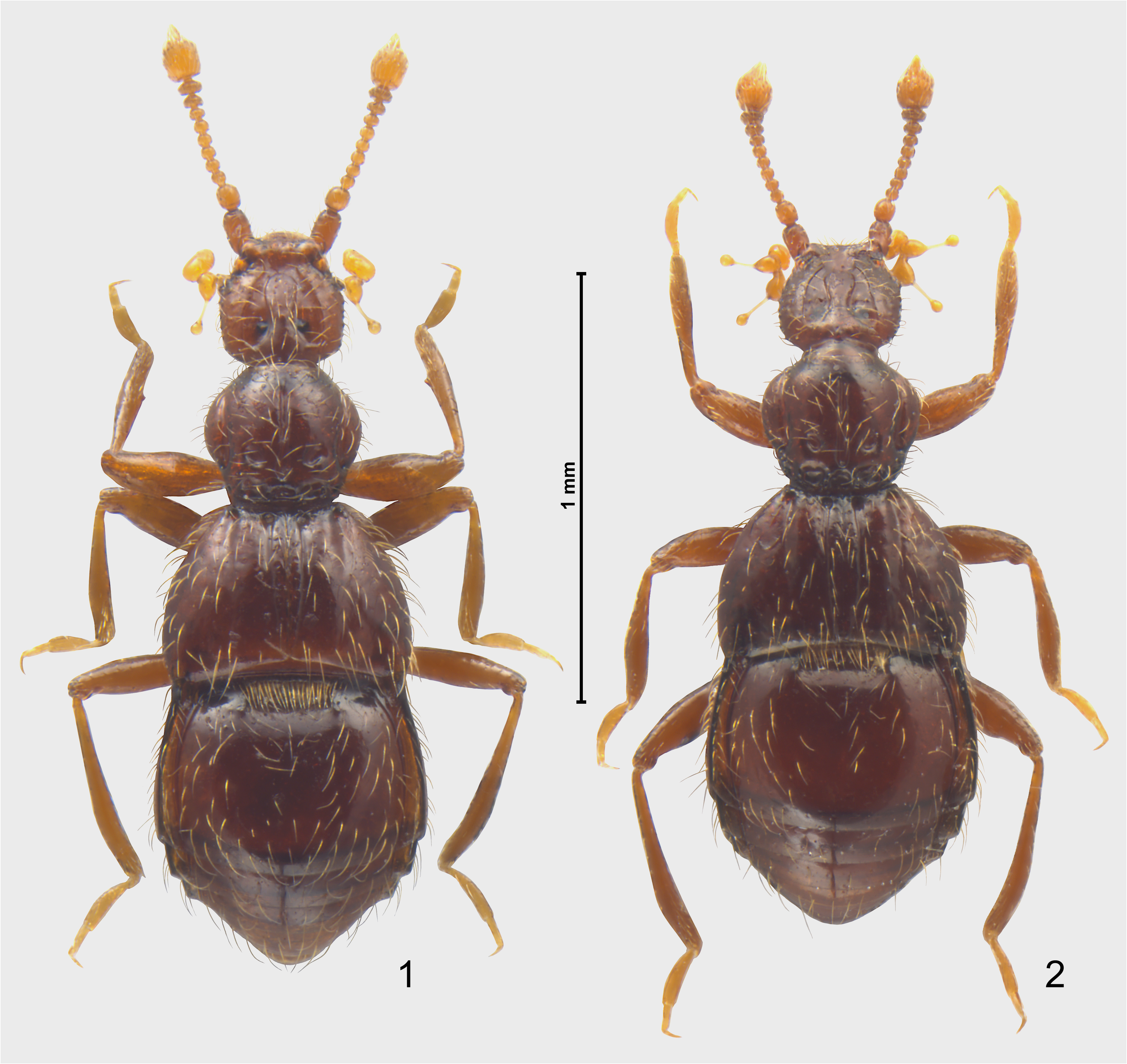
Description. Body ( Figs 1, 2 View FIGURES ) elongate, moderately slender, moderately convex, with distinct constrictions between head and prothorax and between prothorax and elytra.
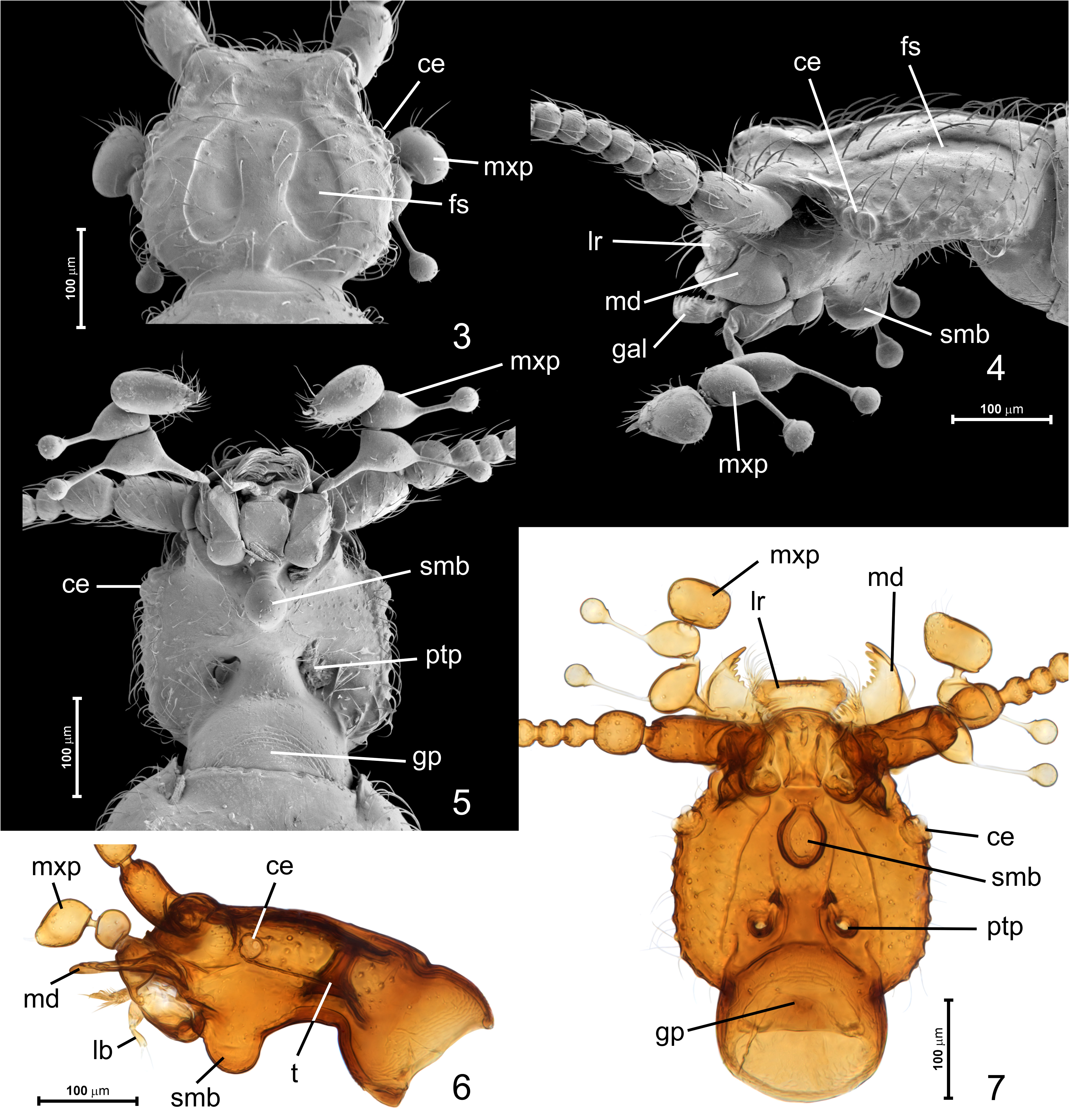
Head ( Figs 3‒7 View FIGURES 3–7 ) approximately cordiform, weakly elongate, broadest posteriorly, strongly flattened in posterior half. Temples round, distinctly longer than eyes; vertex and frons confluent, lacking frontal fovea and dorsal tentorial pits (but dorsal tentorial arms are fused with vertex on inner surface of head capsule ( Fig. 6 View FIGURES 3–7 ; t )), head with broad and shallow V-shaped vertexal and transverse rectangular frontal sulcus ( Fig. 3 View FIGURES 3–7 ; fs), both confluent; compound eyes ( Figs 3–7 View FIGURES 3–7 ; ce) minute and situated in front of middle, slightly below median coronal plane of head capsule. Gular sutures lacking; posterior tentorial pits ( Figs 5, 7 View FIGURES 3–7 ; ptp) large, circular, widely separated, situated in front of transverse impression demarcating neck region ventrally; submental region strongly transverse, with prominent oval submental bulge ( Figs 4‒7 View FIGURES 3–7 , 10 View FIGURES 8–13 ; smb) strongly projecting ventrally; hypostomal ridge ( Fig. 10 View FIGURES 8–13 ; hr) short, not reaching margin of submental bulge.
Antennae ( Figs 8–9 View FIGURES 8–13 ) composed of eleven antennomeres, slender, antennal insertions broadly separated. Scape cylindrical and elongate, longer than pedicel, antennal club trimerous and indistinctly demarcated, antennomere 11 elongate, symmetrical, rapidly narrowing in distal region to pointed apex, lacking cavities and clusters of basiconic sensilla.
Labium ( Fig. 10 View FIGURES 8–13 ) with elongate mentum ( Fig. 10 View FIGURES 8–13 ; mn), which is inversely subtrapezoidal, broadest near anterior third, with sinuate lateral margins; prementum short, transverse, lacking ligula; labial palpi ( Fig. 10 View FIGURES 8–13 ) trimerous, palpomere 1 ( Fig. 10 View FIGURES 8–13 ; lp1) short, nearly annular, palpomere 2 ( Fig. 10 View FIGURES 8–13 ; lp2) strongly elongate and weakly clavate, palpomere 3 setiform, barely discernible, obscured by long outer apical seta of palpomere 2.
Maxilla with large, nearly semicircular cardo ( Fig. 10 View FIGURES 8–13 ; cd); basistipes ( Fig. 10 View FIGURES 8–13 ; bst) triangular, elongate, mediostipes ( Fig. 10 View FIGURES 8–13 ; mst) subtriangular, about as long as basistipes; galea ( Fig. 10 View FIGURES 8–13 ; gal) elongate and with fringe of dense, thick setae along mesal margin; lacinia (not shown) similar to galea but shorter; palpifer elongate. Maxillary palpi ( Figs 4–7 View FIGURES 3–7 ; mxp, 12, 13) shorter than head, palpomere 1 ( Fig. 12 View FIGURES 8–13 ; mxp1) minute, elongate, about twice as long as wide; palpomere 2 ( Fig. 12 View FIGURES 8–13 ; mxp2) pedunculate, rapidly and strongly broadening in distal half, distal region strongly transverse and projecting outwards, with lateral sensory process inserted on conspicuously long and slender petiole broadened distally to form short collar bearing large spherical structure sparsely covered with flattened setae; palpomere 3 ( Fig. 12 View FIGURES 8–13 ; mxp3) non-pedunculate, strongly transverse and projecting outwards, with similar sensory process as palpomere 2; palpomere 4 ( Fig. 12 View FIGURES 8–13 ; mxp4) strongly transverse and projecting inwards, elliptical, wider than each of palpomeres 2 and 3, with apical sensory appendage ( Fig. 12 View FIGURES 8–13 ; sa) rod-like, short and slender, pointed at apex, inserted in socket surrounded by distinctly broadened and raised margin.
Mandibles ( Fig. 3 View FIGURES 3–7 ; md) subtriangular, slightly convex dorsally and concave ventrally, large, slightly asymmetrical, differing in shapes and numbers of mesal row of several teeth gradually reducing in length towards mandibular base, outer mandibular margin with conspicuously long seta; prostheca and mola lacking.
Labrum ( Fig. 11 View FIGURES 8–13 ; lr) transverse, subtrapezoidal, broadest anteriorly, with nearly straight anterior and rounded lateral margins; anteroventral margin with one pair of short peg-like sensilla ( Fig. 4 View FIGURES 3–7 ; pls) inserted on anterior epipharyngeal region.
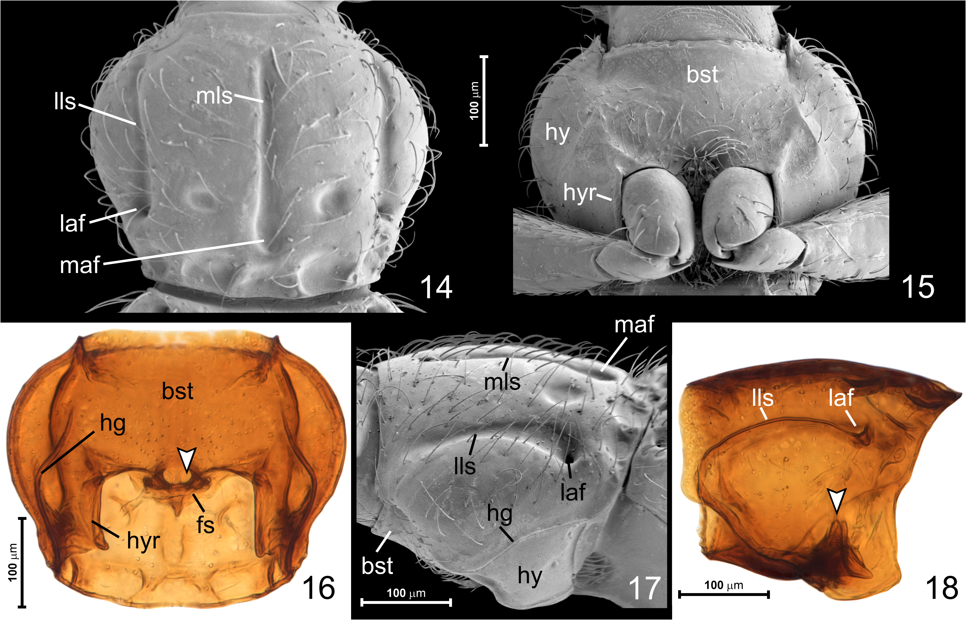
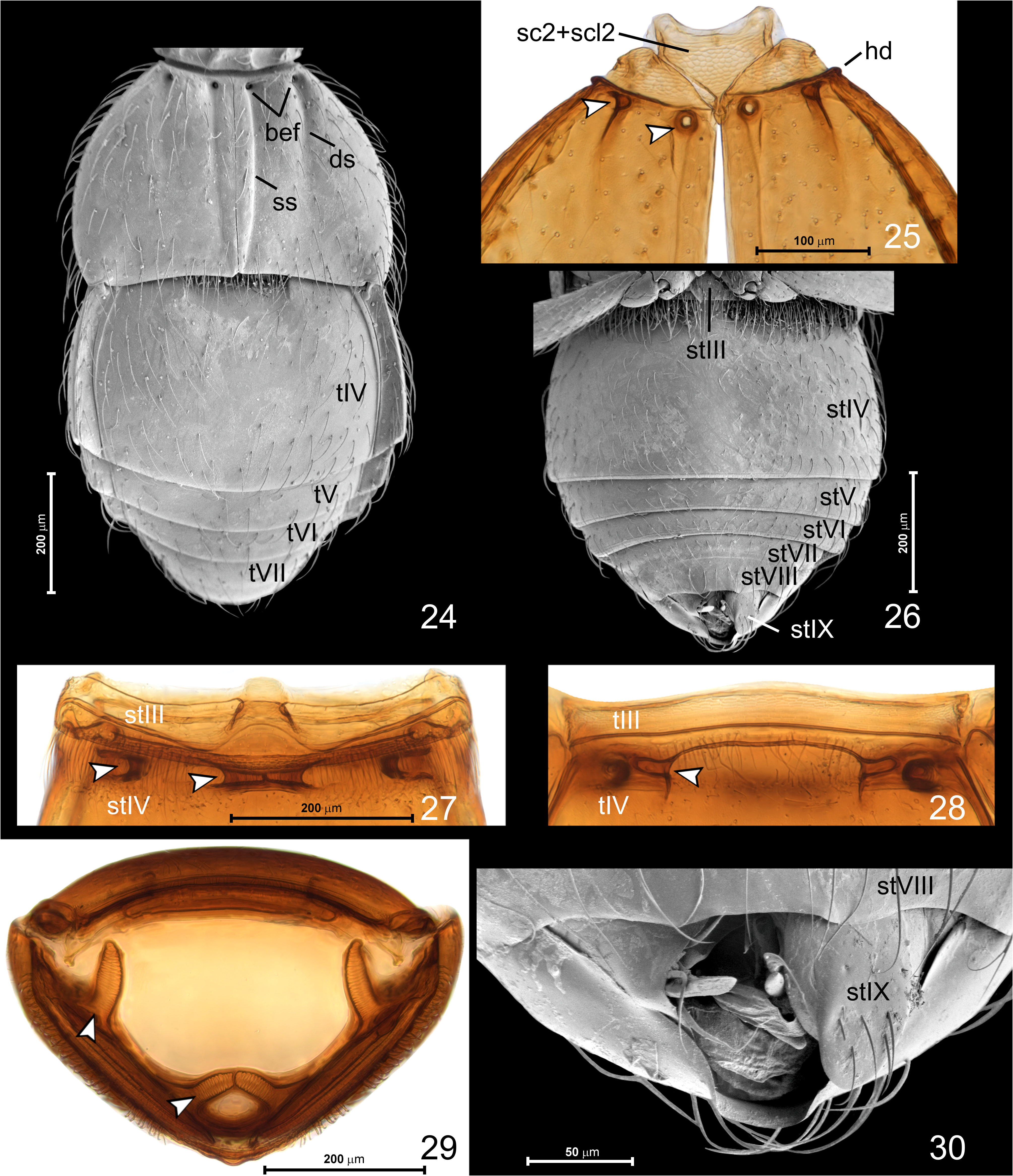
Pronotum ( Fig. 14 View FIGURES 14–18 ) widest distinctly in front of middle; lateral margins strongly rounded in anterior half and indistinctly sinuate in posterior half, anterior pronotal margin only slightly shorter than posterior one, anterior corners poorly marked, strongly obtuse-angled, posterior corners distinct, obtuse-angled; pronotal base arcuate. Pronotal disc with long median ( Figs 14, 17 View FIGURES 14–18 ; mls) and long lateral ( Figs 14, 17 View FIGURES 14–18 ; lls) longitudinal sulci, lateral sulcus posteriorly originating from asetose lateral antebasal fovea ( Figs 14, 17 View FIGURES 14–18 ; laf) with short subcuticular pocket, median antebasal fovea ( Fig. 14 View FIGURES 14–18 ; maf) vestigial, developed as broadening of median longitudinal sulcus and lacking subcuticular pocket; pronotal base with basal and sub-basal pairs of shallow but distinct suboval impressions, and with median impression adjacent to posterior margin.
Prosternum ( Figs 15–18 View FIGURES 14–18 ) fused with hypomera, but with short grooves on anterior margin representing probable rudiments of notosternal sutures; hypomera ( Figs 15, 17 View FIGURES 14–18 ; hy) broad and posteriorly weakly concave, each with shallow hypomeral groove ( Figs 16, 17 View FIGURES 14–18 ; hg) clearly visible in lateral view; basisternal region ( Figs 15‒16 View FIGURES 14–18 ; bst) much longer than very short coxal region (= furcasternum); prosternum with pair of submedian posterior foveae near middle of posterior basisternal margin, foveal pockets directed dorsally and slightly laterally from body midline ( Figs 16, 18 View FIGURES 14–18 ; arrowheads); anterior prosternal margin weakly crenulate; prosternal process indistinct, not separating procoxae. Hypomera with hypomeral ridges ( Figs 15, 17 View FIGURES 14–18 ; hyr) demarcating conspicuously narrow, parallel-sided inner (adcoxal) regions.
Mesonotum ( Fig. 25 View FIGURES 24–30 ) with mesoscutum and mesoscutellum fused ( Fig. 25 View FIGURES 24–30 ; sc2+scl2) forming subtriangular scutellar shield, only its minute tip projects posteriorly beyond transverse ridge demarcating basal articulating lobe and discal region of elytra, so that scutellar shield is not visible between elytral bases in intact specimens.
Metanotum (not shown) strongly shortened and weakly sclerotized due to loss of wings.
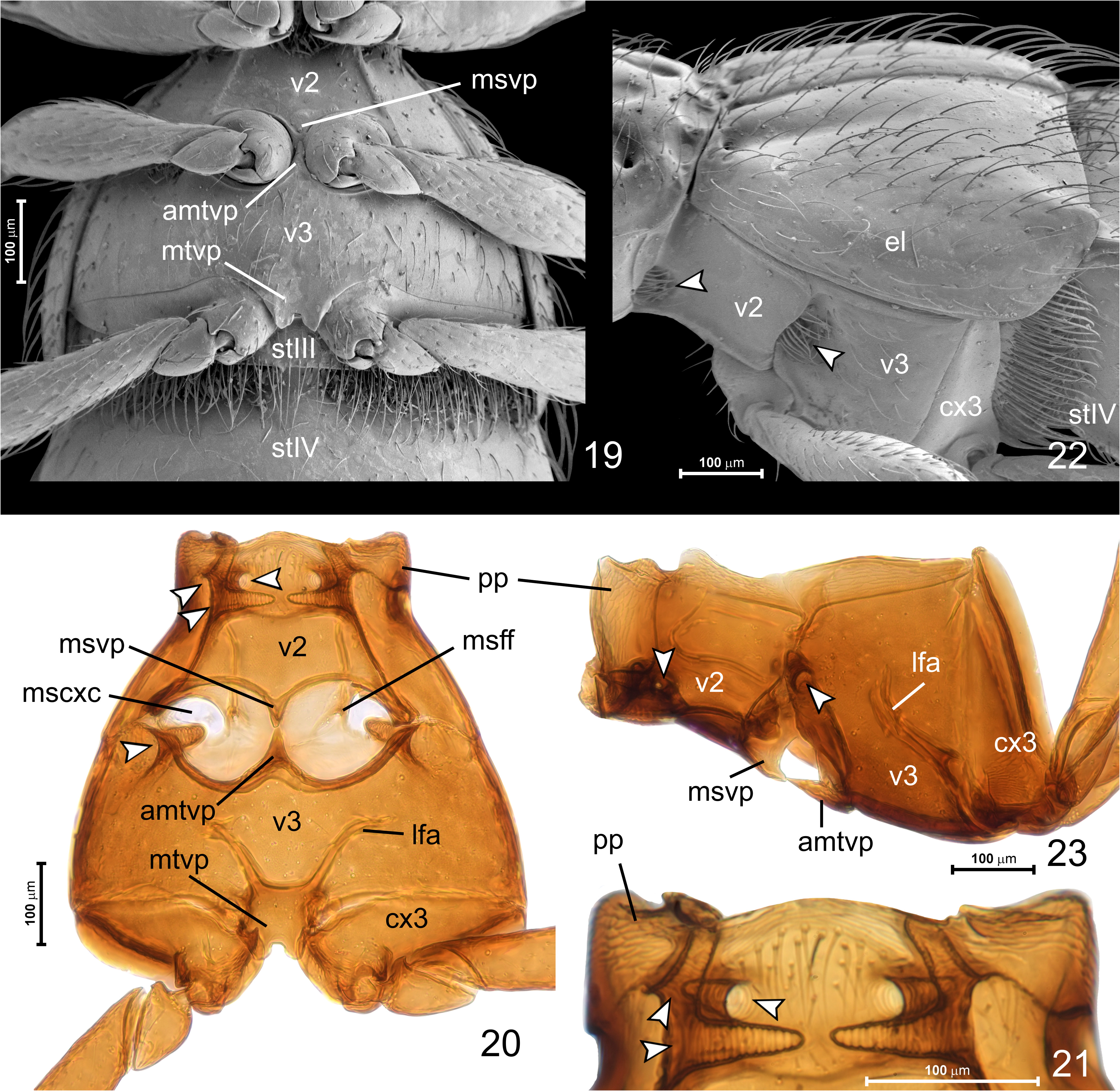
Mesoventrite ( Figs 19–23 View FIGURES 19–23 ; v 2 View FIGURES ) broadening posteriorly, with massive prepectus ( Figs 20–23 View FIGURES 19–23 ; pp); mesoventrite with three pairs of foveae in its anterior half ( Figs 20, 21 View FIGURES 19–23 , arrowheads): one sublateral fovea directed laterally with ventral opening, one lateral fovea directed anteromesally, and one lateral fovea directed mesally, lateral foveae share one large laterally visible setose opening ( Fig. 22 View FIGURES 19–23 , upper arrowhead). Median subtrapezoidal area of mesoventrite demarcated laterally by complete ridges. Mesocoxae narrowly separated by elongate, subtriangular mesoventral process ( Figs 19, 20, 23 View FIGURES 19–23 ; msvp) with rounded apex reaching middle of mesocoxae, meeting, but not fused with anterior tip of anterior metaventral process ( Figs 19, 20, 23 View FIGURES 19–23 ; amtp), both processes flat. Division between mesoand metaventrite marked with narrow sutures lateral to mesocoxal cavities (visible in Figs 20, 22, 23 View FIGURES 19–23 ), but division obliterated within mesocoxal rests.
Metaventrite ( Figs 19, 20, 22, 23 View FIGURES 19–23 ; v 3 View FIGURES 3–7 ) strongly transverse and broadening posteriorly; posterior margins of mesocoxal rests carinate; metaventrite with pair of lateral setose foveae directed anteromesally ( Fig. 20, 22, 23 View FIGURES 19–23 ; lower arrowhead); metaventral intermetacoxal process ( Figs 19, 20 View FIGURES 19–23 , mtvp) broad and clearly separating metacoxae, with posteromedian emargination.
Metendosternite with broad and subquadrate lamellate stem and short lateral furcal arms ( Fig. 20 View FIGURES 19–23 ; lfa) diverging anteriorly.
Elytra ( Figs 22 View FIGURES 19–23 , 24, 25 View FIGURES 24–30 ) together subtrapezoidal, dorsally flattened, each elytron with two asetose basal foveae ( Figs 24 View FIGURES 24–30 ; bef, 25; arrowheads), minute humeral denticle ( Fig. 25 View FIGURES 24–30 , hd), short discal stria ( Fig. 24 View FIGURES 24–30 , ds), and entire sutural stria ( Fig. 24 View FIGURES 24–30 , ss). Posterior elytral margin slightly concave and lacking fringe of modified setae.
Abdomen ( Figs 19 View FIGURES 19–23 , 24, 26–30 View FIGURES 24–30 ) broadest near middle, weakly convex dorsally and strongly convex ventrally, with well-developed broad paratergites on segments IV–VI. Tergite IV distinctly longer than remaining dorsally visible tergites (i.e., V–VII) combined, with transverse mediobasal impression laterally demarcated by short discal carinae, with one pair of basolateral foveae each adjacent to mesal margin of discal carina and with subcuticular pocket directed laterally ( Fig. 28 View FIGURES 24–30 ; arrowhead). Sternite III ( Figs 19 View FIGURES 19–23 , 26 View FIGURES 24–30 ; stIII) short and partly exposed only between metacoxae, with dense fringe of long setae along posterior margin; sternite IV ( Fig. 26 View FIGURES 24–30 ; stIV) about as long as remaining visible sternites (i.e., V–VIII in female and V–IX in male) combined, with two pairs of foveae: submedian pair with subcuticular pockets directed mesally and touching at midline ( Figs 27, 29 View FIGURES 24–30 , right arrowhead), and sublateral pair with pockets directed slightly mesally ( Fig. 27 View FIGURES 24–30 ; left arrowhead) and strongly dorsally ( Fig. 29 View FIGURES 24–30 ; left arrowhead). Male with sternite VIII ( Fig. 30 View FIGURES 24–30 ; stVIII) slightly modified, with trisinuate posterior margin; male sternite IX with large and elongate median penial plate ( Fig. 30 View FIGURES 24–30 ; stIX), lateral portions subtriangular and shorter than penial plate, weakly visible in intact specimens.
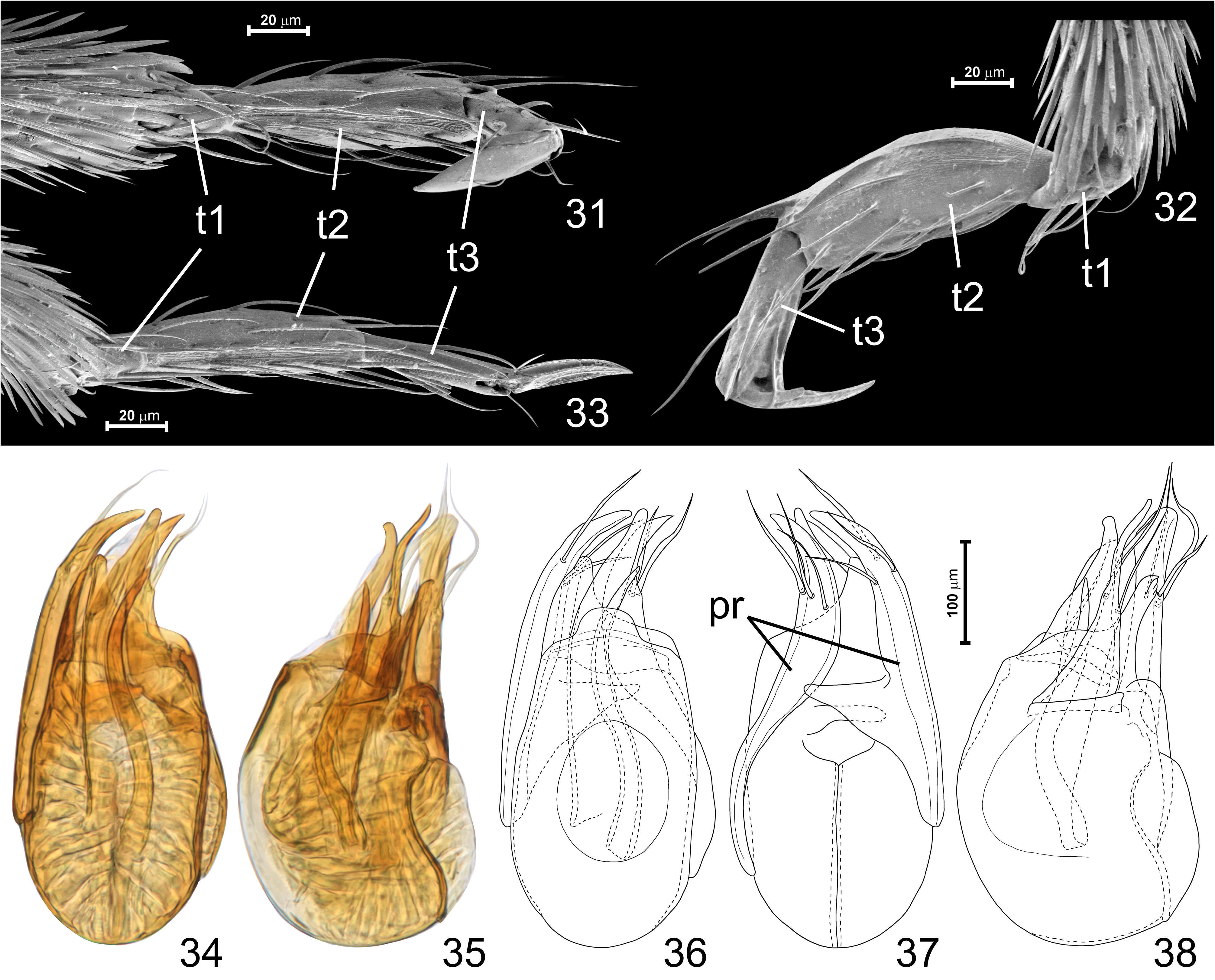
Legs ( Figs 1, 2 View FIGURES , 15 View FIGURES 14–18 , 19, 20, 22, 23 View FIGURES 19–23 , 26 View FIGURES 24–30 , 31–33 View FIGURES31–38 ) long and slender, all femora slightly clavate, tibiae and tarsi slender; tarsomere 1 ( Figs 31–33 View FIGURES31–38 ; t 1 View FIGURES ) minuscule, tarsomere 2 ( Figs 31–33 View FIGURES31–38 ; t 2 View FIGURES ) longer than tarsomere 3 ( Figs 31–33 View FIGURES31–38 ; t 3 View FIGURES 3–7 ); tarsal claw only slightly shorter than tarsomere 3; protarsi shorter than half length of tibiae, meso- and metatarsi longer than half length of tibiae.
Aedeagus ( Figs 34–38 View FIGURES31–38 ) elongate, with weakly sclerotized walls and asymmetric median lobe bearing oval dorsal diaphragm; endophallic sclerites elongate and asymmetric, parameres ( Fig. 37 View FIGURES31–38 , pr) asymmetric, long and broad, with subapical setae.
Sexual dimorphism. Males with thickened protarsomere 2, submedian ventral tooth on protibiae and sternite IX exposed and tripartite.
Etymology. The name Percussiopalpus refers to the peculiar maxillary palps with sensory processes shaped like percussion mallets, typical of Thaumastocephalini . Gender masculine.
Distribution. Northwestern Anatolia, Turkey (Balıkesir Province).
Species included. Percussiopalpus inusitatus Jałoszyński & Hlaváč.
Remarks. Characters given in the diagnosis all clearly differentiate this genus from Thaumastocephalus ; major differences are also compiled in Table 1 View TABLE .


No known copyright restrictions apply. See Agosti, D., Egloff, W., 2009. Taxonomic information exchange and copyright: the Plazi approach. BMC Research Notes 2009, 2:53 for further explanation.