
Andrena (Planiandrena) sella Wood, 2022
|
publication ID |
https://doi.org/ 10.5852/ejt.2022.843.1947 |
|
publication LSID |
lsid:zoobank.org:pub:C03BE897-EFE2-4CCD-913A-723792CDF050 |
|
DOI |
https://doi.org/10.5281/zenodo.7237888 |
|
persistent identifier |
https://treatment.plazi.org/id/EDCCBC04-DFE5-46BD-92C6-2EA819162D4C |
|
taxon LSID |
lsid:zoobank.org:act:EDCCBC04-DFE5-46BD-92C6-2EA819162D4C |
|
treatment provided by |
Felipe |
|
scientific name |
Andrena (Planiandrena) sella Wood |
| status |
sp. nov. |
Andrena (Planiandrena) sella Wood sp. nov.
urn:lsid:zoobank.org:act:EDCCBC04-DFE5-46BD-92C6-2EA819162D4C
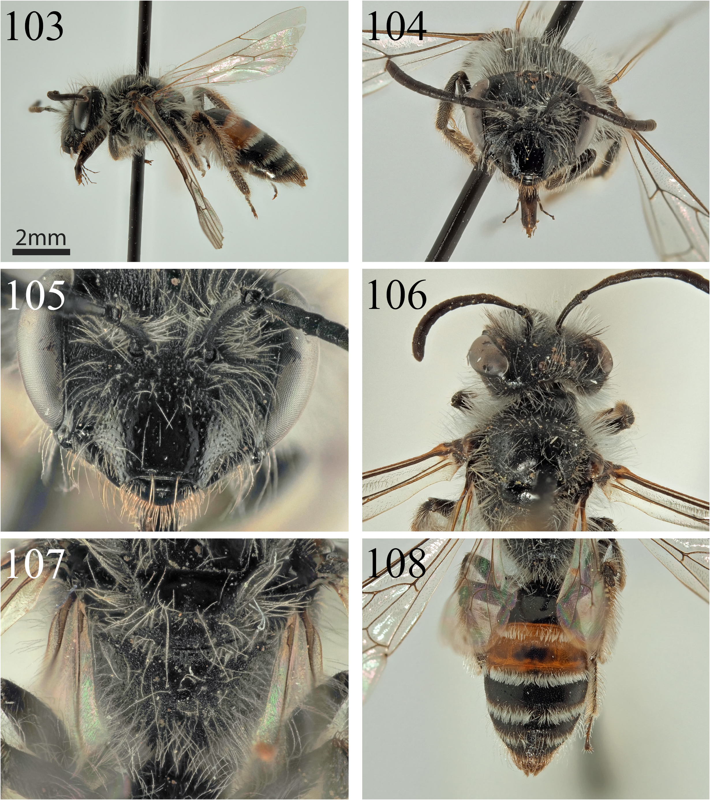
Figs 103–108 View Figs 103–108
Diagnosis
Andrena sella sp. nov. differs from the two previously described species of Planiandrena . It has the same very broad process of the labrum ( Fig. 105 View Figs 103–108 ) and narrow foveae that tightly follow the inner margin of the compound eye, but the propodeal triangle is noticeably narrower ( Fig. 107 View Figs 103–108 ), slightly depressed, and there is no obviously raised area centrally. The propodeal triangle does, however, still have basal rugae, a feature present in A. huma sp. nov. and absent in A. flagrans sp. nov. Because the shape of the head is so extremely similar, A. sella is confidently placed in the Planiandrena . It can be instantly separated from all described species of Planiandrena because the apical margin of T1 and the entirety of T2 are red marked ( Fig. 108 View Figs 103–108 ). Structurally, it can be separated by the combination of the shape of the propodeal triangle, which is slightly depressed and medially narrowed (broad and unnarrowed in A. huma and A. flagrans ), by the clypeus, which is domed and highly polished over the majority of its area ( Fig. 105 View Figs 103–108 ) (shiny only in the apical ⅓ in A. huma and A. flagrans ), and by the terga, which are shiny but clearly punctate throughout, both on the discs and the marginal areas (at least basally shagreened in A. huma , without shagreen but also with only occasional punctures in A. flagrans ).
Etymology
From the Latin noun ‘ sella ’, meaning ‘saddle’, in reference to the red-banded abdomen.
Material examined
Holotype IRAN • ♀; Yasouj, Deli Bajak, Sepidar ; [30.518° N, 51.495° E]; 2181 m a.s.l.; 23 Apr. 2021; E. Rostami leg.; OÖLM. GoogleMaps
Description
Female
MEASUREMENTS. Body length 8.5 mm ( Fig. 103 View Figs 103–108 ).
HEAD. 1.3 times as wide as long ( Fig. 104 View Figs 103–108 ). Clypeus dark, domed, with clear rounded punctures, punctures laterally separated by 0.5–1 puncture diameter; punctures becoming sparse and irregular centrally and apically, punctures here separated by 1–6 puncture diameters ( Fig. 105 View Figs 103–108 ). Clypeus surface laterally and basally with narrow area of slightly raised microreticulation, weakly shining; clypeus strongly polished and shining over majority of remaining area. Process of labrum short, rounded trapezoidal, four times as wide as long. Face, gena, vertex, and scape with long whitish hairs, frons and gena dorsolaterally with some intermixed black hairs, no hairs exceeding length of scape. Gena exceeding width of compound eye; ocelloccipital distance small, ½ diameter of lateral ocellus. Foveae narrow dorsally, occupying ⅓of space between compound eye and lateral ocellus, narrowed below at level of antennal insertions; filled with dark brown hairs. Antennae dark, A3 exceeding A4+5, shorter than A4+5+6.
MESOSOMA. Scutum with dense granular microreticulation, weakly shining, with large shallow punctures, punctures separated by 2–3 puncture diameters ( Fig. 106 View Figs 103–108 ). Scutellum with shagreen restricted to lateral areas, smooth and shining over majority of area, with scattered punctures. Pronotum with hint of humeral angle, inconspicuous. Mesepisternum and lateral and dorsolateral faces of propodeum with granular shagreenation and fine network of raised reticulation, very weakly shining. Propodeal triangle slightly depressed, internal surface with weakly shining granular microreticulation; basally with network of short raised longitudinal rugae, centrally longer and extending into centre of propodeal triangle; propodeal triangle therefore defined by depressed surface area and change in surface sculpture ( Fig. 107 View Figs 103–108 ). Mesosoma with long whitish hairs, equalling length of scape, propodeal corbiculae with long white plumose hairs, internal surface with long white simple hairs. Legs uniformly dark, with whitish to light brownish pubescence; tibial scopae white with intermixed brown hairs dorsobasally. Femoral scopae and flocculus white. Hind tarsal claws with inner tooth. Hind tibial spur parallel-sided. Wings hyaline, stigma dark orange, venation orange to dark orange, nervulus weakly antefurcal; first recurrent vein enters second submarginal cell beyond its middle.
METASOMA. Apical margin of T1 and disc of T2 red marked, with exception of dark mark centrally on T2 ( Fig. 108 View Figs 103–108 ). Remaining terga dark, apical margins of T3–4 lightly lightened yellow-hyaline. Terga with very weak shagreenation, smooth and shining throughout. Terga clearly punctate, T1 with punctures sparse, separated by 3–4 puncture diameters, T2–4 more densely punctate, punctures separated by 1–2 puncture diameters. Tergal discs with extremely scattered short white hairs, T1–4 with dense unbroken apical fringes of white hairs that obscure underlying integument. Fringe of T5 and hairs flanking pygidial plate brown; pygidial plate rounded triangular, broadly flat without obvious structure.
Male
Unknown.
Distribution
Southern Iran (Yasouj).
No known copyright restrictions apply. See Agosti, D., Egloff, W., 2009. Taxonomic information exchange and copyright: the Plazi approach. BMC Research Notes 2009, 2:53 for further explanation.
|
Kingdom |
|
|
Phylum |
|
|
Class |
|
|
Order |
|
|
Family |
|
|
Genus |