
Meriola Banks
|
publication ID |
https://doi.org/10.11646/zootaxa.4936.1.1 |
|
publication LSID |
lsid:zoobank.org:pub:DAC2F49B-EE13-4827-B549-C59B9C43550B |
|
DOI |
https://doi.org/10.5281/zenodo.4672980 |
|
persistent identifier |
https://treatment.plazi.org/id/03AB87CF-FFCB-E34E-FF73-517BFE3AFE3E |
|
treatment provided by |
Plazi |
|
scientific name |
Meriola Banks |
| status |
|
Genus Meriola Banks View in CoL View at ENA
Meriola Banks, 1895: 81 View in CoL ( type species by monotypy: M. decepta Banks, 1895 View in CoL ); Platnick & Ewing, 1995: 8.
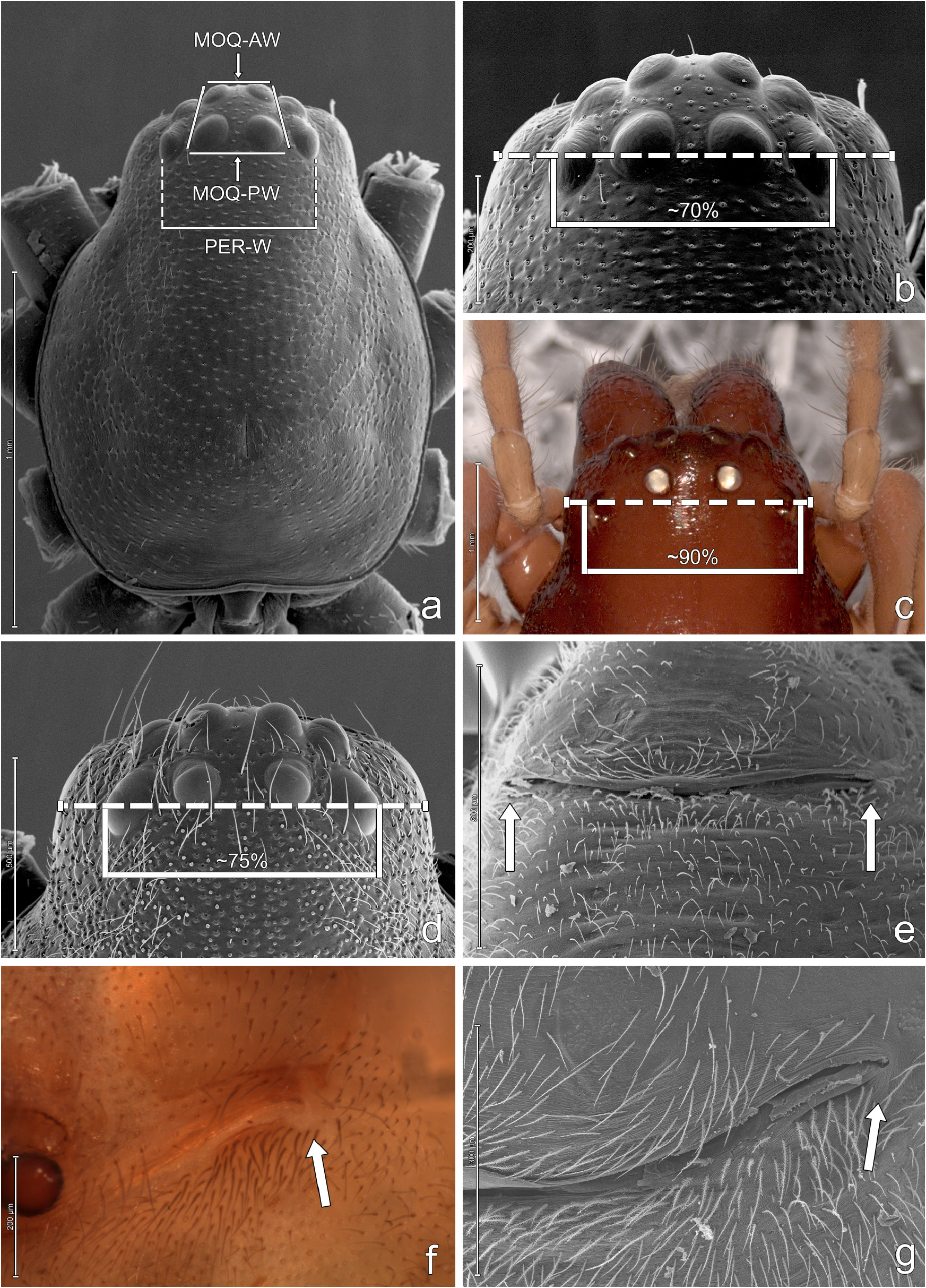
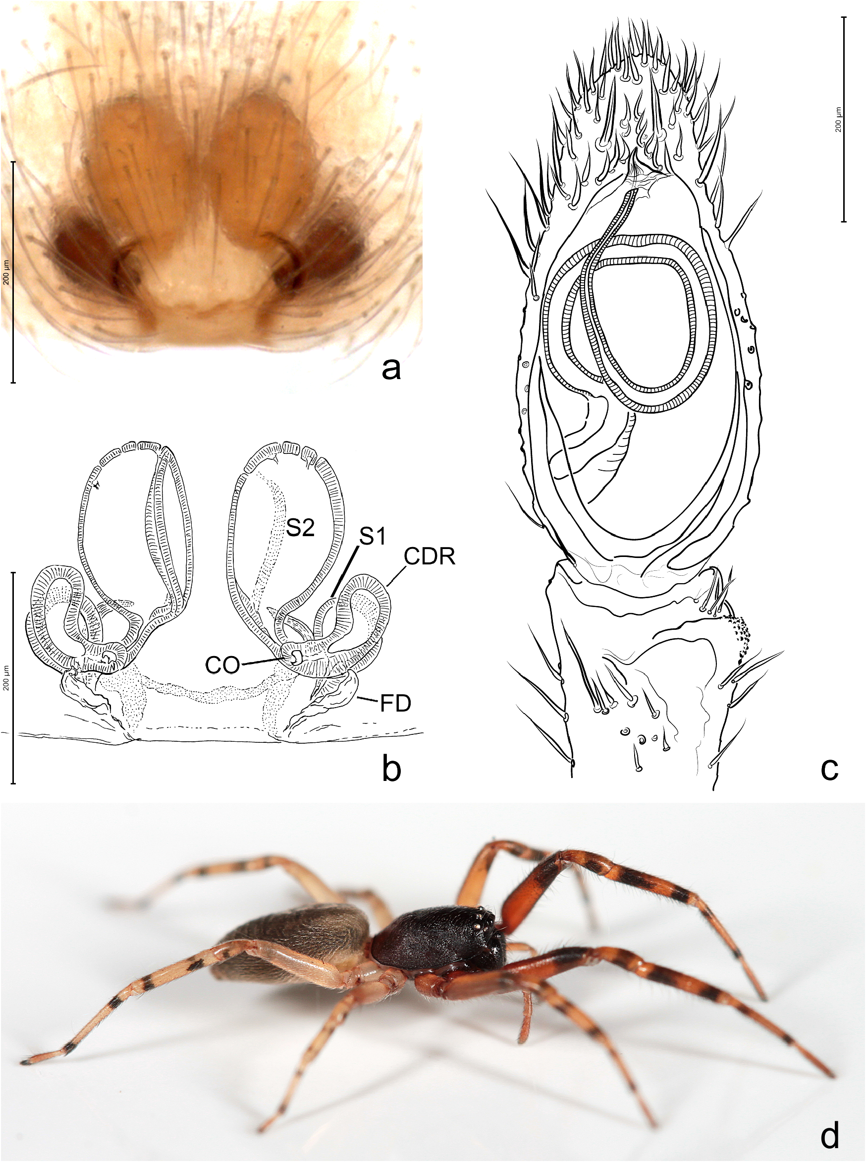
Diagnosis. Members of the genus Meriola resemble the American trachelids currently included in Trachelas and Trachelopachys by having male cuspules and reduced spination, but differ in several respects. The most remarkable characteristics are their almost straight, rather than recurved, posterior eye row ( Fig. 1 View FIGURE 1 a–b), narrower than in the other American genera ( Fig. 1c View FIGURE 1 ), and the elongated, sharply tipped ventral leg cuspules ( Fig. 4c View FIGURE 4 ).
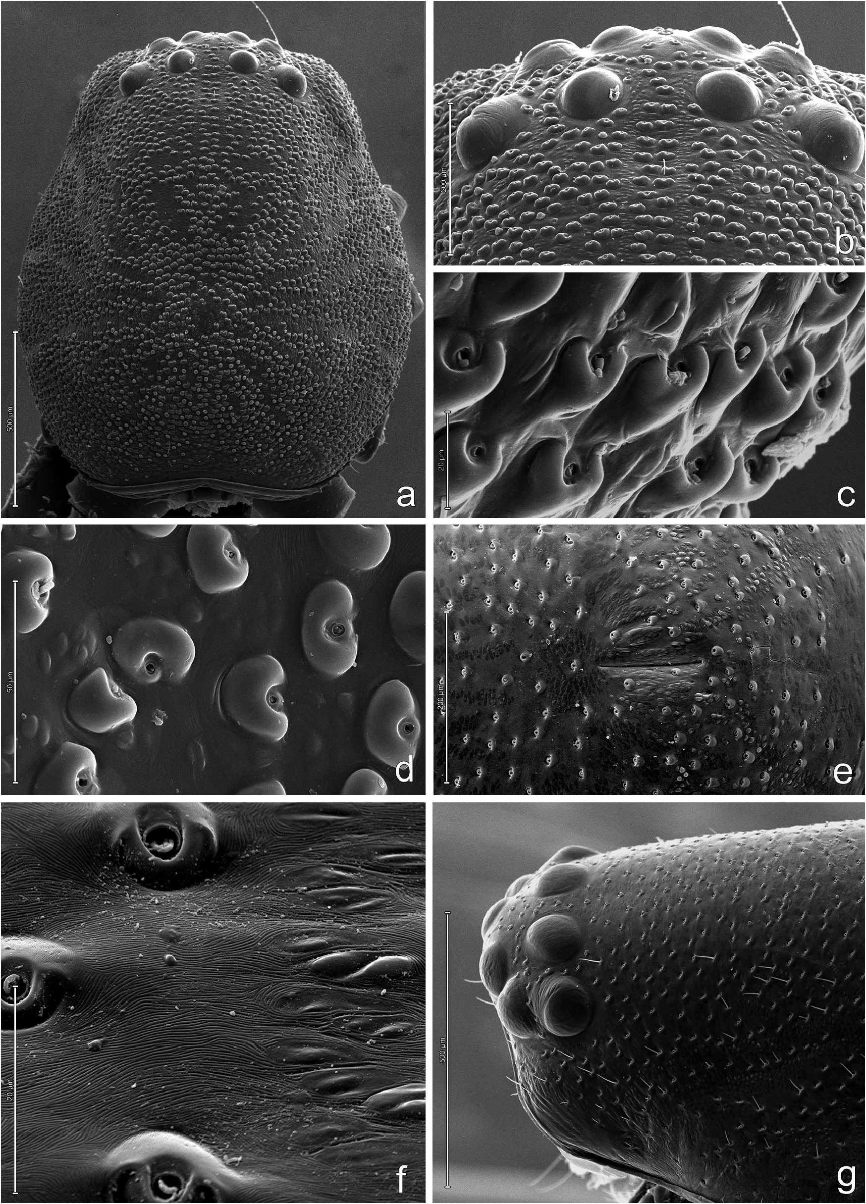
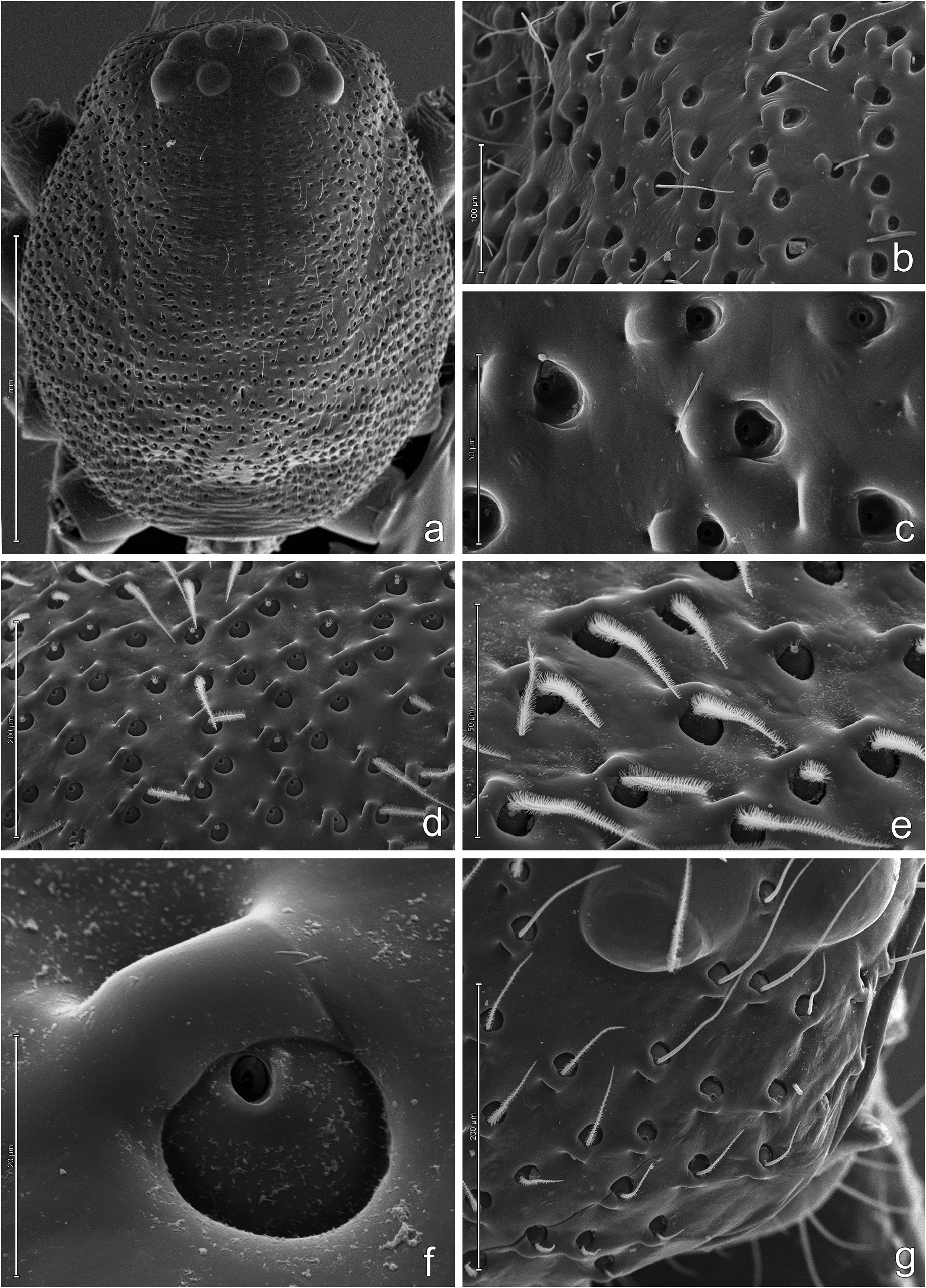
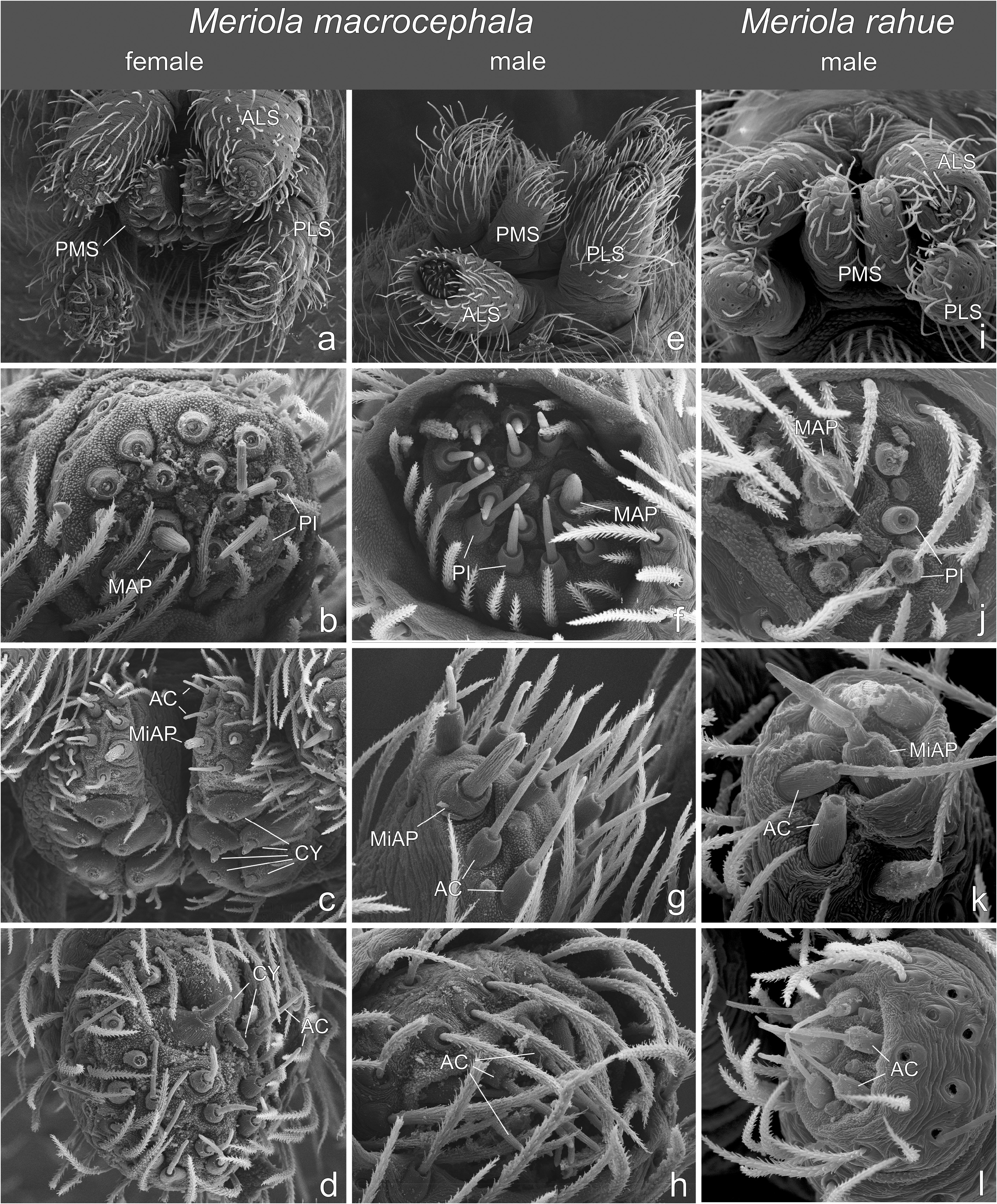
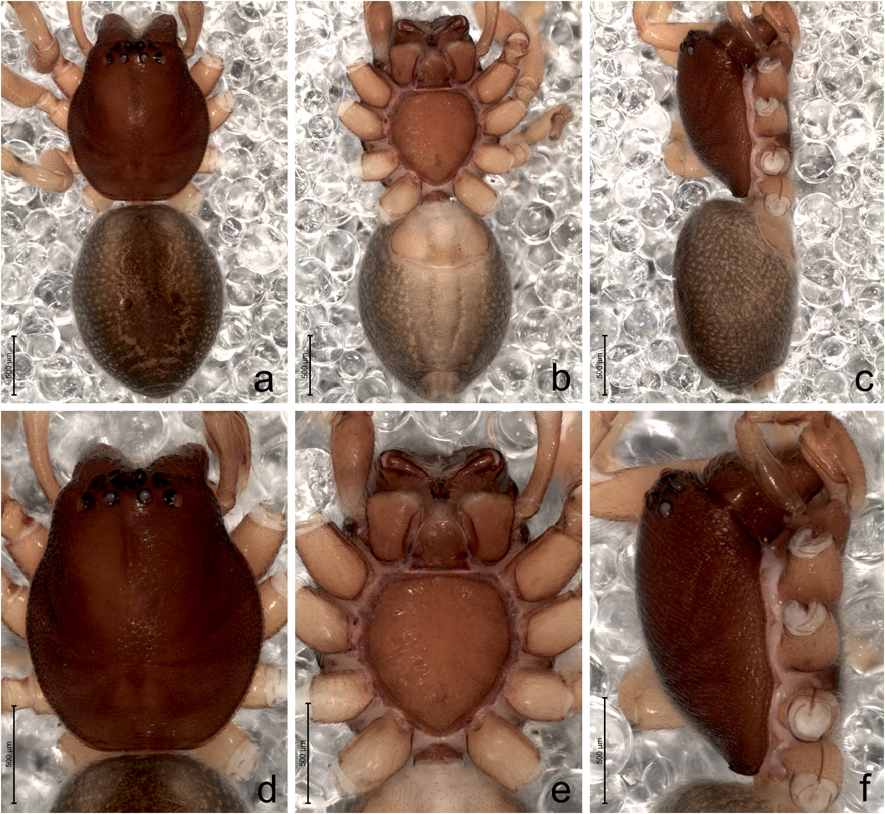
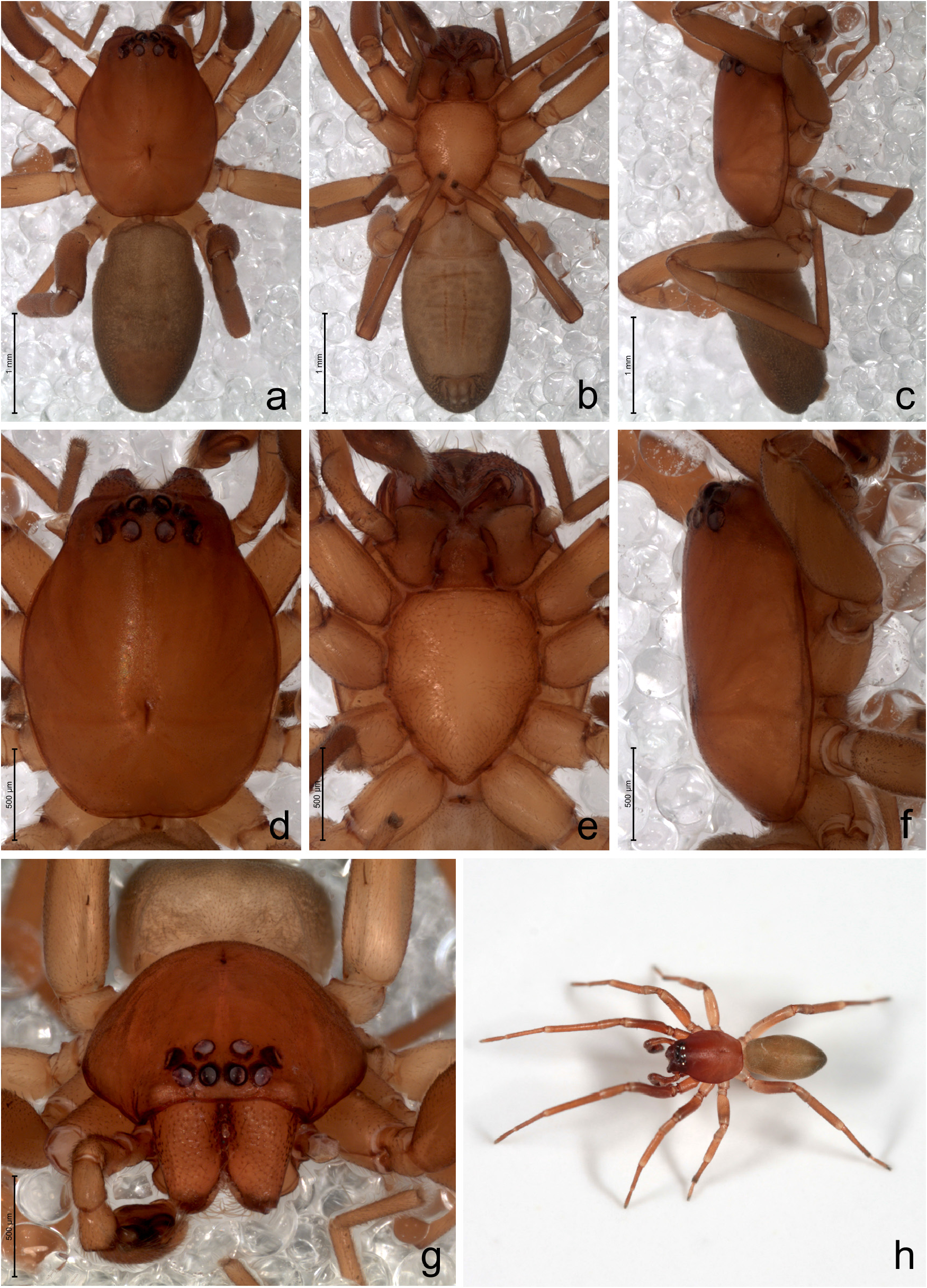
Description. Small to medium sized trachelids (total length 2.52̅6.88). Carapace elongate to oval in dorsal view ( Figs 1a View FIGURE 1 , 2a View FIGURE 2 , 3a View FIGURE 3 ), widest at rear of coxae II, cephalic and thoracic area shiny, with short thin hairs, or bearing tubercles associated with setal sockets ( Figs 2 View FIGURE 2 ̅3). Sockets may be on surface or in depressions on carapace ( Figs 2c View FIGURE 2 ̅d, 2f, 3c, 3f). In some cases, socket presents a cuticular fold on one side, resulting in a tubercle ( Fig. 3e View FIGURE 3 ̅f). These folds can vary in size, distribution and number in different species. Thoracic groove short. Color brownish-orange to dark brown, sometimes lighter posteriorly. Eyes: from above, anterior eye row slightly recurved to recurved, posterior eye row straight to slightly recurved; from front, anterior eye row slightly procurved, posterior eye row procurved ( Fig. 10g View FIGURE 10 ̅h); AME circular, dark, PME circular to oval, with silvery oblique tapeta forming a 90° angle between each other ( Ramírez 2014: character 26), ALE and PLE circular to oval, silvery; eyes nearly equal in size, except some species where AME are slightly smaller; all eyes about evenly spaced; MOQ-AW ± MOQ-PW ( Fig. 1a View FIGURE 1 ). Clypeal height about equal to AME diameter. Chelicerae with three teeth on promargin and two on retromargin. Endites rectangular, their inner margins with distinct longitudinal depression, distal portion of their external edge angulate, parallel; labium nearly trapezoidal, almost as long as its basal width, in general with basal protuberances and constriction near base; sternum oval to heart-shaped, longer than wide, pointed behind coxae IV. Palpal claw without teeth. Precoxal triangles present in both sexes. Legs: spination reduced (see under species description), species-specific except for females of M. balcarce Platnick & Ewing and M. foraminosa Keyserling , both with distal spine on prolateral side of femur I; metatarsi III and IV sometimes with preening combs ( Fig. 4g View FIGURE 4 ). Macrosetae present as cuspules in some males, but absent females and males of M. californica , M. decepta , M. foraminosa , M. lineolata comb. nov., M. mauryi Platnick & Ewing , M. ramirezi and M. tablas Platnick & Ewing ; present in M. macrocephala (Nicolet) comb. nov. females. Cuspules elongated and sharply tipped, on ventral side of tibiae, metatarsi and tarsi, number varying intra- and interspecifically ( Fig. 4c View FIGURE 4 ; compare Figs 56g View FIGURE 56 ̅h with 57h̅i). Leg segments pale brown to yellowish-brown, in some cases with dark rings, or tibiae I darker. Scopulae composed of distally spatulate tenent setae, from distal end of metatarsi to distal tarsi I and II ( Figs 4a, 4c View FIGURE 4 ), absent on III and IV. Females of some species exhibit strong ventral scopula on anterior legs, which start as loosely dense in distal tibiae and become stronger towards tarsus. Claw tufts composed of spatulate setae, with block-like shape of the bases ( Ramírez 2014: figs 72A̅C). The mesal side of the seta, at its insertion, is widely expanded in large blocks with defined vertices, while the ectal side becomes narrow ( Figs 4e, h View FIGURE 4 ). Claws pectinate, claw-tuft clasping mechanism present in structure of teeth appressed together, with claw lever file-claw tuft bases interlocking (observed with SEM in M. penai Platnick & Ewing , M. mauryi , M. fasciata Mello-Leit „o and M. macrocephala ) ( Figs 4b, h View FIGURE 4 ; Ramírez 2014: figs 72A̅C). Tarsal organ capsulate, aperture oval to slit-shaped ( Ramírez 2014: fig. 58O). Trichobothria with transversal ridges on anterior hood, shaft with basal bulbous expansion with bumps ( Fig. 4d View FIGURE 4 , Ramírez 2014: fig. 96D). Abdomen of male sometimes with dorsal faintly slightly sclerotized area, on anterior half ( Fig. 35a View FIGURE 35 ), posterior half ( Figs 62a View FIGURE 62 , 74a View FIGURE 74 ), or most of dorsum ( Fig. 71a View FIGURE 71 ); without epiandrous spigots. Coloration: dorsum grayish-brown or brownish, sometimes with chevrons, reticulations and/or cardiac mark; venter pale, in cases with dark ventral stripe, epigastric area of males more sclerotized. Tracheal spiracle about ALS length, just before spinnerets. Spinnerets ( Fig. 6 View FIGURE 6 ; observed with SEM in M. rahue Platnick & Ewing , M. fasciata , M. macrocephala ): Anterior lateral spinnerets with single major ampullate gland spigot and several piriform gland spigots ( Figs 6b, f, j View FIGURE 6 ). Posterior median spinnerets of females with five large cylindrical gland spigots, as well as single minor ampullate gland spigot and few aciniform gland spigots ( Fig. 6c View FIGURE 6 ; Ramírez 2014: fig. 133G); males with single minor ampullate gland spigot and few aciniform gland spigots ( Figs 6g, k View FIGURE 6 ). The posterior lateral spinnerets bear two cylindrical gland spigots in females and several aciniform gland spigots in both sexes ( Figs 6d, h, i View FIGURE 6 ; Ramírez 2014: fig. 114D). Genitalia: Male palp with retrolateral tibial apophysis ( Fig. 5f View FIGURE 5 ); copulatory bulb with simple looping sperm duct, median apophysis absent, embolus long and slender to short and thick ( Figs 5e View FIGURE 5 , 21b View FIGURE 21 , 63c View FIGURE 63 ). Epigyne with median field of variable size, lateral lobes in general with sclerotized ridges ( Fig. 5a View FIGURE 5 ), primary spermatheca usually small, identified by origin of fertilization duct, which is close to epigastric furrow, secondary spermathecae present, sometimes larger than primary spermatheca, identified by gland ducts, with additional expansion of copulatory duct, here referred to as copulatory duct receptacle (CDR) ( Figs 5b, c View FIGURE 5 ; 24b View FIGURE 24 ).
Distribution. All species occur in temperate to subtropical areas of the New World, with most of the diversity in southern South America ( Peru, Bolivia, Brazil, Chile and Argentina). There are two native species in North America, one ( M. californica ) occurring only on the west coast, while the second ( M. decepta ) is found throughout most of U.S.A. and southeast Canada. In addition, an introduced species native to South America ( M. arcifera ) occurs in California. There are as well isolated records of M. foraminosa in Venezuela and Ecuador, which may be the result of poor collecting efforts, and of M. decepta in Guatemala, Colombia, Peru and Brazil, probably due to anthropogenic introductions.


No known copyright restrictions apply. See Agosti, D., Egloff, W., 2009. Taxonomic information exchange and copyright: the Plazi approach. BMC Research Notes 2009, 2:53 for further explanation.
|
Kingdom |
|
|
Phylum |
|
|
Class |
|
|
Order |
|
|
Family |
Meriola Banks
| González, María E., Grismado, Cristian J. & Ramírez, Martín J. 2021 |
Meriola
| Platnick, N. I. & Ewing, C. 1995: 8 |