
Neodiplopeltula incisa ( Southern, 1914 ), 2018
|
publication ID |
https://doi.org/ 10.5852/ejt.2018.458 |
|
publication LSID |
lsid:zoobank.org:pub:16C565AB-22A6-496F-AE95-0A876066F958 |
|
DOI |
https://doi.org/10.5281/zenodo.3843749 |
|
persistent identifier |
https://treatment.plazi.org/id/03AC5D37-6848-FFEE-FE7C-574DBBBEFCC7 |
|
treatment provided by |
Valdenar |
|
scientific name |
Neodiplopeltula incisa ( Southern, 1914 ) |
| status |
|
Neodiplopeltula incisa ( Southern, 1914) gen et. comb. nov.
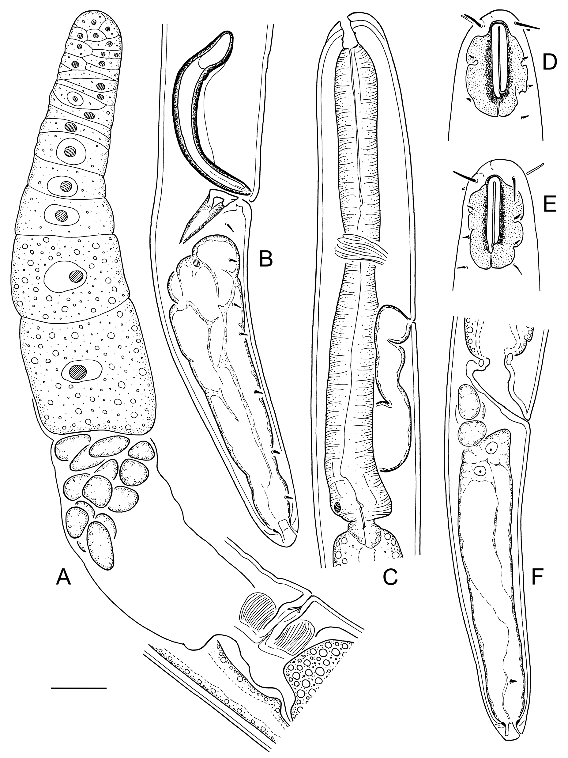
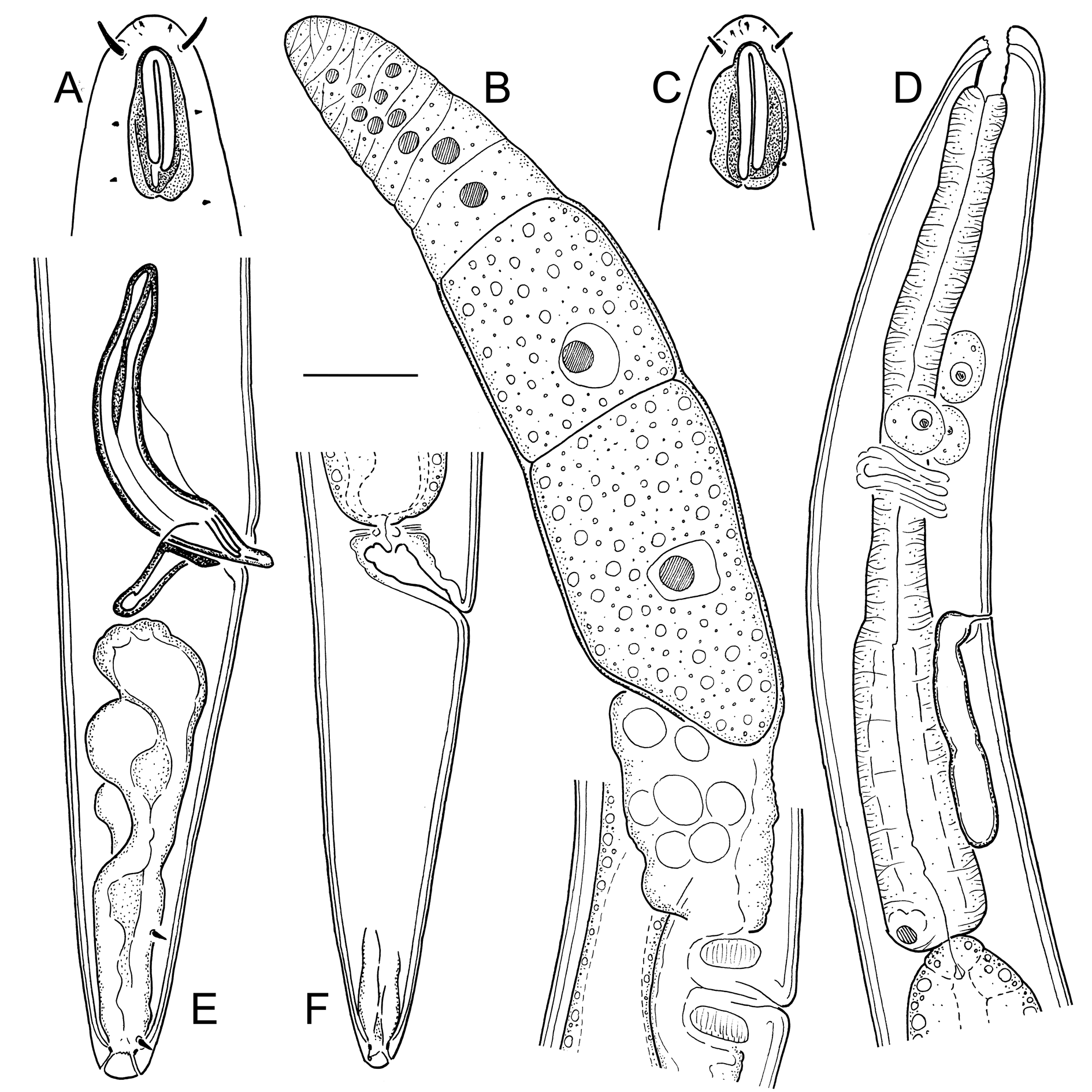
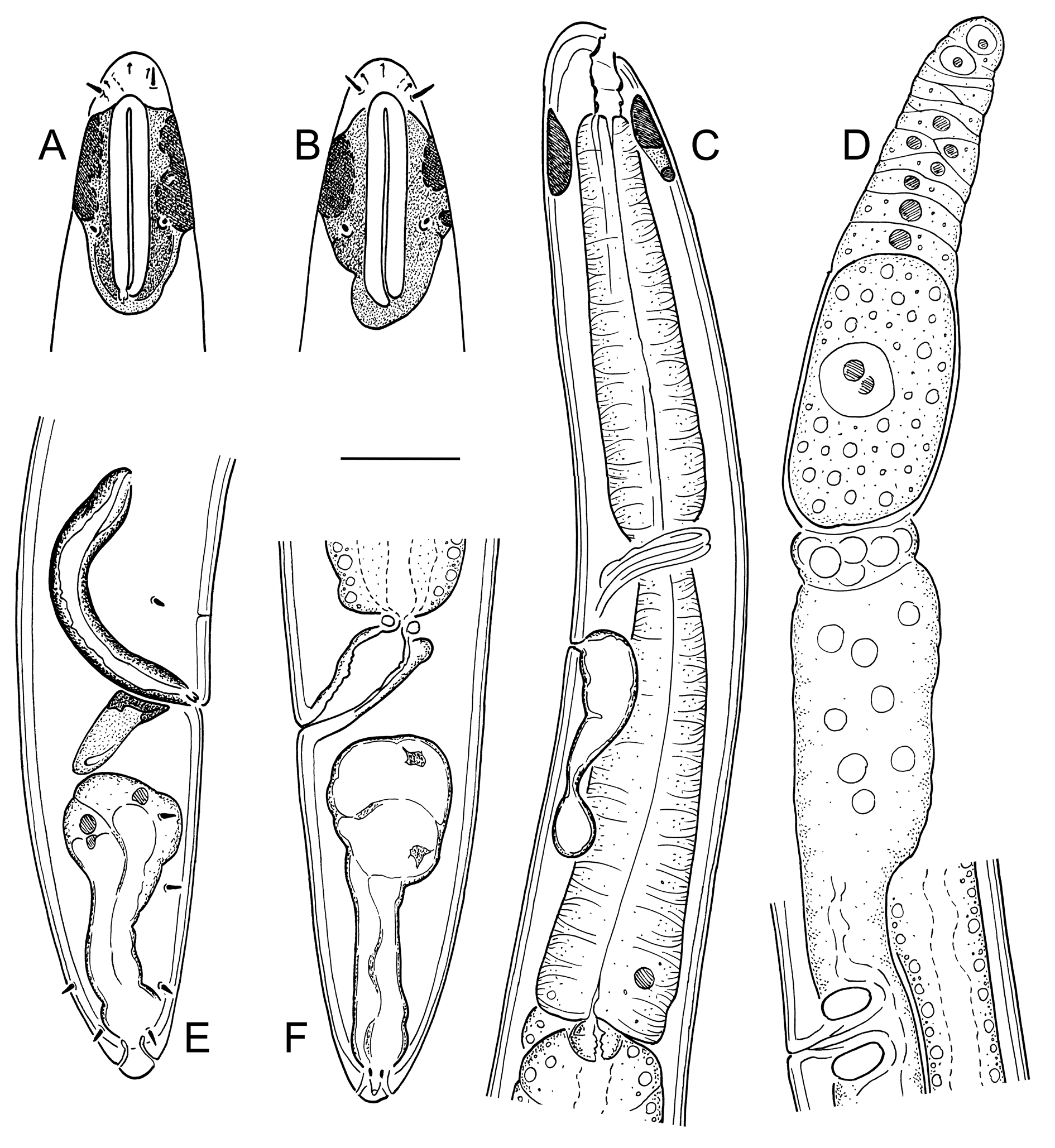
Figs 1–4 View Fig View Fig View Fig View Fig ; Table 2
Diagnosis (based on combined data)
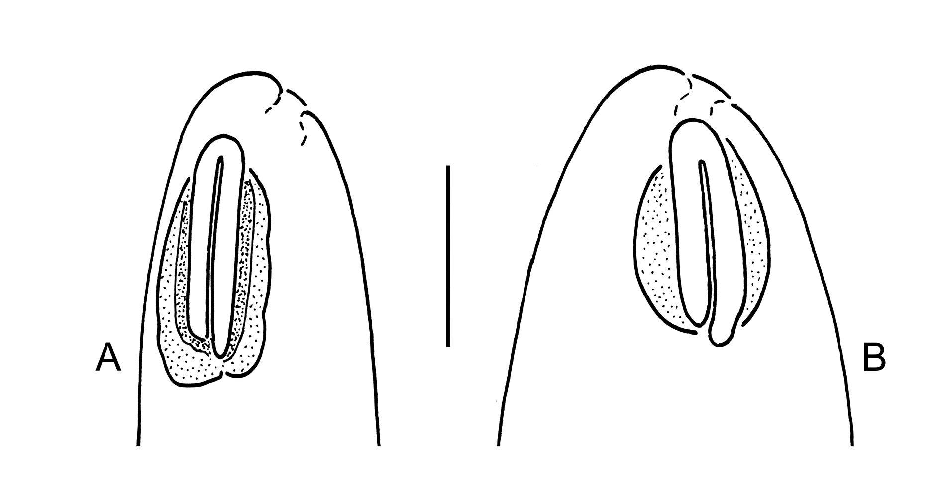
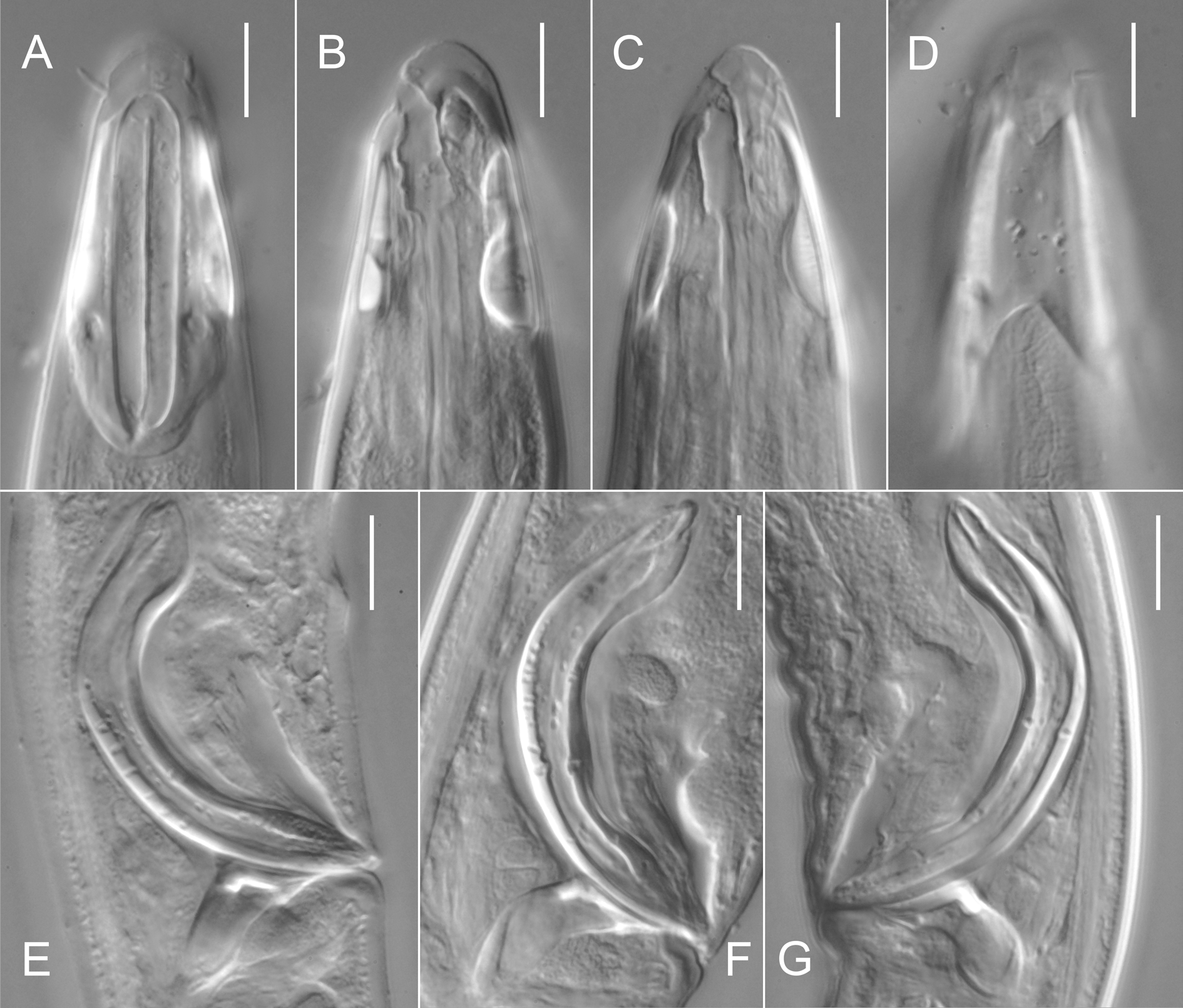
Neodiplopeltula incisa gen. et comb. nov. is characterised by a 1.52–2.24 mm long body; refractive plates underlying cephalic cuticle around amphids present, irregular in shape, not joined, 28–33 µm long and 21–25 µm wide; cephalic setae 8–14 µm long; amphidial fovea 22–28 µm long and 5.5–7 µm wide; oral opening 1–3 µm from anterior end, posterior to cephalic setae bases; secretory-excretory pore located opposite to posterior part of pharynx; tail 2.5–3.9 anal body diameters long; vagina with epiptygmata; spicules 66–79 µm long.
Material examined
SWEDEN: 4 ♀♀, 3 ♂♂, Skagerrak, 58°12′37″ N, 11°18′53″ E, shells and sand at a deep of 15–22 m, 10 Oct. 2012, O. Holovachov leg. (SMNH-169255); 1 ♀, 1 ♂, Skagerrak, 58°20′21″ N, 11°12′42″ E, coarse shell sand at a deep of 14–17 m, 19 Aug. 2014, O. Holovachov leg. (SMNH-169258).
Description
Adult
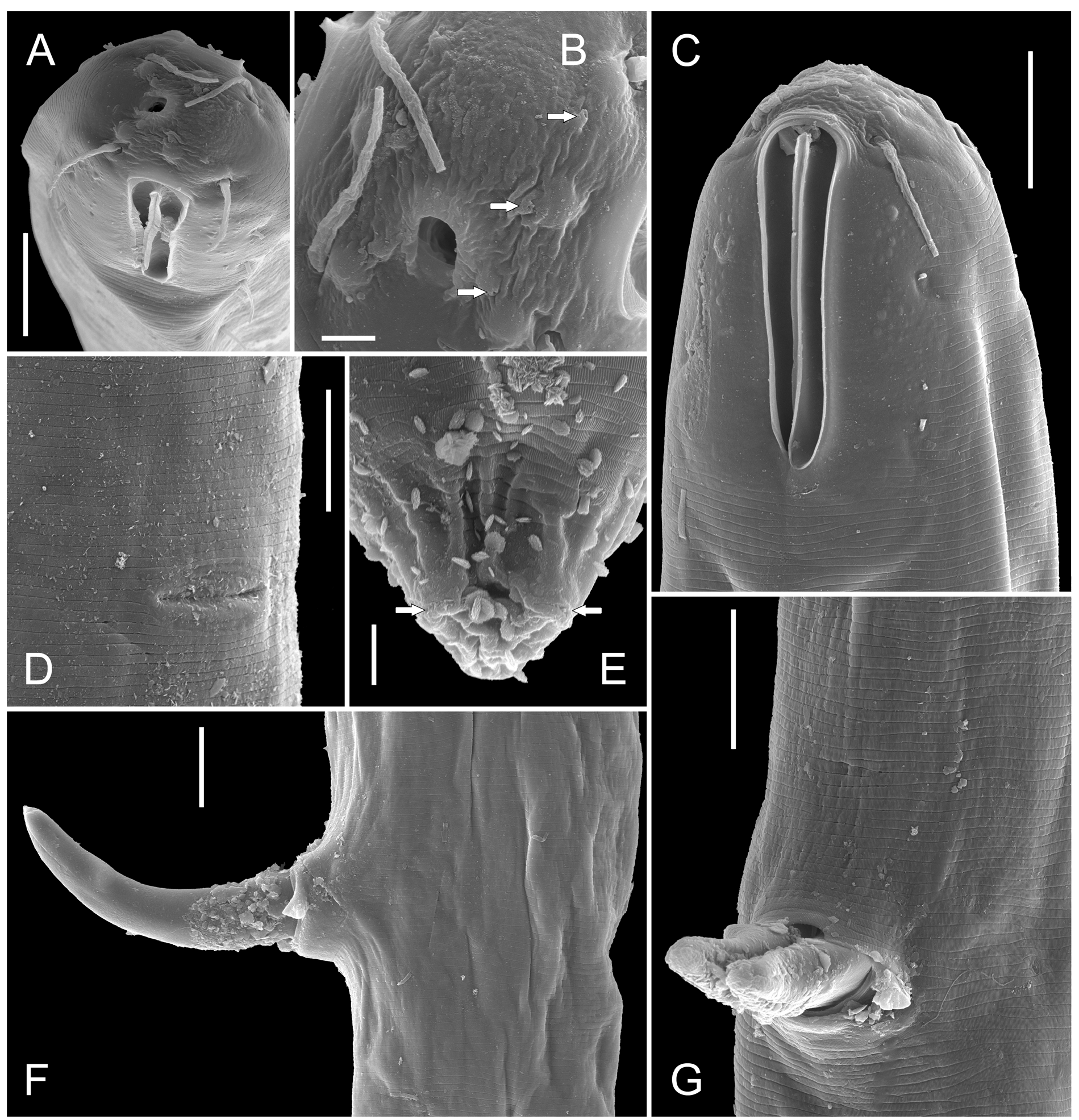
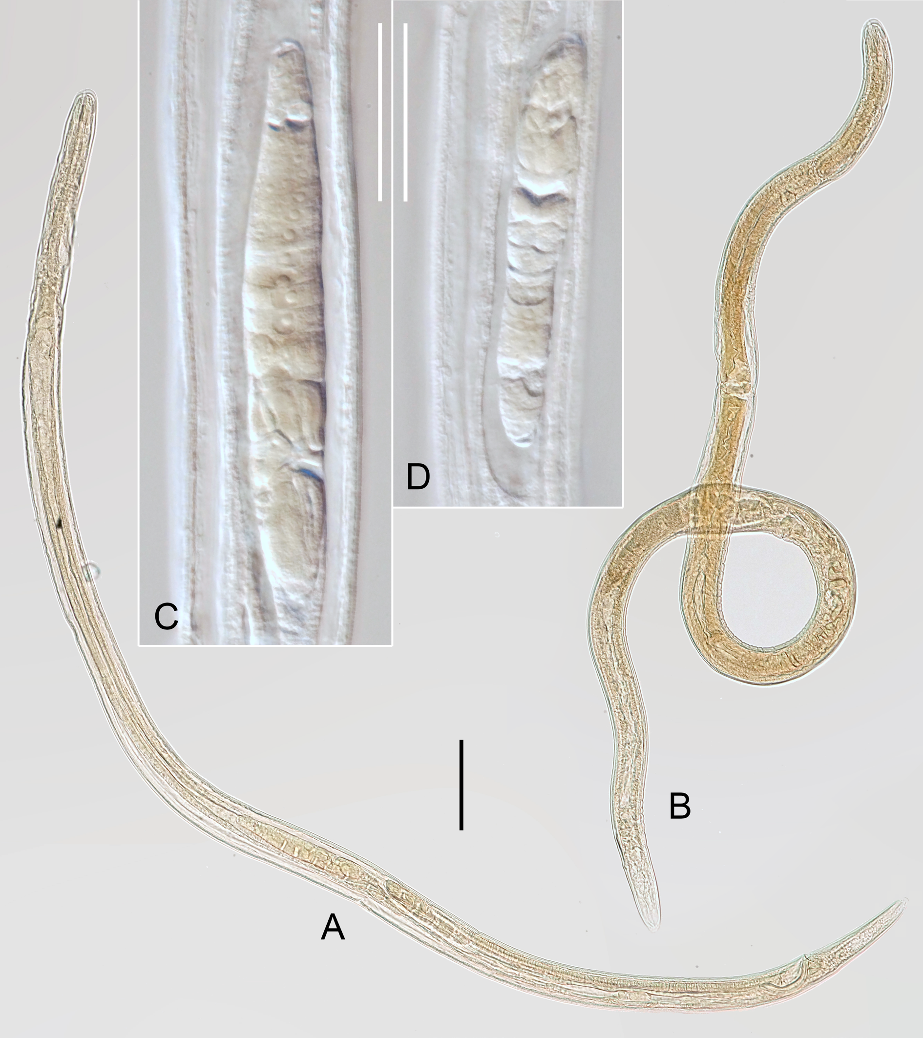
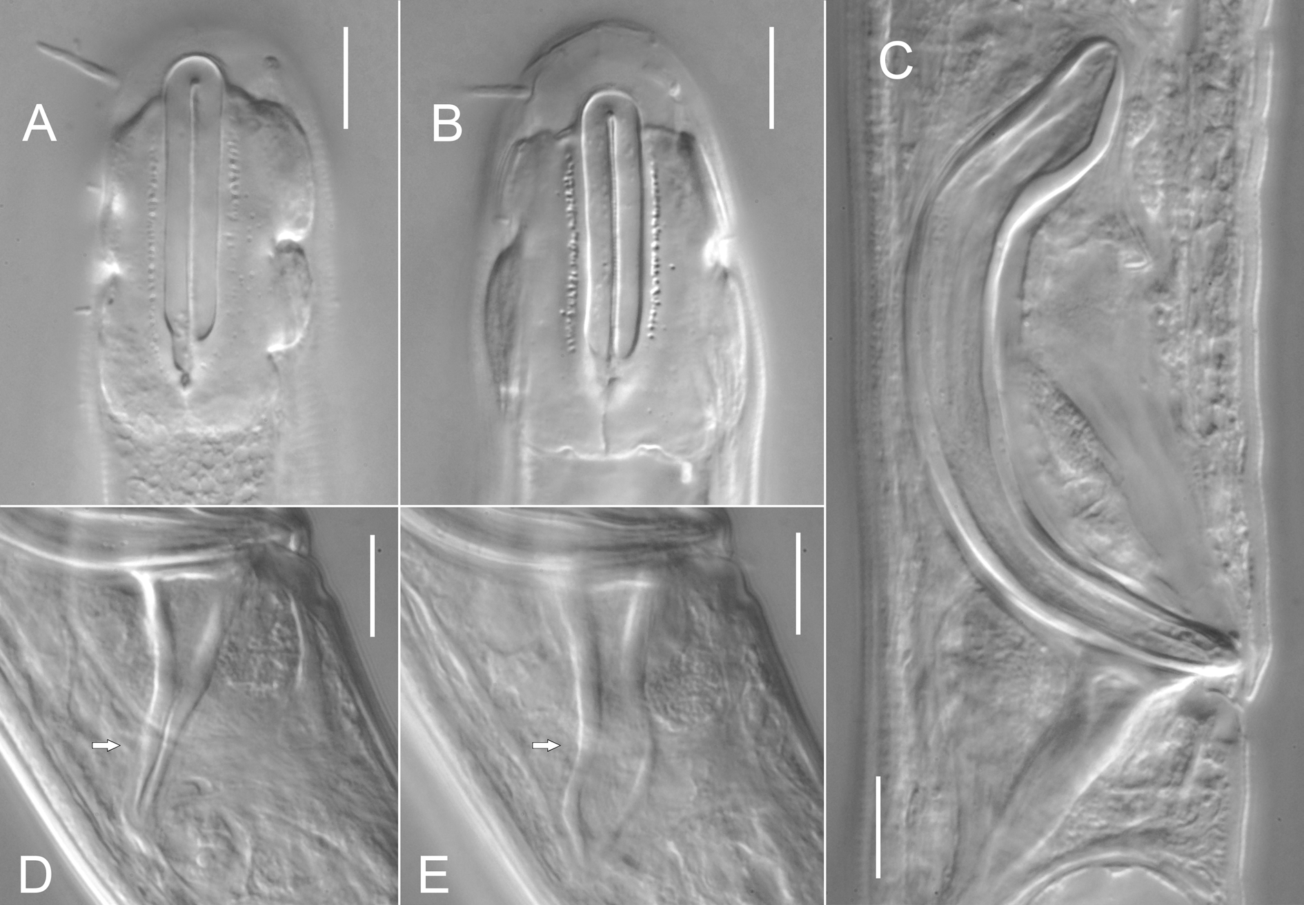
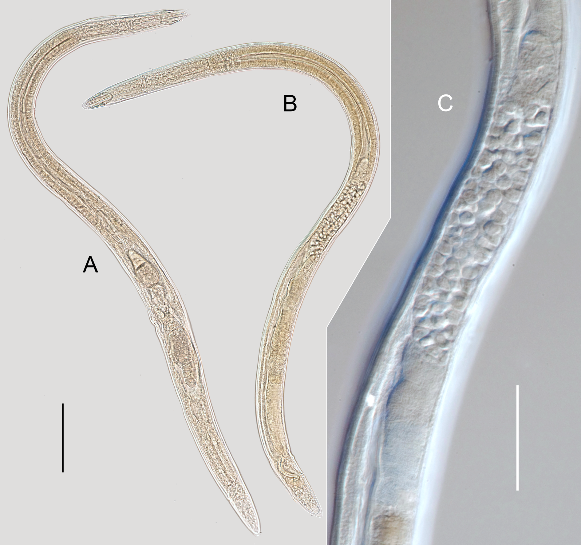
Body cylindrical, posteriorly tapering in tail region, straight or weakly ventrally curved upon fixation. Cuticle finely transversely striated along entire body as seen under SEM (striation can be observed under LM but very fine and shallow and cannot be measured with confidence), except for visually smooth labial region and terminal part of tail; longitudinal striation not observed under the light microscope, but distinct under the scanning electron microscope, covers cuticle over entire body length. Somatic sensilla visible along pharyngeal region (cervical setae, see below) and on tail. Labial region bluntly rounded, lips fused. Refractive plates underlying cephalic cuticle around amphids present (periamphideal), extending from level of anteriormost edge of amphid posteriorly some distance behind posteriormost edge of amphid; refractive plates irregular in shape (with incised edges), not connected with each other on ventral and dorsal sides. Inner labial sensilla small pore-like, located on anterior surface of lips, discernible under SEM only. Outer labial sensilla papilliform, located on anterior surface of lips, clearly visible under SEM only. Cephalic sensilla setiform, bases of dorsosublateral setae located posterior to oral opening. Cervical sensilla papilliform, arranged in four sublateral rows starting at level with middle of amphid and ending at level with posterior edge of refractive plate, two or three per row. Amphids similar in shape and size between sexes: amphidial fovea inverted U-shape with dorsal branch 0.5–3.5 µm longer than ventral branch. Oral opening shifted towards dorsal side of body. Stoma barrel-shaped: cheilostom broad; gymnostom barrel-shaped, with weakly cuticularised walls; stegostom short conoid, its lining uniform with lining of pharynx. Pharyngeal tubes absent. Pharynx subcylindrical, muscular, with evenly distributed myofilaments, gradually expanding towards posterior end; not subdivided in distinct sections; pharyngeal lumen uniform in thickness along entire pharynx length; valves absent. Cardia ovoid, almost entirely embedded into intestinal tissue. Secretory-excretory system present; secretory-excretory pore located along ventral body line opposite to posterior 3/5 th of pharynx; secretory-excretory duct very short, leading from pore to ampulla; renette cell small, its body adjacent and ventral to posterior part of pharynx. Tail cylindro-conical with bluntly rounded terminus. Caudal glands opening via three separate subterminal openings, spinneret absent.
Female
Reproductive system didelphic, amphidelphic, ovary branches outstretched and symmetrical, on opposite sides of intestine. Anterior genital tube 248–280 µm long, situated to either right (n = 1) or left (n = 4) of intestine; posterior genital tube 224–259 µm long, situated to either left (n = 1) or right (n = 4) of intestine. Vulva a transverse slit, located posterior to midbody. Vagina straight, with developed sphincter muscle surrounding proximal part and distinct epiptygmata in distal part; pars refringens vaginae absent. Sack-like spermatheca present, filled with oval spermatozoa in fertilized specimens. Rectum short.
Male
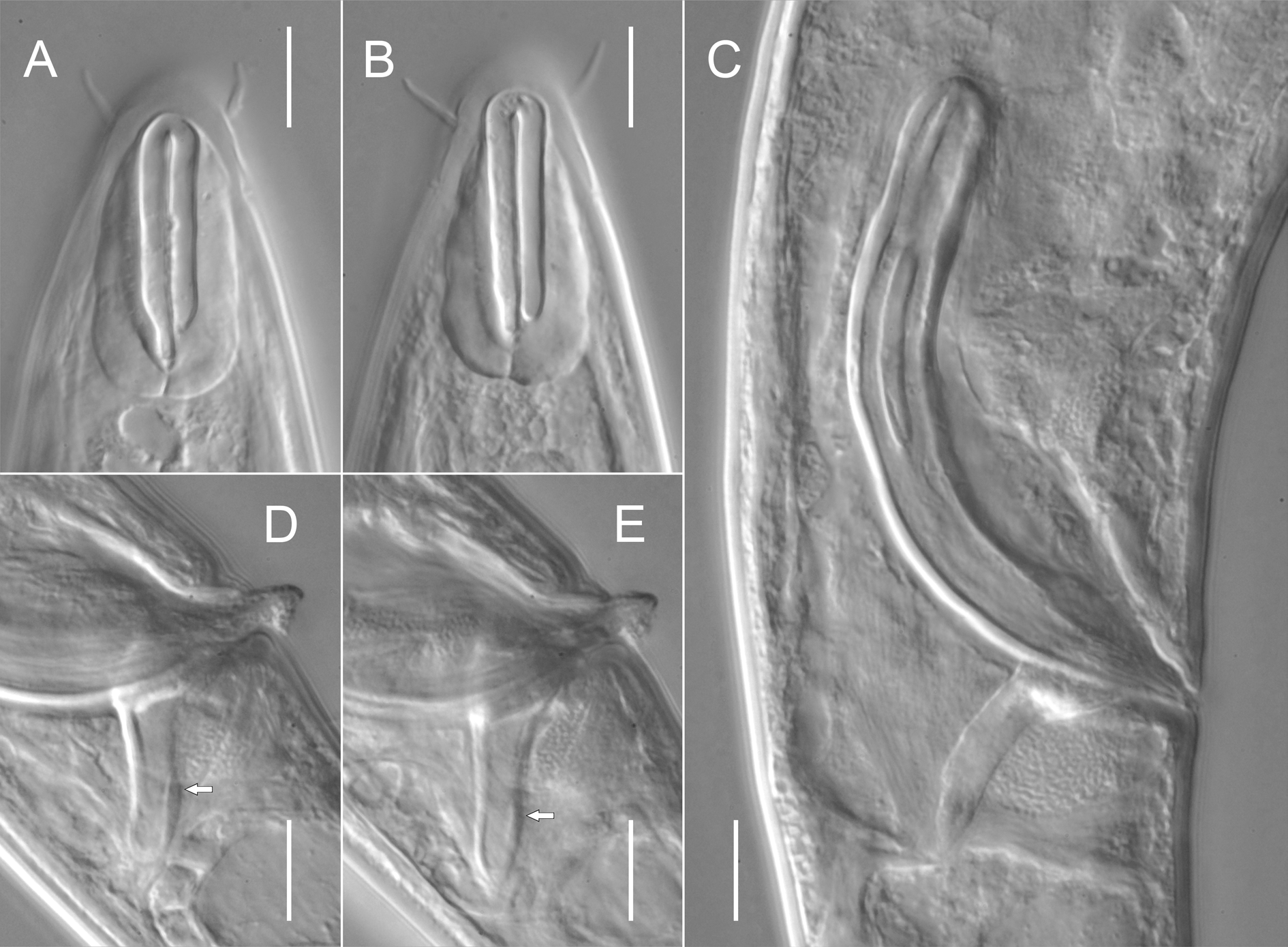
Reproductive system diorchic, testes opposed; anterior testis 162–286 µm long, outstretched and posterior testis 117–162 µm long, reflexed. Spicules paired and symmetrical, strongly curved, with ovoid manubrium and subcylindrical shaft. Gubernaculum plate-like, with pair of strong, closely set caudal apophyses variable in shape ( Fig. 4 View Fig C–E). Caudal setae present, several ventrosublateral and dorsosublateral pairs arranged in rows along entire tail length (often difficult to observe).
Remarks
Despite the fact that the original description by Southern (1914) is rather brief, the present population closely resembles the type specimens in most morphological and morphometric features, including the shape of the refractive plates underlying the amphids (described as “shield shaped” with “front and lateral walls are notched”). Spicules are slightly shorter in the type specimens (66 µm vs 75–79 µm in recent specimens).
Neodiplopeltula barentsi ( Steiner, 1916) gen. et comb. nov. Figs 5–7 View Fig View Fig View Fig , 14–15 View Fig View Fig ; Table 3
Diagnosis (based on combined data)
Neodiplopeltula barentsi gen. et comb. nov. is characterised by a 0.94–1.67 mm long body; refractive plates underlying cephalic cuticle around amphids present, elongated ovoid in shape, not joined, 23.5– 32 µm long and 12–16 µm wide; cephalic setae 3.5–7.5 µm long; amphidial fovea 21–32 µm long and 4–7 µm wide; oral opening 1–2 from anterior end, posterior to cephalic setae bases; secretory-excretory
pore located opposite to posterior part of pharynx; tail 1.6–3.3 anal body diameters long; vagina with epiptygmata; spicules 45–73 µm long.
Material examined
SWEDEN: 1 ♀, 1 ♂, Skagerrak, 58°20′06″ N, 11°09′24″ E, muddy sediment at a deep of 53 m, 9 Aug. 2011, O. Holovachov leg. (SMNH-169264); 1 ♀, 1 ♂, Skagerrak , 58°22′19″ N, 11°04′55″ E, muddy sediment at a deep of 55– 70 m, 9 Aug. 2011, O. Holovachov leg. (SMNH-169266); 3 ♀♀, 1 ♂, Gullmarn Fjord near Östersidan , 58°15′09″ N, 11°27′54″ E, shells, gravel sand and mud, 14 Aug. 2014, O. Holovachov leg. (SMNH-169268); 1 ♀ lectotype of Diplopeltis asymmetricus, Öresund , west of Valgrundet , at a deep of 30– 36 m, Zostera and Echinocardium , 7 Aug. 1926 ( SMNH Type-9015).
GREENLAND: 1 ♀, holotype of Diplopeltis ovalis, Godhavn , mud, 17 Aug. 1926, Reisinger and Steinböck leg. ( NHMD 102171) .
Description
Adult
Body cylindrical, posteriorly tapering in tail region, straight or weakly ventrally curved upon fixation. Cuticle finely transversely striated along entire body, except for visually smooth labial region and terminal part of tail (striation can be observed under LM but very fine and shallow and cannot be measured with confidence); longitudinal striation not observed. Somatic sensilla visible along pharyngeal region (cervical sensilla, see below) and on tail. Labial region bluntly rounded, lips fused. Refractive plates underlying cephalic cuticle around amphids present (periamphideal), extending from level of anteriormost edge of amphid posteriorly some distance behind posteriormost edge of amphid; refractive plates elongated ovoid in shape (plate edges not incised), not connected with each other on ventral and on dorsal sides. Inner labial sensilla indistinct. Outer labial sensilla papilliform, located on anterior surface of lips. Cephalic sensilla setiform, bases of dorsosublateral setae located posterior to oral opening. Cervical sensilla papilliform, arranged in four sublateral rows starting at level with middle of amphid and ending at level with posterior edge of refractive plate, one or two per row. Amphids similar in shape and size between sexes: amphidial fovea inverted U-shape with dorsal branch usually 0.5–2.0 µm longer than ventral branch (in two specimens ventral branch 0.5–1.0 µm longer than dorsal branch). Oral opening shifted towards dorsal side of body. Stoma barrel-shaped: cheilostom broad; gymnostom subcylindrical, with weakly cuticularised walls; stegostom short conoid, its lining uniform with lining of pharynx. Pharyngeal tubes absent. Pharynx subcylindrical, muscular, with evenly distributed myofilaments, gradually expanding towards posterior end; not subdivided in distinct sections; pharyngeal lumen uniform in thickness along entire pharynx length; valves absent. Cardia ovoid, entirely embedded into intestinal tissue. Secretoryexcretory system present; secretory-excretory pore along ventral body line opposite to 3/5 th of pharynx; secretory-excretory duct very short, leading from pore to ampulla; renette cell small, its body adjacent and ventral to posterior part of pharynx. Tail conoid with bluntly rounded terminus. Caudal glands opening via three separate subterminal openings, spinneret absent.
Female
Reproductive system didelphic, amphidelphic, ovary branches outstretched and symmetrical, on opposite sides of intestine. Anterior genital tube 179–269 µm long, situated to either right (n = 3) or left (n = 2) of intestine; posterior genital tube 173–286 µm long, situated to either left (n = 3) or right (n = 2) of intestine. Vulva transverse slit, located posterior to midbody. Vagina straight, with developed sphincter muscle surrounding proximal part and distinct epiptygmata in distal part; pars refringens vaginae absent. Sacklike spermatheca present, filled with oval spermatozoa in fertilized specimens. Rectum short.
Male
Reproductive system diorchic, testes opposed; anterior testis 145–314 µm long, outstretched and posterior testis 117–207 µm long, reflexed. Spicules paired and symmetrical, strongly curved, with weakly defined elongated manubrium and shaft, cylindrical along most of its length. Gubernaculum plate-like, with pair of strong, closely set caudal apophyses variable in shape ( Fig. 7 View Fig C–E). Caudal setae present, several ventrosublateral and dorsosublateral pairs arranged in rows along entire tail length (often difficult to observe).
Remarks
The original description of this species by Steiner (1916) is rather short, with a few measurements given. The present population has a shorter pharynx and tail compared to the type specimen, but in all other respects both match very well, including the position of the oral opening and the shape of the underlying refractive plates.
Neodiplopeltula onusta ( Wieser, 1956) gen. et comb. nov. Figs 8–10 View Fig View Fig View Fig ; Table 4
Diagnosis (based on combined data)
Neodiplopeltula onusta gen. et comb. nov. is characterised by a 0.99–1.48 mm long body; refractive plates underlying cephalic cuticle around amphids present and joined, 32.5–48 µm long and 18.5–24 µm wide; cephalic setae 3–6.5 µm long; amphidial fovea 32–44 µm long and 5–8 µm wide; oral opening 2–5 from anterior end, at level with cephalic setae bases; secretory-excretory pore located opposite to posterior part of pharynx; tail 1.4–2.3 anal body diameters long; vagina with epiptygmata; spicules 52–77 µm long.
Material examined
SWEDEN: 1 ♀, Skagerrak, 58°19′15.6″–20.9″ N, 10°29′33.5″–34.0″ E, soft bottom at a deep of 352–374 m, 10 Sep. 2012, “Inventering Bratten” leg. (SMNH-169284); 1 ♂, Skagerrak, 58°23′00.8″– 22′00.8″ N, 10°20′28.8″–38.3″ E, soft bottom at a deep of 390–428 m, 10 Sep. 2012, “Inventering Bratten” leg. (SMNH-169280); 1 ♂, Skagerrak, 58°28′21.2″–19.2″ N, 10°29′35.6″–43.6″ E, soft bottom at a deep of 248–316 m, 11 Sep. 2012, “Inventering Bratten” leg. (SMNH-169281); 3 ♀♀, 1 ♂, Skagerrak, 58°27′36.7″–43.3″ N, 10°32′52.0″–59.4″ E, soft bottom at a deep of 232–240 m, 12 Sep. 2012, “Inventering Bratten” leg. (SMNH-169282 – SMNH-169283).
Description
Adult
Body cylindrical, posteriorly tapering in tail region, straight or weakly ventrally curved upon fixation. Cuticle finely transversely striated along entire body, except for visually smooth labial region and terminal part of tail (striation can be observed under LM but very fine and shallow and cannot be measured with confidence); longitudinal striation not observed.Somatic sensilla visible along pharyngeal region (cervical setae, see below) and on tail. Labial region bluntly rounded, lips fused. Refractive plates underlying cephalic cuticle around amphids present (periamphideal), extending from level of anteriormost edge of amphid posteriorly some distance behind posteriormost edge of amphid; refractive plates connected with each other on ventral and on dorsal sides. Inner labial sensilla indistinct. Outer labial sensilla papilliform, located on anterior surface of lips. Cephalic sensilla setiform, bases of dorsosublateral setae located at level with oral opening. Cervical sensilla papilliform, arranged in four sublateral rows at level with amphid, one or two per row. Amphids similar in shape and size between sexes: amphidial fovea inverted U-shape with dorsal branch 1.0–2.0 µm longer than ventral branch. Oral opening shifted towards dorsal side of body. Stoma subcylindrical: cheilostom broad; gymnostom barrel-shaped, with weakly cuticularised walls; stegostom short conoid, its lining uniform with lining of pharynx. Pharyngeal tubes absent. Pharynx subcylindrical, muscular, with evenly distributed myofilaments, gradually expanding towards posterior end; not subdivided in distinct sections; pharyngeal lumen uniform in thickness along entire pharynx length; valves absent. Secretory-excretory system present; secretory-excretory pore located along ventral body line opposite to 3/5 th of pharynx; secretory-excretory duct very short, leading from pore to ampulla; renette cell small, its body adjacent and ventral to posterior part of pharynx. Tail conoid with bluntly rounded terminus. Caudal glands opening via three separate subterminal openings, spinneret absent.
Female
Reproductive system didelphic, amphidelphic, ovary branches outstretched and symmetrical, on opposite sides of intestine. Anterior genital tube 109–180 µm long, situated to either right (n = 3) or left (n = 1) of intestine; posterior genital tube 109–145 µm long, situated to either left (n = 3) or right (n = 1) of intestine. Vulva transverse slit, located posterior to midbody. Vagina straight, with developed sphincter muscle surrounding proximal part and distinct epiptygmata in distal part; pars refringens vaginae absent. Sack-like spermatheca present, filled with oval spermatozoa in fertilized specimens. Rectum short.
Male
Reproductive system diorchic, testes opposed; anterior testis outstretched and posterior testis reflexed (poorly discernible in most specimens due to highly granular and dark content of overlapping intestine and cannot be measured). Spicules paired and symmetrical, strongly curved, with elongated ovoid manubrium and subcylindrical shaft. Gubernaculum plate-like, with pair of strong closely set caudal apophyses variable in shape ( Fig. 10 View Fig E–G). Caudal setae present, several ventrosublateral and dorsosublateral pairs arranged in rows along entire tail length.
Remarks
The single female used by Wieser (1956) to describe this species has a slightly broader labial region and longer cephalic setae than the population from Sweden, but the specimens described under the name Diplopeltula tchesunovi fill this gap. The position of the oral opening is at level with the cephalic setae bases and the shape of the refractive plates underlying the cephalic cuticle around the amphids (joined along the dorsal and ventral body sides) in both type female and recent specimens are identical and confirm the conspecificity.

| SMNH |
Department of Paleozoology, Swedish Museum of Natural History |
No known copyright restrictions apply. See Agosti, D., Egloff, W., 2009. Taxonomic information exchange and copyright: the Plazi approach. BMC Research Notes 2009, 2:53 for further explanation.