
Satsumaocnus kaiyomarui, Yamana & Thandar & Hayashibara & Setiamarga, 2022
|
publication ID |
https://doi.org/ 10.11646/zootaxa.5209.2.7 |
|
publication LSID |
lsid:zoobank.org:pub:A4C30358-A40E-46EB-8FC6-C0119AFD20D7 |
|
DOI |
https://doi.org/10.5281/zenodo.7331178 |
|
persistent identifier |
https://treatment.plazi.org/id/D34C1889-8E71-4965-90B4-5B724DF38C53 |
|
taxon LSID |
lsid:zoobank.org:act:D34C1889-8E71-4965-90B4-5B724DF38C53 |
|
treatment provided by |
Plazi |
|
scientific name |
Satsumaocnus kaiyomarui |
| status |
sp. nov. |
Satsumaocnus kaiyomarui sp. nov. Yamana, Thandar, & Setiamarga
[New Japanese name: Satsuma-kinko]
( Figs 2 View FIGURE 2 , 3 View FIGURE 3 , 4 View FIGURE 4 , and 5; Table 1 View TABLE 1 and 2 View TABLE 2-2 )
Etymology. The specific name kaiyomarui is the genitive case of the R/V Kaiyo Maru of Fisheries Agency of Japan.
Diagnosis. As for the genus.
Material examined. Holotype, WMNH-2018-INV-3 (contracted length 62 mm, width 17 mm). Paratypes, 10 individuals: WMNH-2018-INV-4—only contracted measurements (length x breadth) recorded (69 mm x 24 mm); WMNH-2018-INV-5 (58 mm x 20 mm); WMNH-2018-INV-6 (42 mm x 12 mm); WMNH-2018-INV-10 (28 mm x 9 mm); WMNH-2018-INV-11 (23 mm x 8 mm); WMNH-2018-INV-12 (14 mm x 16 mm); WMNH-2018-INV-13 (23 mm x 6 mm). Three observed paratypes were also used for molecular analysis reported previously ( Yamana et al., 2019): WMNH-2018-INV-7 (27 mm x 11 mm; Genbank accession numbers LC 425500 for COI, LC 425503 for H3); WMNH-2018-INV-8 (21 mm x 9 mm; Genbank accession numbers LC 425501 for COI, LC 425504 for H3); WMNH-2018-INV-9 (19 mm x 7 mm; Genbank accession numbers LC 425502 for COI, LC 425505 for H3)
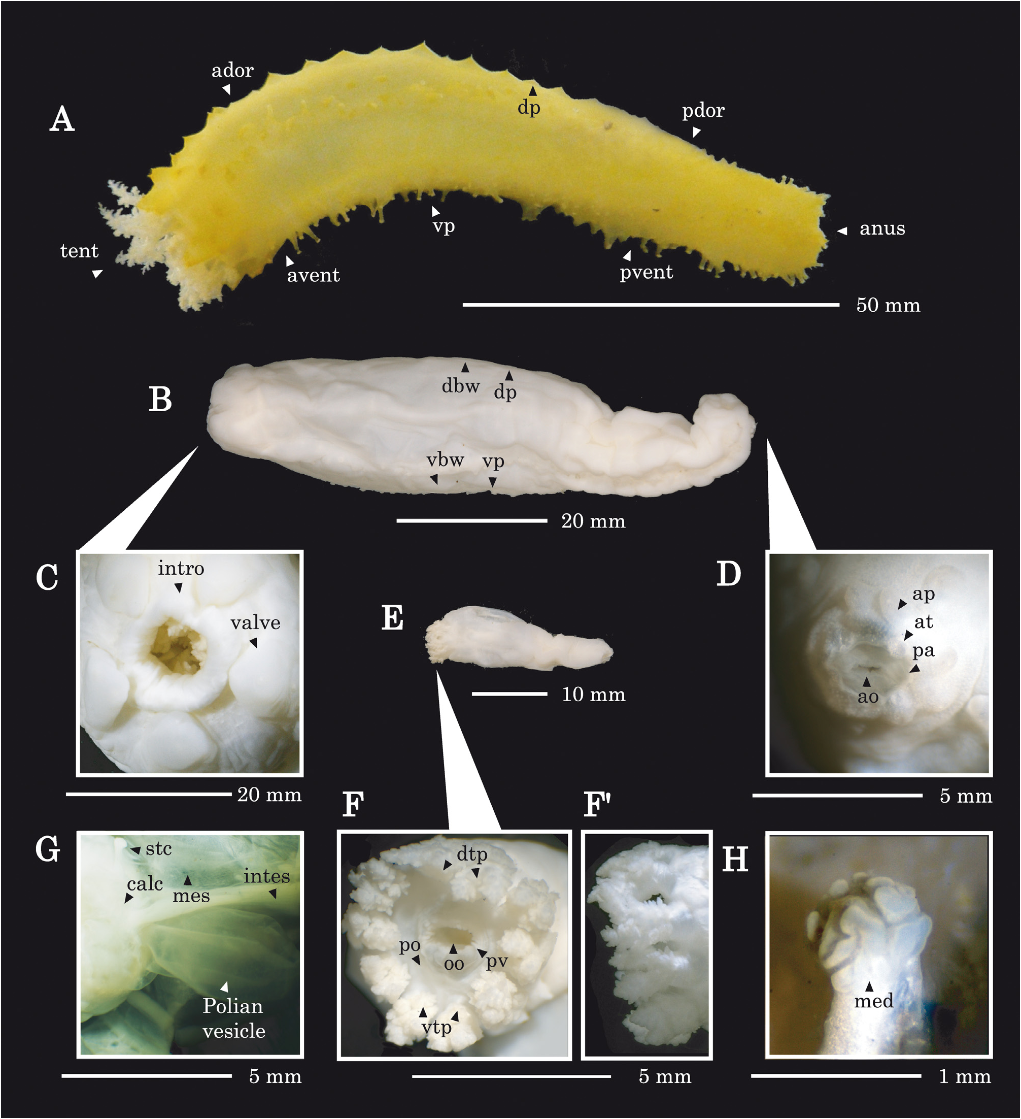
Holotype description. Body form straight, posteriorly tapering. Live coloration yellow. dorsal podia short, papillae-like, restricted to radii in two longitudinal rows; ventral podia (pedicels), retractile, restricted to radii in three apparently zig-zag rows, absent anteriorly. In preserved state, anterior end expanded and posterior conspicuously contracted. Body wall soft, thin in anterior (<0.5 mm), thick in posterior (>1.0 mm); preserved coloration pure white ( Fig. 2A, B View FIGURE 2 ); contracted length 62 mm, width 17 mm (see Table 1 View TABLE 1 ). Tentacles 10, dendritic, equal length, all tentacles arranged in a single circle ( Fig. 2F View FIGURE 2 ), each with two tufts, subdivided basally ( Fig. 2 View FIGURE 2 F’). Body form cylindrical, with five oral valves ( Fig. 2C View FIGURE 2 ) and five radially arranged anal teeth, each accompanied by a pair of anal papillae lying in a circle outside of the anal teeth ( Fig.2D View FIGURE 2 ). Tentacles and introvert are of same coloration as the adjoining body wall.
Calcareous ring low, stout, radial and interradial elements compact, not forming a mosaic and lacking posterior prolongations ( Fig. 3 View FIGURE 3 ). All plates sagittate, each with a posterior depression and anterior projection. The mid-ventral element R 1 and adjoining two interradial elements IR1 and IR2 not fused, both IR1 and IR2 narrower than RI, both with sharper anterior projection ( Fig. 3 View FIGURE 3 ) but mid-dorsal interradial plate (IR5) with deepest posterior depression. Retractor muscles originate from respective longitudinal muscle at 20–30% of body length from anterior end, more anterior dorsally, and insert on anterior projection ( Fig. 3 View FIGURE 3 ) of corresponding radial plate.
Polian vesicle single, mid-ventral in position, in line with RI, fusiform in shape, narrowing distally (distal end 1/6–1/3 of vesicle length); stone canal also single, mid-dorsal in position, in line with IR5; madreporite brain-/coralshaped, on the left side of anterior portion of the dorsal mesentery ( Table 1 View TABLE 1 , Fig. 2G, H View FIGURE 2 ). Gonadal stolon attached anteriorly, gonad in two tufts, one on each side of dorsal mesentery, most tubules short, branched. Respiratory trees paired, larger tree situated on right side of body cavity, reaching about half body length. Smaller tree situated on left side, length approximately 1/5–1/4 of the right one. Small intestine extends from mid-dorsal region in line with IR5, turns towards the right dorsal interradius in line with IR4, passes along IR4 to about 3/4 the body length and then extends anteriad to middle of body along interradius (IR5) before opening into the cloaca.
Body wall ossicles as an external layer of small (22–35 µm) x-framed hourglass ossicles ( Fig. 4F, G View FIGURE 4 , Table 2 View TABLE 2-2 ) and an inner layer of sparsely distributed, large (123–154 µm) dendriform rods ( Fig. 4 F, G View FIGURE 4 , Table 2 View TABLE 2-2 ), sometimes forming delicate plates. Buttons, thick plates, x-shaped ossicles and/or rosettes absent. Tentacle ossicles mostly large, thick rods (55–643 µm), distally branched or perforated by few small holes ( Fig. 4A View FIGURE 4 , Table 2 View TABLE 2-2 ). Peri-oral ossicles, sparse, occurring as small (39–49 µm) typical, simple cups and large (169–369 µm) spinous rods ( Fig. 4B View FIGURE 4 , Table 2 View TABLE 2-2 ). Ossicles of pharyngeal villi as large (78–251 µm) rods, with few distal perforations and/or branches, sometimes also with a medial third branch/arm ( Fig. 4C View FIGURE 4 , Table 2 View TABLE 2-2 ). Introvert ossicles also as small (26–31 µm), x-framed hourglasses ( Fig. 4D View FIGURE 4 , Table 2 View TABLE 2-2 ). Oral valve ossicles as mostly small (24–34 µm) hourglasses and medium to large (101–358 µm), often distorted or rod-derived supporting plates at tip of valve ( Fig. 4E View FIGURE 4 , Table 2 View TABLE 2-2 ). Ossicles of dorsal podia mostly small (24–34 µm) hourglasses and small to large (85–175 µm), irregular supporting plates at tip ( Fig. 4H View FIGURE 4 , Table 2 View TABLE 2-2 ). Ossicles of ventral podia, mostly small (24–39 µm) hourglasses, medium-sized (57–149 µm) terminal supporting plates and large (451–514 µm) endplate ( Fig. 4I View FIGURE 4 , Table 2 View TABLE 2-2 ). Peri-anal ossicles as small (16–32 µm) hourglasses. Anal papillae with medium-sized (85–175 µm), irregular supporting plates ( Fig. 4J View FIGURE 4 , Table 2 View TABLE 2-2 ). Ossicles were also detected in the basal part of ovary as medium-sized (78–163 µm), distally branched spinous or dendriform rods ( Fig. 4K View FIGURE 4 , Table 2 View TABLE 2-2 ).
Variations in paratypes. Body form in life straight or slightly U-shaped, straight in preserved state. Contracted dimensions of the largest paratype (WMNH-INV-2018-4) 69 mm x 24 mm ( Table 1 View TABLE 1 ). All paratypes with 10 equal tentacles in a single circle ( Fig. 2 F View FIGURE 2 ), each tentacle basally divided into two large tufts. Except for five small specimens (WMNH-INV- 2018-9–13), the posterior end of all specimens is strongly contracted. Polian vesicle always single, in RI, stone canal also single in IR5. calcareous ring low, stout, without posterior prolongations; oral valves present. All specimens possess abundant x-framed hourglasses in body-wall, but simple cups and scarce spinous rods in perioral skin lacking in some individuals ( Table 2 View TABLE 2-2 ). Hourglasses absent in the introvert of several specimens ( Table 2 View TABLE 2-2 ). In all specimens dendriform rods sparser in ventral body wall. Gonadal tubules ( Table 1 View TABLE 1 ) absent in some specimens (perhaps due to immaturity). Other ossicles of the paratypes consistent with those of the holotype.
Distribution. Known only from type locality.
Ecology/behaviour. All individuals were observed basally attached with their posterior end, or this end was embedded in the sediment (pebbles and coral debris) ( Fig. 6A, B View FIGURE 6 ). Most specimens were observed with their tentacles extended into the water column while feeding. ( Fig. 6C View FIGURE 6 ). Sometimes animals were half-embedded in the sandy gravel bottom. Other movements were not observed ( Fig. 6D View FIGURE 6 ).
Cup: a Length, a long side of lectangle rim; b Breadth, a short side of lectangle rim; c High, a distance between upper edge of upside rim and belower edge of downside rim. In all three directions, spinneret and/or process were omitted from the measurement. Among the three directions, one vertical direction was omitted from the measurement.


| COI |
University of Coimbra Botany Department |
| R |
Departamento de Geologia, Universidad de Chile |
No known copyright restrictions apply. See Agosti, D., Egloff, W., 2009. Taxonomic information exchange and copyright: the Plazi approach. BMC Research Notes 2009, 2:53 for further explanation.