
Rudogorites simonei, Polak & Mulaomerović, 2021
|
publication ID |
https://doi.org/10.11646/zootaxa.5061.3.8 |
|
publication LSID |
lsid:zoobank.org:pub:25C44022-4BE9-453B-9D96-359A00A18EC1 |
|
DOI |
https://doi.org/10.5281/zenodo.5700153 |
|
persistent identifier |
https://treatment.plazi.org/id/03AC87B6-3A0D-FFE5-FF52-BF48FC484EF4 |
|
treatment provided by |
Plazi |
|
scientific name |
Rudogorites simonei |
| status |
sp. nov. |
Rudogorites simonei View in CoL sp. nov.
Figs. 1 View FIGURE 1 , 2, 3, 6, 7, 9 View FIGURES 2–10 , 11, 12, 15, 16 View FIGURES 11–16 , 17, 18, 21, 24 View FIGURES 17–24 , 25–30 View FIGURES 25–30 , 31 View FIGURE 31 .
Type locality: Lijina Pećina ( Lijina cave ) ( Katastar speleoloških objekata Bosne i Hercegovine, Ev. Br. 5391) which is located North East of the village of Gunjani and 4.4 km South East of town Kreševo in Kreševo municipality. The cave is approximately 20 km West of Sarajevo in Central Bosnia and Herzegovina at a proximal altitude 700 m a.s.l. (Coordinates: 43.831N / 18.075 E) GoogleMaps .
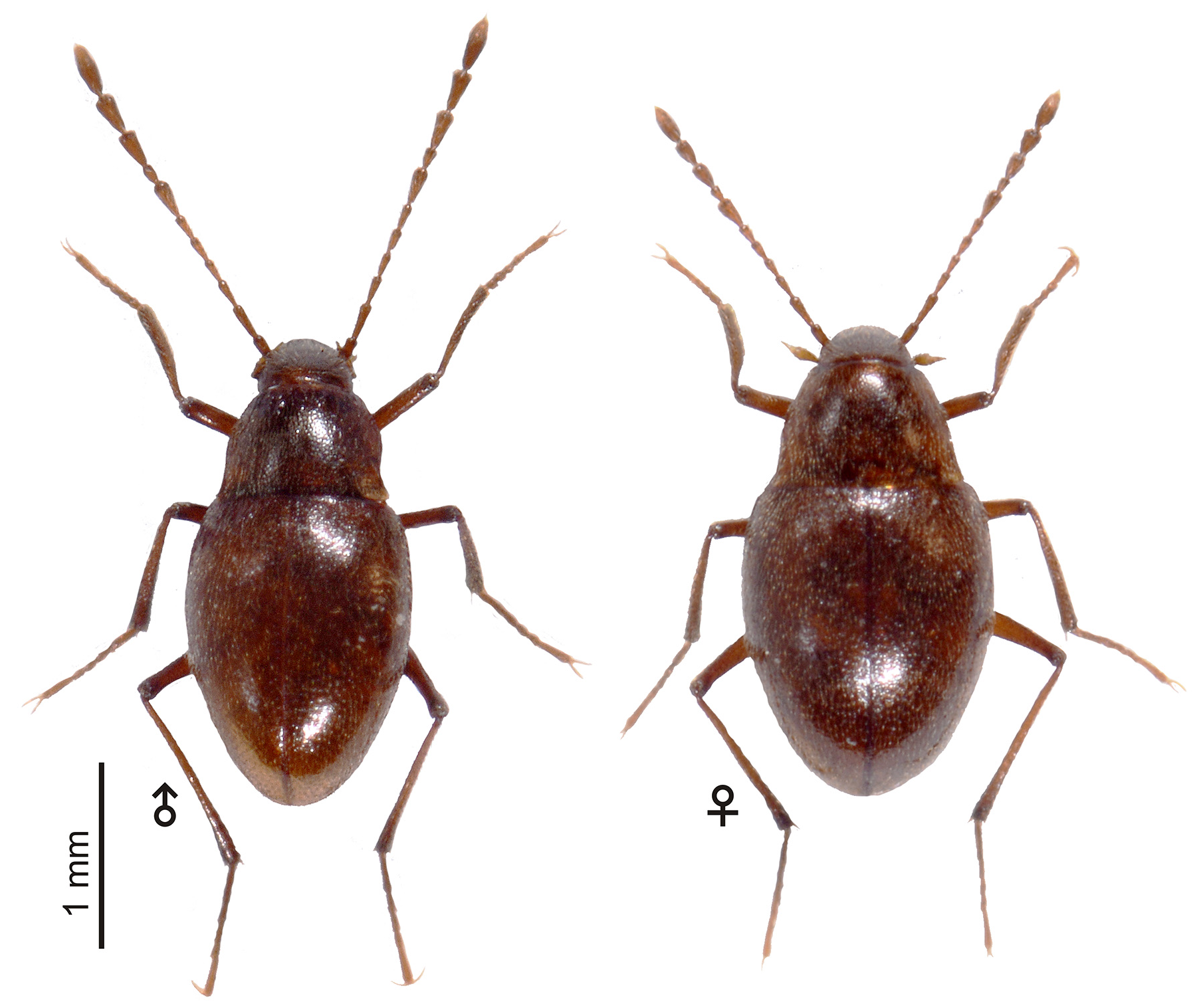
Description. Size: Total body length (BL) 2.37–2.52 mm (M= 2.43, N= 4) in ♂♂ and 2.34–3.53 mm (M= 2.44, N= 4) in ♀♀. Color: Reddish brown, moderately shiny, antenna and legs slightly paler ( Fig. 1 View FIGURE 1 ).
Head: Eyes atrophied, head dorsally cowered by sparse erected setae, occipital carina atrophied. Maxillary apical palpomere (4 th) prolonged conical with slightly concave lateral edges, long of 2/3 of preapical palpomere length, preapical palpomere sub-conical, swollen with convex lateral edges. Mandible regularly curved short and armed with variable number of 1 to 3 short teeth. Antenna inserted on the middle third of head length.
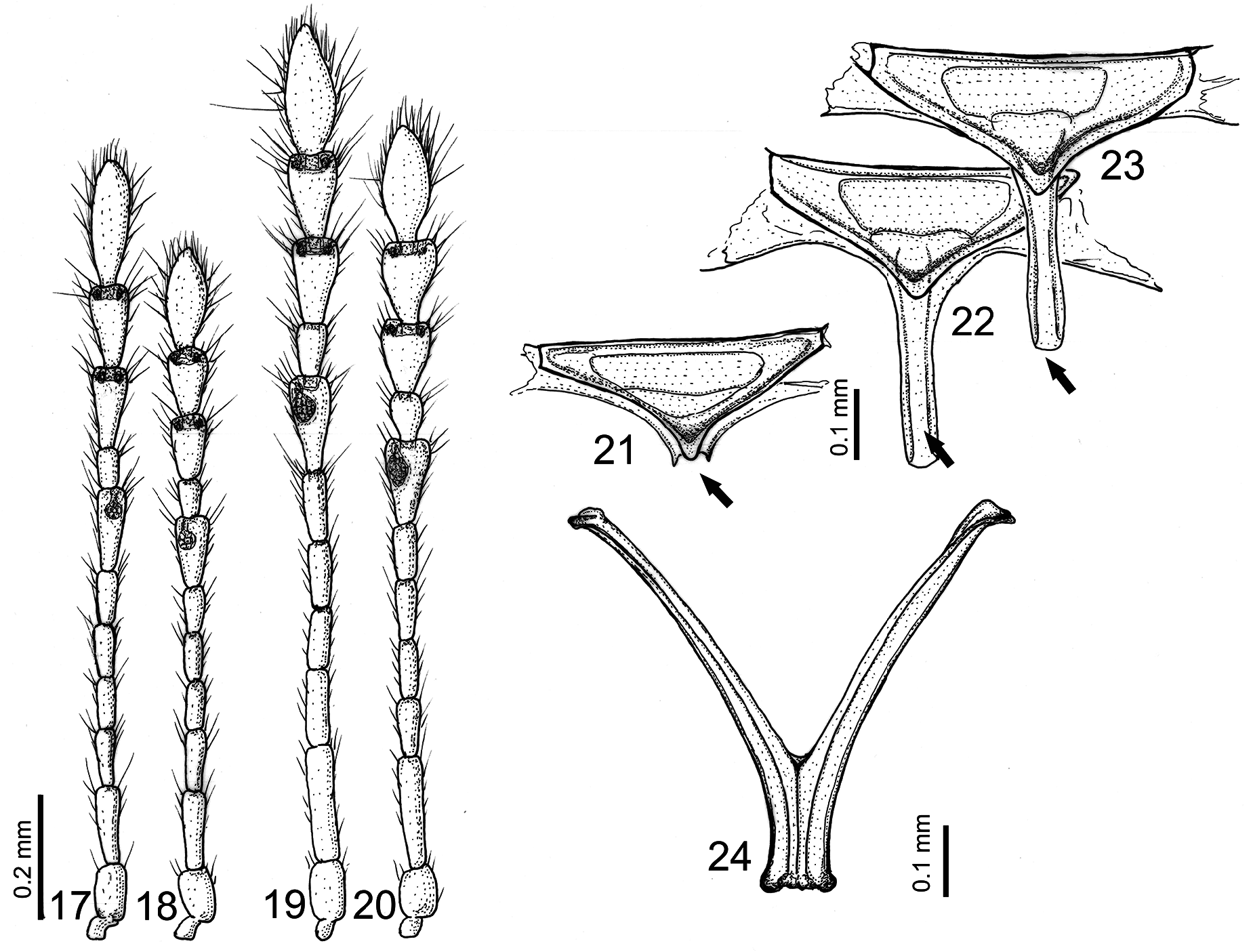
Antenna: ( Figs. 1 View FIGURE 1 , 17, 18 View FIGURES 17–24 ) Antenna total length (AL): 1.75–1.99 mm (M= 1.86, N= 4) in ♂♂ and 1.30–1.49 mm (M= 1.40, N= 4) in ♀♀. AL/BL: 0.74–0.79 in ♂♂ and 0.56–0.59 in ♀♀, AL/ PL: 2.62–2.88 in ♂♂ and 2.24– 2.20 in ♀♀. First antennomere distinctly shorter but not twice as short as second antennomere. Second antennomere distinctly longer than third, which is slightly longer than following 4 th to 6 th antennomeres which are sub-equally long, 7 th, 9 th and 10 th antennomeres prolonged and sub-equally long, 8 th antennomere the shortest, 11 th antennomere the longest, elliptical. All antennomeres longer than wide .
AmL ♂♂: 0.13–0.14; 0.19–0.20; 0.14–0.15; 0.11–0.12; 0.12–0.13; 0.12–0.13; 0.19–0.20; 0.10–0.11; 0.19–0.20; 0.20–0.020; 0.27–0.227 (N= 3).
AmL ♀♀: 0.12–0.13; 0.18–0.19; 0.12–0.13; 0.10–0.11; 0.11–0.12; 0.11–0.12; 0.16–0.17; 0.09–0.10; 0.14–0.15; 0.14–0.15; 0.22–0.23 (N= 3).
AL/AmL (in %) ♂♂: 7.26; 10.61; 7.82; 6.70; 7.26; 6.70; 11.17; 5.59; 10.61; 11.17; 15.08.
AL/AmL (in %) ♀♀: 7.69; 11.54; 8.33; 7.05; 7.69; 7.05; 10.90; 5.77; 9.62; 9.62; 14.74.
AmL/AmW ♂♂: 1.63; 3.17; 2.80; 2.40; 2.60; 2.40; 2.86; 1.67; 2.71; 2.86; 3.38.
AmL/AmW ♀♀: 1.50; 3.00; 2.60; 2.20; 2.40; 2.20; 2.13; 1.80; 1.88; 1.67; 2.56.
Vesicular gland organ (Hamman’s organ) in 7 th antennomere voluminous, spherical with short but distinct vestibulum (duct) opened to atrium. Sensory glands on distal edge of 9 th and 10 th antennomere present as hemi-spherical anteriorly opened vesicles.
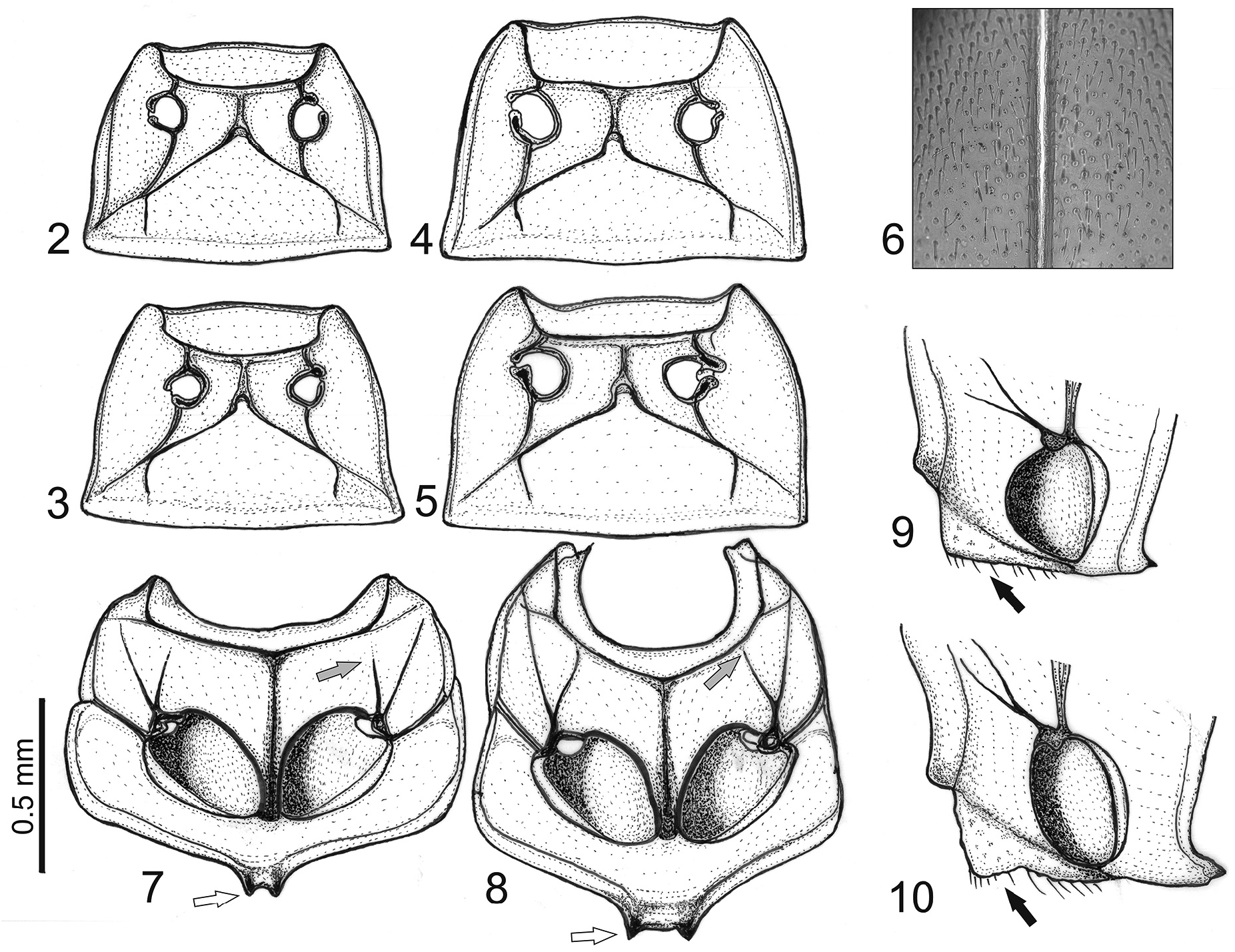
Pronotum: Transverse, wider than long, distinctly narrower than elytra width, making the habitus of sub-pholeuonoid aspect ( Fig.1 View FIGURE 1 ). Lateral edge in dorsal observation ( Figs. 2, 3 View FIGURES 2–10 ) slightly sinuate concave, bell-like form, maximum width on posterior edge. Dorsal face strongly pubescent, with uniform distribution of short semierect setae.
PL: 0.60–0.69 mm (M= 0.63, N= 5) in ♂♂ and 0.58–0.68 mm (M= 0.64, N= 6) in ♀♀; PaW: 0.49–0.54 mm (M= 0.51, N= 2) in ♂♂ and 0.53–0.55 mm (M= 0.54, N= 3) in ♀♀; PcW: 0.77–0.83 mm (M= 0.81, N= 5) in ♂♂ and 0.79–0.89 mm (M= 0.83, N= 6) in ♀♀; PpW: 0.87–0.89 mm (M= 0.88, N= 5) in ♂♂ and 0.93–0.97 mm (M= 0.96 N = 6) in ♀♀; PL /PpW on average M= 0.72 in ♂♂ and M= 0.67 in ♀♀.
Thorax: Mesocoxal cavities separated by a keel, not confluent. Mesosternal keel (carena mesosternale) well developed, elevated and flattened, lanceolate, posteriorly reduced and thickened, extended to the posterior edge of coxal cavities ( Figs. 7, 9 View FIGURES 2–10 ). In lateral view keel is elevated with ventral edge straight, not dentate and entire from thorax colum to the posterior edge of mesocoxal cavities ( Fig. 9 View FIGURES 2–10 ). Mesosternal intercoxal apophysis bifid, short and narrow, equally long as wide ( Fig. 7 View FIGURES 2–10 ). Suture between mesosetrnum and mesepisternum is partly atrophied on its anterior side, not extending to the colum ( Fig. 7 View FIGURES 2–10 ). Metendosternite (furca or Crowson’s organ) “V” shaped with dorsal arms twice as long as stalk ( Fig. 24 View FIGURES 17–24 ). Metatergal apparatus with medial longitudinal groove (posterior process) strongly reduced, rudimentary and oversized by scutellum posterior beak ( Fig. 21 View FIGURES 17–24 ). Scutellum triangular, wide than long, mostly glabrous.
Elytra: Elytra elongated-oval with maximum width on medial part of elytra ( Fig. 1 View FIGURE 1 ). Sparsely pubescent with uniform short and semi erected, not recumbent setae ( Figs.1 View FIGURE 1 , 6 View FIGURES 2–10 ). Elytral punctation strong with large, deep and widely spaced foveolated punctures, not aligned in transversal strigae. Each seta growing from the bottom of a depressed puncture ( Fig. 6 View FIGURES 2–10 ). Sutural carina absent. Elytra distinctly wider in ♀♀ than in ♂♂. EL: 1.49.– 1.69 mm (M= 1.58, N= 3) in ♂♂ and 1.50–1.74 mm (M= 1.61, N= 3) in ♀♀; EW: 1.14–1.23 mm (M= 1.18, N= 3) in ♂♂ and 1.16–1.25 mm (M= 1.20, N= 3) in ♀♀; ratio EL/EW: 1.40 in ♂♂ and 1.35 in ♀♀.
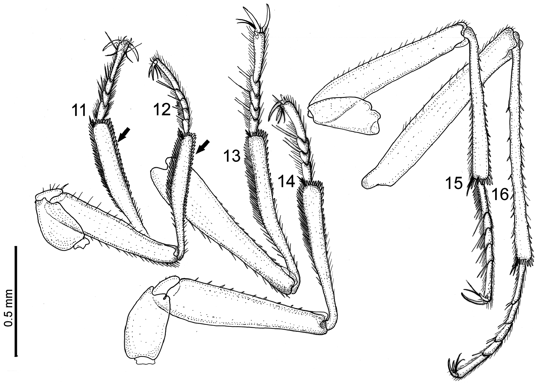
Legs: Moderately long, not retractile, similar to other species of the Apholeuonus phyletic group ( Figs. 1 View FIGURE 1 , 11–16 View FIGURES 11–16 ). Claws long, regularly curved, slender and sharp in all legs. Tarsal empodium between the claws with one long bifid empodial seta (distinctly split in two at the base) in all legs ( Figs. 11, 12, 15, 16 View FIGURES 11–16 ). Male protarsi five-segmented ( Fig. 12 View FIGURES 11–16 ) and female protarsi four-segmented ( Fig. 11 View FIGURES 11–16 ); PtarL: 0.41–0.42 mm in ♂♂ and 0.36–0.39 mm in ♀♀; PtarW: 0.04–0.05 mm in ♂♂ and 0.04 mm in ♀♀, not distinctly dilated in both sexes. Male protarsomeres equipped with dense, terminally hook-like adhesive (tenant) setae ( Fig. 12 View FIGURES 11–16 ).
Protibial internal and ventral apical spur short and tridentate or polydentate. Protibia armed with ring of apical flattened spines that continues to the externo-lateral row of strong spines forming a comb. These comb spines are flattened, short and wide, almost triangular and stretching in straight continuous line on entire two thirds of protibia length. Protibia characteristically curved and the widest at the middle ( Figs. 11, 12 View FIGURES 11–16 ). PtibL: 0.62–0.63 mm in ♂♂, 0.59–0.61 in ♀♀; PtibW: 0.076 –0.079 mm in ♂♂, 0.077 –0.083 mm in ♀♀, widest at the middle of tibia length, 0.066 –0.069 mm in ♂♂, 0.070 –0.074 mm in ♀♀ wide at the apex.
Mesotarsi and metatarsi five-segmented in both sexes ( Figs. 15, 16 View FIGURES 11–16 ). Mesotibia and metatibia internal and ventral apical spur prolonged and multi-toothed. Mesotibia and metatibia ring of apical spines prolonged and of approximately equal length, forming a basket. Externo-lateral edge of mesotibia and metatibia armed with weak spines scattered without order. All femora distinctly expanded at apex ( Figs. 1 View FIGURE 1 , 11, 12, 15, 16 View FIGURES 11–16 ).
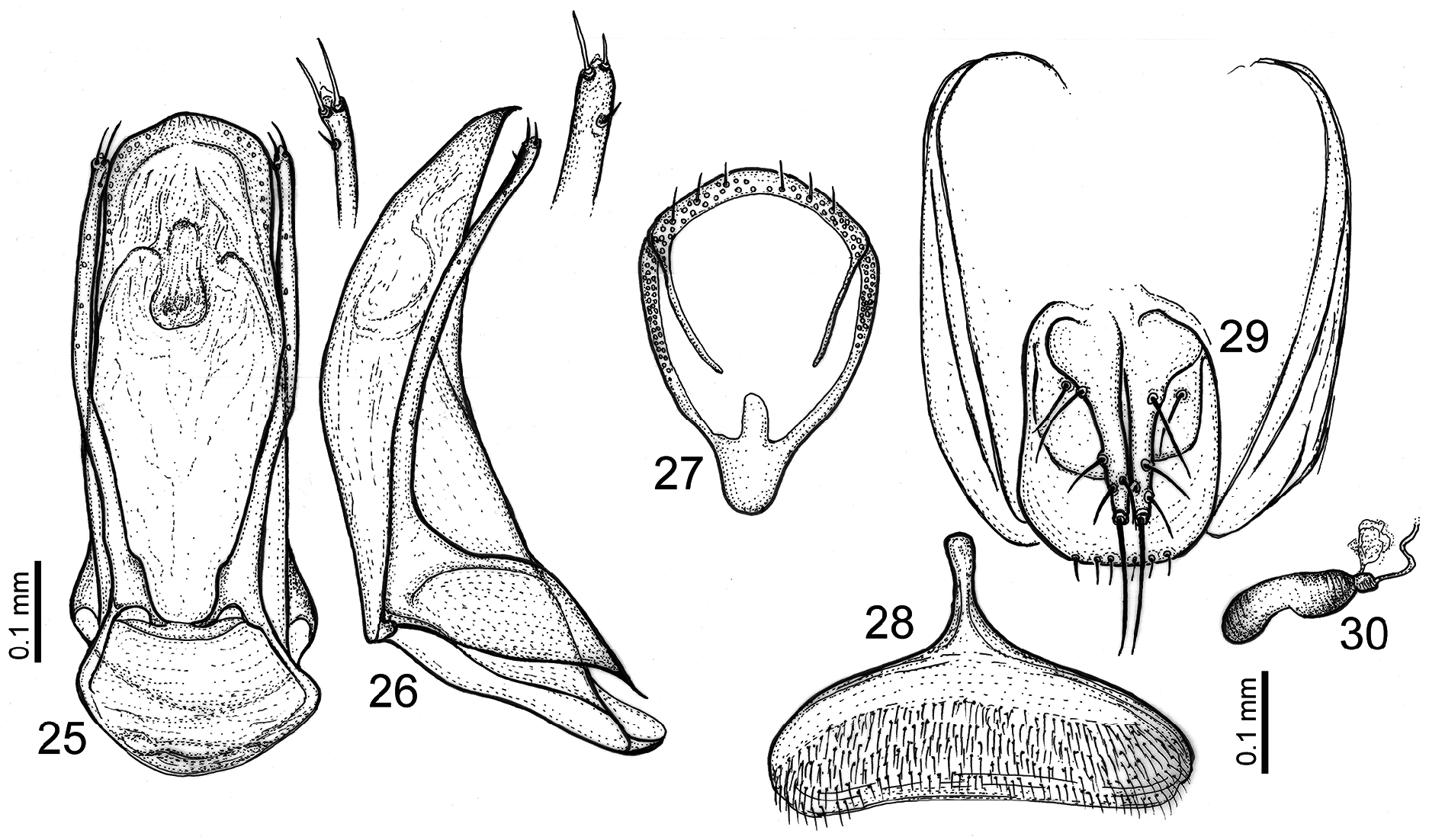
Male genitalia: Aedeagus 0.64–0.65 mm long, broad and truncate with median lobe more or less equally wide all of its length in dorsal view ( Fig. 25 View FIGURES 25–30 ). In lateral view ( Fig. 26 View FIGURES 25–30 ) median lobe tubular and strongly curved in the middle and with sharply deflexed apex. Internal sac of aedeagus without strong sclerotized structures only on its medio apical side; some weakly sclerotized or partly hyaline structures visible. No basal “Y” sclerotized structure present. Basal lamina of median lobe wide and basally invaginated backwards. Ventral lamina of tegmen parallel with median lobe and extended along basal lamina. Parameres (lateral styles) straight, mostly evenly wide all its length, parallel with median lobe, shorter than median lobe, with two apical long setae and one subapical (interno-lateral) short seta ( Figs. 25, 26 View FIGURES 25–30 ). Paramere apex with small terminal hyaline membranous appendage. Male genital segment (IX sternite, urosternite) in lateral view reduced to a chitinous ring. In dorsal view ( Fig. 27 View FIGURES 25–30 ) with prolonged, rounded ventral lobe and small prolonged process on posterior side of its sternal part and with prolonged narrow lateral stylets on its tergal part.
Female genitalia: Female ventrite VIIIth ( Fig. 28 View FIGURES 25–30 ) strongly pubescent on proximal part with anterior stout and prolonged apophysis (spiculum ventrale). Genital segment (urite IXth) well sclerotized and developed ( Fig. 29 View FIGURES 25–30 ). Sub-gonocoxite with one stylus, gonocoxite with one basal, one medial and two subapical styles, apical gonostylus with one the longest stylus. Spermatheca ( Fig. 30 View FIGURES 25–30 ) 0.148 mm long and 0,042 mm wide, sausage-like or short subbilobed, slightly more sclerotize on its both lobes. Hyaline gland attached at the proximal part of spermatheca in small sclerotized nodule.
Differential diagnosis. So far, only Rudogorites simonei is known from this genus. It can be easily separated from only similar species of genus Charonites (see description of genus).
Etymology. The species name simonei is dedicated in honour of Simone Milanolo (Sarajevo), a well-known karstologist and speleologist, who first explored the Lijina Pećina cave and collected most of the specimens of this new species.
Distribution. The new species is known only from its type locality, the cave “Lijina Pećina” near the village Gunjani in area known as Bosansko Rudogorje in Central Bosnia. The cave is situated in an isolated strip of limestone rock of Devon age, 20 km long and only a few km wide, that stretches in the direction SE-NW from Lepenica river up to Vrbas river spring. This karstic strip of limestone is superficially completely isolated from the rest of the Dinaric karstic massifs on its western side by layers of Triassic sandstones, Permian sandstones and various Silurian non-karstic rocks ( Jovanović et al. 1962 -1967; Sofilj & Živanović 1965 -1971). Two caves are known in this area, the Lijina cave that was formed at the contact of Devonian limestone rich in barite and quartz keratophyre ore and the Dusina cave, which was formed at the contact of Devonian limestone and quartz porphyry.
Ecology. Specimens were collected from the bottom of the cave, among the rock debris filled with soil, about 200 m from its entrance in complete darkness and without superficial meteorological influence. No other precise meteorological data are available. Besides the leptodirine beetles, the sphodrine ground beetle Laeomostenus cavicola and troglomorphic centipede Lithobius cf. stygius ( Kos 2021) have been collected in the cave.
The cave consists of one meander developing at three different levels. Two levels are dry, while the lower one is crossed by a small stream. The spring is located just a few tens of meters below the cave entrance. The cave was visited several times by cavers from the Centre for Karst and Speleology from Sarajevo during spring of 2016 and 2017. Although the explorations are not yet completed, about 800 m of the cave have been surveyed so far. Many channels have already been partially visited, however are still to be mapped, so the total length of the cave can be expected to be close to 1 km.
The paint signs on the cave wall in the initial part of the cave testifies that the cave was already partially explored, although there is no evidence of any written record in the literature. The cave was also used by the prehistoric inhabitants in the Iron Age, since the remains of pottery and traces of fire were found deep inside the cave. In the Ottoman period the cave served as a place of solitude, probably belonging to a dervish order, as shown by the remains of a wall and carved holes for beams.
Type series. Holotype ♂, glued to a white card, pinned dry, aedeagus dissected and preserved immersed in DMHF on a smaller transparent board pinned under the card-mounted specimens, labeled : HOLOTYPE | Rudogorites simonei sp. nov. ♂ | Polak & Mulaomerović det., [rectangular red label, printed]. Second label: Bosnia and Herzegovina: Lijina Pećina, Gunjani, Kreševo; 1.5.2016, Milanolo, S. & Prelić, M. leg. [rectangular white label, printed]—( NMBiH) .
Paratypes: specimens glued to white card, pinned dry, labeled:
PARATYPE | Rudogorites simonei sp. nov. Polak & Mulaomerović det. [rectangular yellow label, printed]. Second label: [rectangular white label, printed], 1 ♀, 16.8.2016, Milanolo, S. leg., same locality than the holotype; pinned dry, not dissected—( NMBiH) ; 1 ♂ and 1 ♀, 5.2.2017, Milanolo, S. leg., same locality than the holotype; pinned dry, not dissected—( PMSL) . 1 ♂ and 1 ♀, 5.2.2017, Milanolo, S. leg., same locality than the holotype; pinned dry, not dissected—( NMPO) . Specimens dissected (body parts, aedeagus, genital segments, protarsi, and antenna) preserved in in Solakryl BMX (Xylene diluting) media on glass microscope slides. 2 ♂♂ and 3 ♀♀, 16.8.2016, Milanolo, S. leg., same locality as the holotype—( NMPO) .

| PL |
Západoceské muzeum v Plzni |
| PMSL |
Slovenian Museum of Natural History (Prirodosloveni Muzej Slovenije) |
No known copyright restrictions apply. See Agosti, D., Egloff, W., 2009. Taxonomic information exchange and copyright: the Plazi approach. BMC Research Notes 2009, 2:53 for further explanation.
|
Kingdom |
|
|
Phylum |
|
|
Class |
|
|
Order |
|
|
Family |
|
|
SubFamily |
Cholevinae |
|
Tribe |
Leptodirini |
|
Genus |