
Bradynectes ensifer, Smith Iii & Schärer, 2021
|
publication ID |
https://doi.org/ 10.11646/zootaxa.4927.4.10 |
|
DOI |
https://doi.org/10.5281/zenodo.4557448 |
|
persistent identifier |
https://treatment.plazi.org/id/03AC87C3-FFBF-DA67-38D5-7F20FA763C3F |
|
treatment provided by |
Plazi |
|
scientific name |
Bradynectes ensifer |
| status |
sp. nov. |
Bradynectes ensifer View in CoL , n. sp.
The specific epithet (L. “sword-bearer”) refers to the curved stylet, as the late Dr. R.M. Rieger’s helping-name for this species was “säbel” (derived from the German word “Säbel” for “saber”). Type material was deposited at the Smithsonian Institution National Museum of Natural History ( NMNH) as follows: Holotype—a serially sectioned specimen ( USNM 1642033); Paratypes—one compressed specimen mounted in lactophenol for the stylet ( USNM 1642031), two sets of serial sections ( USNM 1642032 & USNM 1642034); type locality (34.650075, -77.104558). Material examined: three specimens studied alive and photographed (images deposited at http:// macrostomorpha.info under specimen numbers MTP JS 31, MTP JS 32, and MTP JS 33—the first, mounted in lactophenol as the above-mentioned paratype); three sets of serial sections [images deposited at http:// macrostomorpha.info under specimen numbers MTP JS 52 (paratype, sagittal), MTP JS 53 (holotype, sagittal) and MTP JS 54 (paratype, frontal)]; drawings by Rieger of specimens collected in Bogue Inlet (June 17, 1974) and at New River Inlet, NC (June 14, 1972); and one wholemount slide prepared by Rieger of the specimen from New River Inlet.
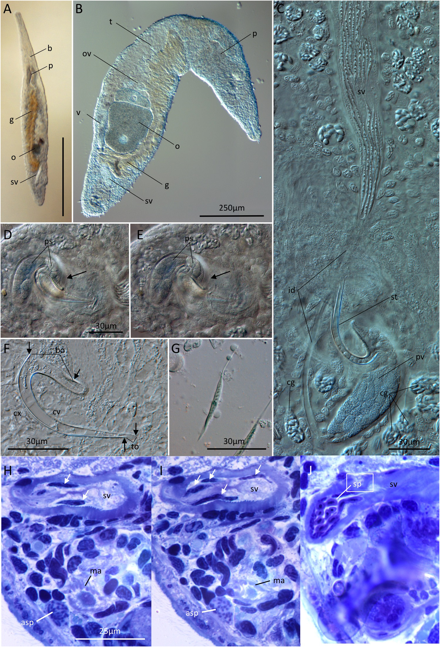
In transmitted light, free-swimming animals were yellowish-brown, 1–1.5 mm long ( Fig. 1A View FIGURE 1 ) and narrow anteriorly, with a pronounced widening at the level of the pharynx. Animals moved by slow ciliary gliding or by a leech-like alternate attachment of the anterior and posterior ends. The unpaired testis and ovary lay on the right-hand side of the body ( Fig. 1B View FIGURE 1 ). The brain was penetrated by rhammite-tracts of the frontal glands; the cell-bodies of these lay at about the level of the germinal zone of the testis. The pharynx led into a sac-like gut that was brown to golden-brown in life and extended posteriorly beyond the maturing oocyte ( Fig. 1B View FIGURE 1 ). The testis was voluminous ( Fig. 1B View FIGURE 1 ); a short vas deferens led from the testis to a muscular seminal vesicle ( Figs. 1 View FIGURE 1 A–C), from which an intervesicular duct ( Fig. 1C View FIGURE 1 ) led to a prostatic vesicle (vesicula granulorum) with two types of secretion ( Figs. 1D,E View FIGURE 1 ). On the prostatic vesicle was mounted a stylet with a rounded base and a long, curved, tapering portion ending in a point with a small oblique opening facing the convex side ( Figs. 1 View FIGURE 1 D–F), and, possibly, a longitudinal slit or a long fold on the convex surface near the tip ( Fig. 1F View FIGURE 1 ). The stylet ( Fig. 1F View FIGURE 1 ) had the following dimensions (see also Table 1 View TABLE 1 ): base opening = 30 µm, convex side = 95µm, concave side = 81 µm, tip opening= 4.4 µm (average of three specimens examined in squeeze preparation). A thickened portion of the musculature surrounding the prostatic vesicle inserted on the anterior extension of the base of the stylet ( Figs. 1D,E View FIGURE 1 , arrows). The proximal portion of the male antrum was unciliated and received glands with a small granular secretion ( Fig. S1A View FIGURE 1 , ag). Distally, the male antrum was ciliated (for more details see deposited images of specimens studied alive). Cement glands ( Figs. 1C, S1A,F View FIGURE 1 ) were located in an arc surrounding the male gonopore, and opened on the ventral surface. The female tract comprised a single ovary located medial to and near the posterior end of the testis ( Fig. 1B View FIGURE 1 ). Both male and female gonads originated ventrally. We were unable to detect female ducts in our live material or our sections, nor was there evidence in sections of a sperm-receiving organ or bursa-like tissue in the posterior extension of the gut (as mentioned by Rieger 1971, for B. sterreri , p.226; see his figures 4 and 6f). However, both the holotype and one paratype exhibited what may be a bundle of allosperm in the posterior part of the body ( Fig. 1H,I View FIGURE 1 ); the threadlike material in these bundles is approximately the same diameter of the nucleus in a mature sperm (comp. Fig. 1H & I View FIGURE 1 to J). Sperm released from the seminal vesicle by squeezing measured 60µm in length (average of seven sperm from one mature specimen), with a nucleus that appeared serpentine in DIC illumination ( Fig. 1G View FIGURE 1 ).
Although the layout of the reproductive system in Bradynectes ensifer is generally similar to that in the five previously described members of the genus ( Tyler et al 2006 –2016, Janssen et al 2015), it differs substantially in the shape of its copulatory stylet, which is a nearly closed, curved tube with a small angled opening directed at the convex side. In contrast, the existing species possess a stylet that was described by Rieger (1971) as resembling “a wide-open boot with no sole”. It would appear that one of these stylet forms could be derived from the other by great elongation and then curving of the shank of the “boot”, to produce a curved tube, along with reduction in the size of the distal opening (or possibly, the closing of the opening into a slit). As they lack female ducts, it is likely that Bradynectes spp. copulate by injecting sperm via hypodermic insemination ( Janssen et al 2015), and the thickened musculature attached to the anterior base of the copulatory stylet in B. ensifer could serve to drive the tip of the protracted stylet into the partner when the musculature surrounding the prostatic vesicle contracts. The existence of what may be a bundle of allosperm in the posterior parenchyma of two of our serially-sectioned specimens provides tentative evidence that copulation in this species involves hypodermic sperm injection.
Rieger apparently noted the existence of this species from Bogue Inlet in North Carolina, USA, over 40 years ago. Among his research material stored at the Institute of Zoology, Innsbruck, Austria is a drawing of a Bradynectes species with the helping-name “säbel” from Bogue Inlet, N.C. dated June 16, 1974 ( Fig. S1 View FIGURE 1 ). The drawings of stylet and sperm correspond closely in shape to those of our species. The scale-bar on Rieger’s drawing, added in ink at some point after the drawing was made, would make the stylet and sperm of his specimen approximately 20% and 30% smaller, respectively, than the stylet and sperm measured in our specimens ( Table 1 View TABLE 1 ). However, comparison of the added scale bar with the “canonical” scales for Wild M20 microscopes used in the Rieger lab at that time suggests that the scale added to the drawing actually represents 50 µm, instead of the 40 µm indicated by the tick-marks, rendering the size-differences mentioned above insignificant ( Table 1 View TABLE 1 ). In either case, we are confident that Rieger’s drawing of a specimen from the same site as ours corresponds to the species we have described above. Finally, Tyler’s figure of the stylet and sperm from his Bradynectes sp. ( Tyler, 1975) indicate that both of these structures were approximately the same size as those illustrated in Rieger’s draw-ing—stylet: base opening = 20µm, convex side = 58µm, concave side = 70µm, tip opening = 4µm; sperm 35–38µm ( Table 1 View TABLE 1 ).
The likely existence of a second, similar species is posed by an earlier Rieger drawing of a Bradynectes species designated with the helping-name “säbel” collected from the New River Inlet, N.C. dated June 14, 1972 ( Fig. S2). Although this specimen exhibits a similar stylet morphology to that in B. ensifer , notes on the drawing indicate that the animal was only 0.6mm– 0.7mm long, or about half the size of our species. A single wholemount slide prepared by Rieger bears the same collection number and helping name, and, as best as can be seen, the stylet in this wholemount is roughly the same size as the stylet in the drawing. Working backward from the canonical scales referred to above, the length of the sperm in this species is 27µm, close to the 22–23µm indicated on this drawing. Using the stated length of the sperm (23µm) for calibration, the drawing of the squeezed specimen using the 20x objective measures 670µm, exactly in the range given for body length in the notes. Using the same calibration, measurements of the stylet in this drawing (100x objective) give: base opening= 13µm, convex side= 25µm, concave side= 32µm, tip opening= 2µm, so considerably smaller than our specimens ( Table 1 View TABLE 1 ). Specimens of Bradynectes collected from this site and fixed for electron microscopy were used by Doe (1981) in his doctoral thesis work.
It appears that Rieger referred to the New River Inlet species as Bradynectes “short-stylet” and to the Bogue Banks species (discovered later) as Bradynectes “long-stylet” (S. Tyler, pers. com.). Given the size difference and the fact that this smaller species has not been collected again in the intervening 40+ years, we do not formally assign the specimen depicted in Figure S2 and preserved in Rieger’s wholemount or the Bradynectes sp. studied by Doe to our new species.
No known copyright restrictions apply. See Agosti, D., Egloff, W., 2009. Taxonomic information exchange and copyright: the Plazi approach. BMC Research Notes 2009, 2:53 for further explanation.