
Labiobaetis academicus Kaltenbach, Surbakti & Kluge, 2021
|
publication ID |
https://doi.org/ 10.14203/treubia.v48i1.4020 |
|
publication LSID |
lsid:zoobank.org:pub:94A84853-BB44-4940-A439-BBD9D8A7D2B9 |
|
DOI |
https://doi.org/10.5281/zenodo.5643881 |
|
persistent identifier |
https://treatment.plazi.org/id/0806CF31-FAAA-460C-99A2-D89D8481BA2C |
|
taxon LSID |
lsid:zoobank.org:act:0806CF31-FAAA-460C-99A2-D89D8481BA2C |
|
treatment provided by |
Valdenar |
|
scientific name |
Labiobaetis academicus Kaltenbach, Surbakti & Kluge |
| status |
sp. nov. |
Labiobaetis academicus Kaltenbach, Surbakti & Kluge sp. nov.
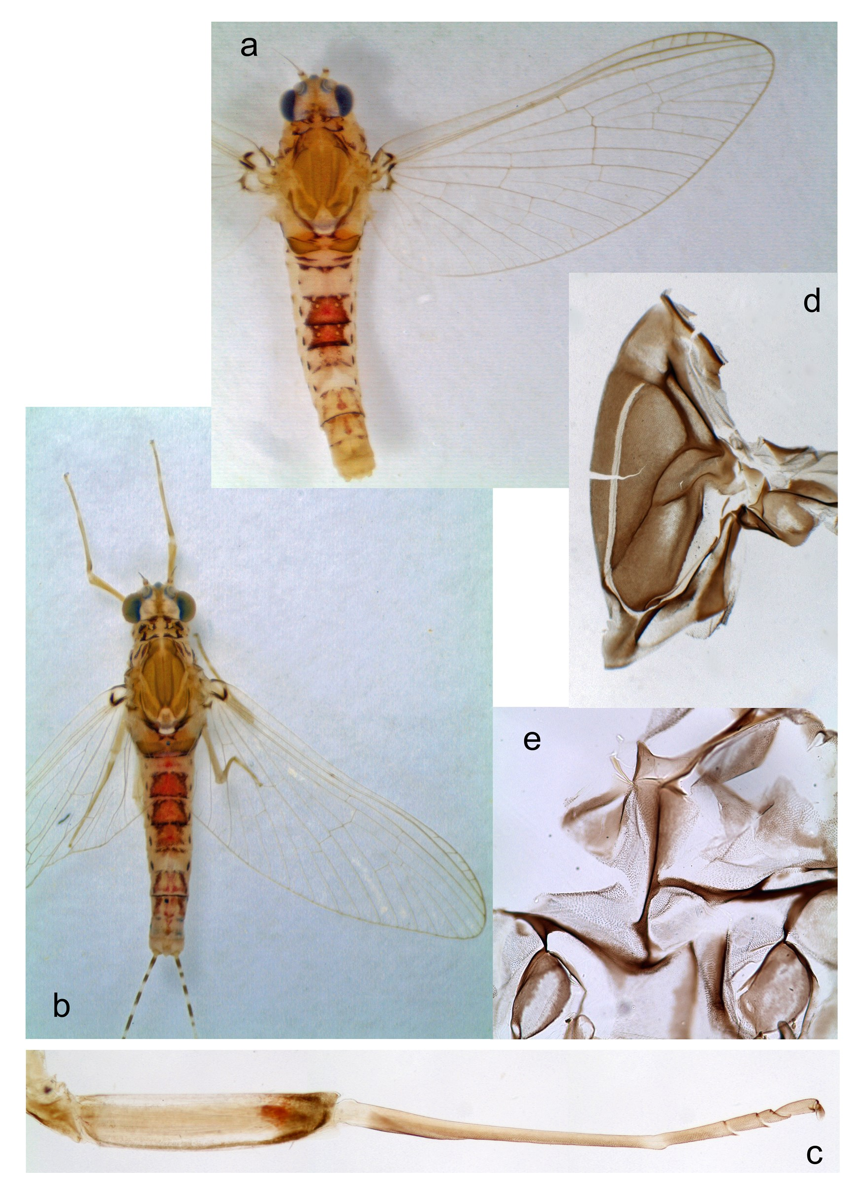
Figures 1–3 View Figure 1 View Figure 2 View Figure 3 , 5–8 View Figure 5 View Figure 6 View Figure 7 View Figure 8
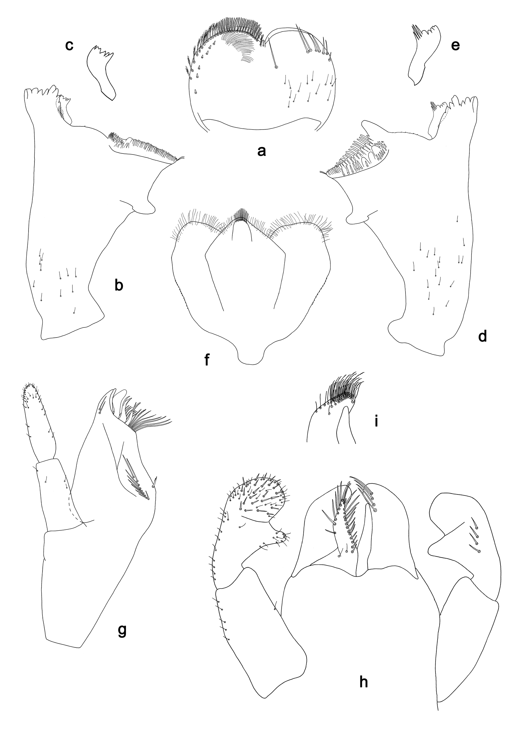
Differential diagnosis. Larva. Following combination of characters differentiate the new species from L. claudiae and L. stagnum : A) maxilla medially with 7–9 medium to long, spineJlike setae; B) shape of labial palp segment II as Fig. 1h View Figure 1 ; segment III slightly pentagonal; C) shape of tergalius IV as Fig. 2d; F View Figure 2 ) paraproct distally not expanded, with 20–25 stout, marginal spines.
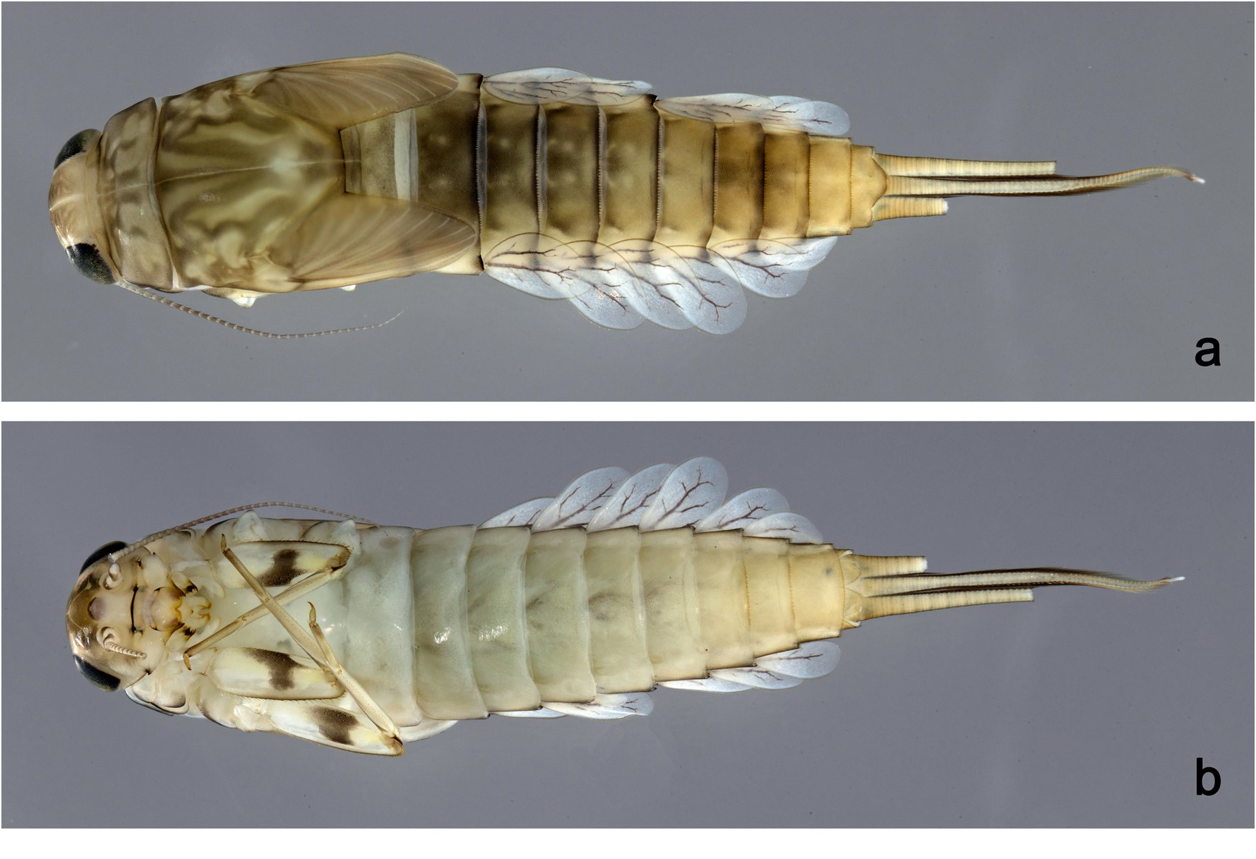
Description. Larva ( Figs 1–3 View Figure 1 View Figure 2 View Figure 3 ). Cerci ca. 2/3 of body length, paracercus ca. 2/3 of cerci length, antenna approx. twice as long as head length.
Coloration. Head, thorax and abdomen dorsally brown, with pattern as in Fig. 3a View Figure 3 . Fore protoptera brown with bright striation. Head, thorax and abdomen ventrally light brown. Legs light brown; femur with a large, distomedial brown spot, dorsal margin and apex brown; tibia in basal part with brown spot, bordered by patellotibial suture. Caudalii light brown.
Antenna ( Fig. 2h View Figure 2 ) with scape and pedicel subcylindrical, without distolateral process at scape.
Labrum ( Fig. 1a View Figure 1 ). Rectangular, length 0.7x maximum width. Distal margin with medial emargination and a small process. Dorsally with medium, fine, simple setae scattered over surface; submarginal arc of setae composed of one plus 5–7 long, simple setae. Ventrally with marginal row of setae composed of lateral and anterolateral long, feathered setae and medial long, bifid, pectinate setae; ventral surface with ca. seven short, spineJlike setae near lateral and anterolateral margin.
Right mandible ( Fig. 1b, c View Figure 1 ). Incisor and kinetodontium fused. Incisor with four denticles; kinetodontium with four denticles, inner margin of innermost denticle with a row of thin setae. Prostheca robust, apically denticulate. Margin between prostheca and mola slightly convex. Tuft of setae at apex of mola present.
Left mandible ( Fig. 1d, e View Figure 1 ). Incisor and kinetodontium fused. Incisor with four denticles; kinetodontium with four denticles. Prostheca robust, apically with small denticles and combJ shaped structure. Margin between prostheca and mola slightly convex, with minute denticles
toward subtriangular process. Subtriangular process long and slender, above level of area between prostheca and mola. Denticles of mola apically constricted. Tuft of setae at apex of mola present.
Both mandibles with lateral margins almost straight. Basal half with fine, simple setae scattered over dorsal surface.
Hypopharynx and superlinguae ( Fig. 1f View Figure 1 ). Lingua approx. as long as superlinguae. Lingua longer than broad; medial tuft of stout setae well developed, short; distal half laterally expanded. Superlinguae distally rounded; lateral margin rounded; fine, long, simple setae along distal margin.
Maxilla ( Fig. 1g View Figure 1 ). GaleaJlacinia ventrally with two simple, apical setae under canines. Inner dorsal row of setae with three dentiJsetae, distal dentiJseta toothJlike, middle and proximal dentiJsetae slender, bifid and pectinate. Medially with one bipectinate, spineJlike seta and 7–9 medium to long, simple setae. Maxillary palp approx. as long as length of galeaJlacinia; 2Jsegmented; palp segment II 1.1x length of segment I; setae on maxillary palp fine, simple, scattered over surface of segments Iand II; apex of last segment constricted, without excavation at inner distolateral margin.
Labium ( Fig. 1h, i View Figure 1 ). Glossa basally broad, narrowing toward apex; shorter than paraglossa; inner margin with ca. 12 short, stout, spineJlike setae increasing in length distally; apex with two long and one short, robust setae; outer margin with six spineJlike setae; ventral surface with few fine, simple, scattered setae. Paraglossa subJrectangular, curved inward; apex rounded; ventrally with three rows of long, robust, distally pectinate setae in apical area and one medium, simple seta in anteromedial area; dorsally with a row of four long, spineJlike setae near inner margin. Labial palp with segment I approx. as long as segments II and III combined. Segment Iventrally with short, fine, simple setae. Segment II with narrow, thumbJlike distomedial protuberance; distomedial protuberance 0.5x width of base of segment III; ventral surface with short, fine, simple setae; dorsally with a row of 3–6 medium, spineJlike, simple setae near outer margin. Segment III slightly pentagonal; length 0.9x width; ventrally covered with short, spineJlike, simple setae and short, fine, simple setae.
Hind protoptera absent.
Legs ( Fig. 2a, b View Figure 2 ). Ratio of foreleg segments 1.3:1.0:0.6:0.2. Femur. Length ca. 3x maximum width. Dorsal margin with a row of 25–32 medium, curved, spineJlike setae; length of setae 0.12x maximum width of femur. Apex rounded, with medium to short,
curved, spineJlike setae. Many stout, lanceolate setae scattered along ventral margin; femoral patch present. Tibia. Dorsal margin with a row of short to medium, stout, spineJlike setae. Ventral margin with a row of short, curved, spineJlike setae, on apex some longer, spineJlike setae and a tuft of fine, simple setae. Anterior and posterior surface scattered with stout, lanceolate setae. Patellotibial suture present on basal 1/2 area. Tarsus. Dorsal margin with a row of short, spineJlike setae. Ventral margin with a row of curved, spineJlike setae. Claw with one row of 10–12 denticles; distally pointed; with 4–5 stripes; subapical setae absent.
Abdomen ( Fig. 2c View Figure 2 ). Surface with dense, irregular rows of UJshaped scale bases. Posterior margin of terga II–IX with triangular spines, approx. as long as wide; spines diminished on middle of posterior margin of tergum IX behind pair of submedian setae. Posterior margin of tergum Xwith longer and narrower spines. Posterior margins of sterna VII–IX with shorter triangular spines.
Tergalii ( Fig. 2d–f View Figure 2 ). Present on segments II–VII. Margin with denticles of different sizes, intercalating both medium and long, fine simple setae. Tracheae extending from main trunk to inner and outer margins. Tergalius IV as long as length of segments Vand VI combined. Tergalius VII as long as length of segments VIII and 1/2 IX combined.
Paraproct ( Fig. 2g View Figure 2 ). Distally not expanded, with 20–25 stout, marginal spines. Surface scattered with UJshaped scale bases and fine, simple setae. Cercotractor with numerous small, marginal spines.
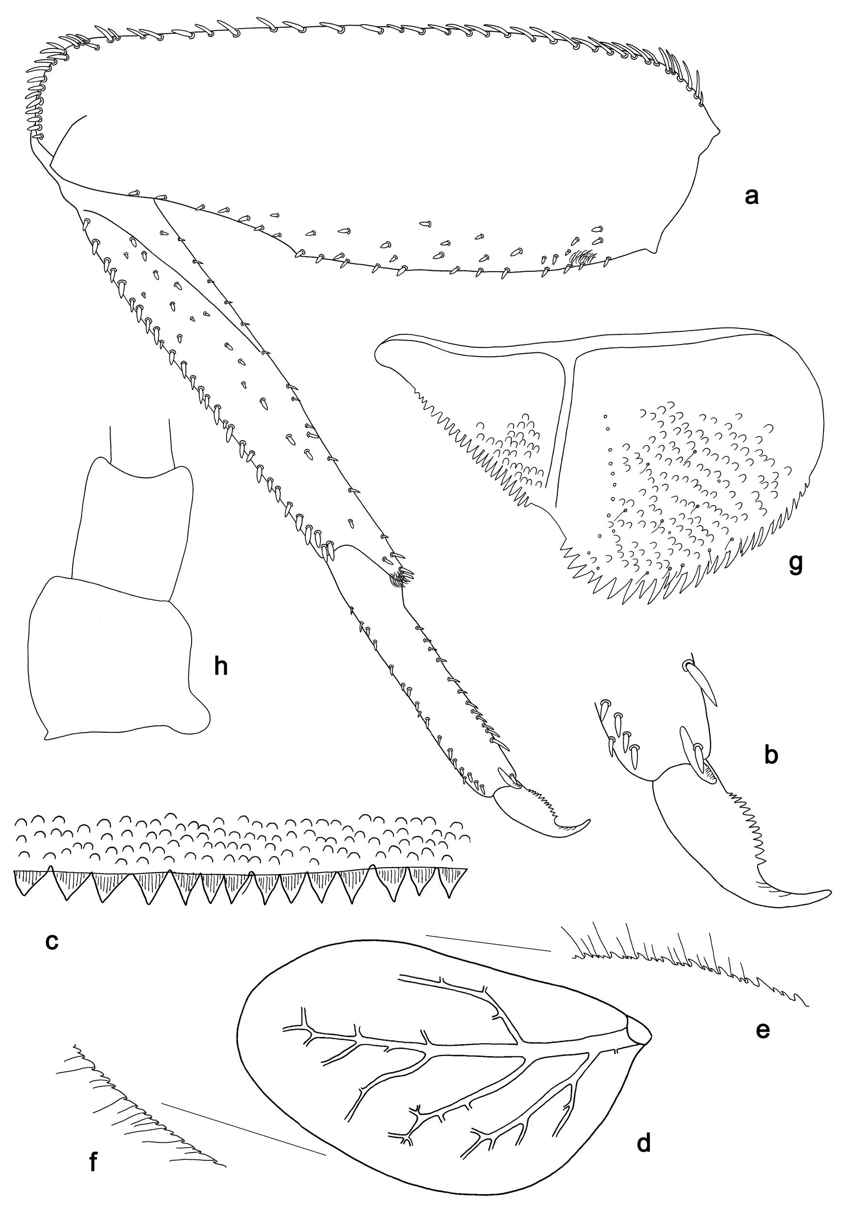
Subimago (both sexes; Fig. 6d, e View Figure 6 ). Cuticular coloration. Pronotum brown with lighter areas. Mesonotum brown with medioparapsidal suture contrastingly colorless, other sutures darker brown ( Fig. 6d View Figure 6 ). Thoracic pleura with brown sclerites and colorless membranes ( Fig. 6e View Figure 6 ). Legs mostly colorless; femur colored with brown at base, outer and inner margins, with indistinct brownish spot in distal part; tibia colored with brown at base. Abdominal terga uniformly light brown, sterna lighter. Cerci uniformly colorless.
Hypodermal coloration. As in imago (see below).
Texture. On all legs of both sexes, all tarsomeres covered with blunt microlepides.
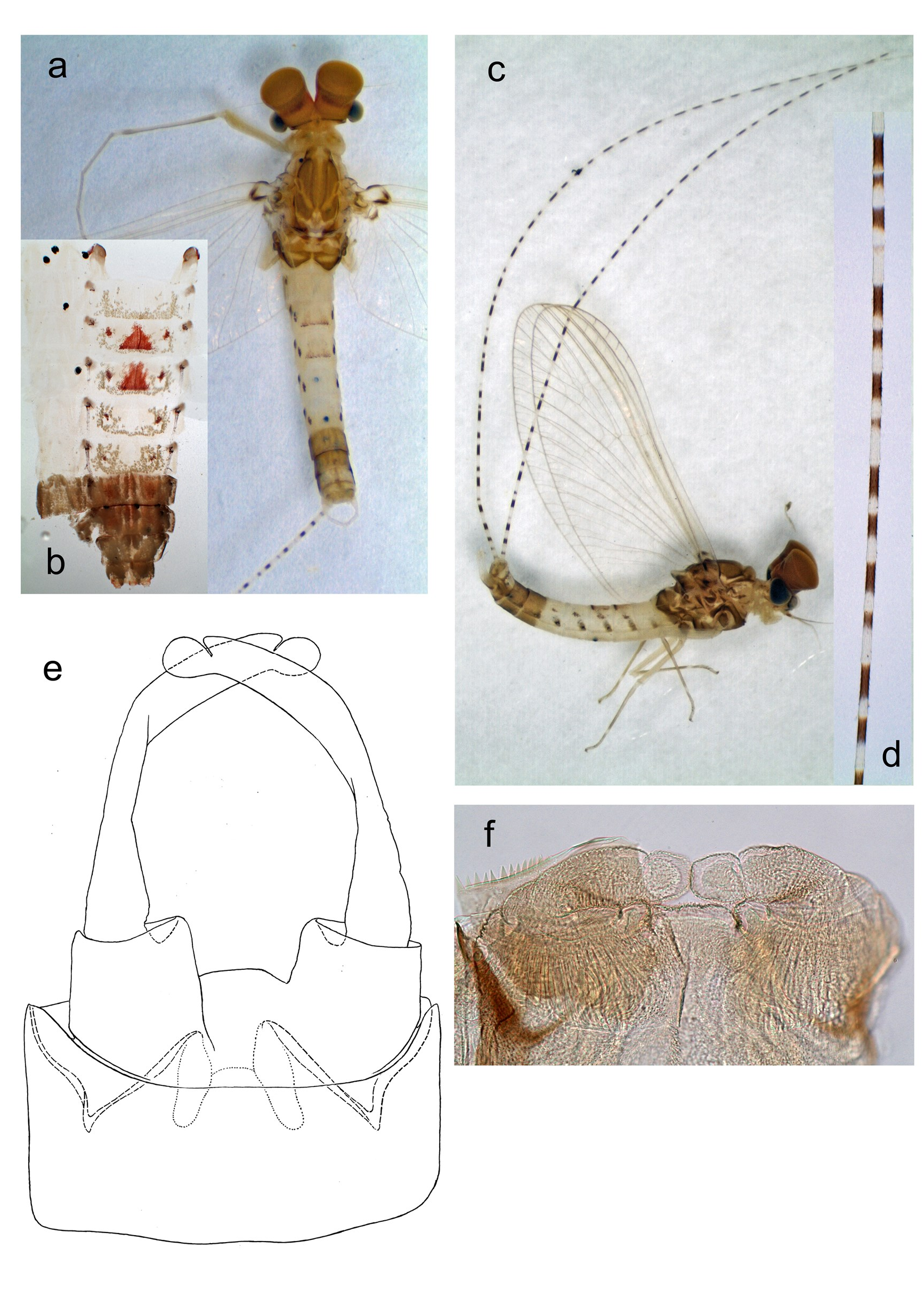
Imago, male ( Fig. 5a–d View Figure 5 , 6c View Figure 6 ). Head ocher. Turbinate eyes high widened apically, orange. Thorax light brown with ocher areas. Wing membrane colorless, veins ocher; costal brace and adjacent areas with contrasting dark brown stripes. Pterostigma with several oblique veins. Hind wing absent. Legs of all pairs with similar coloration ( Fig. 6c View Figure 6 ): femur light ocher, with inner margin and apex bordered with brown, with reddish macula near apex; tibia with base and apex whitish, other part light brownish on inner side, lighter on
outer side; tarsus light brownish. Tarsus of middle and hind legs with two apical spines, on segments 1st+2nd and 3rd. Abdominal segments I–VI whitish, with peculiar maculae: each tergum II–VII with pair of lateral brown maculae on spiracles; each tergum III–VI with pair of sublateral brown maculae; each tergum III–IV with unpaired reddish stripe on posterior margin and with more or less expressed triangular macula arising from this stripe in anterior direction. Abdominal segments VII–VIII uniformly brown. Abdominal terga IX–X lighter. Each segment of cerci white at base and at apex, contrastingly brown at middle; segments with longer and shorter brown areas irregularly alternating ( Fig. 5d View Figure 5 ).
Male genital structure and development. Imaginal genitals as in Fig. 5e View Figure 5 . SternoJ styligeral muscle completely absent. Each gonovectis with small hook at apex. Penial bridge without prominent median projection. Unistyligers cylindrical, with straight inner margins. Gonostylus with 1st segment smoothly narrowed toward apex; 2nd segment slightly widened toward apex; 3rd segment short.
Protogonostyli of male larva represent very shallow convexities of posterior margin of abdominal sternum IX. In mature larva ready to molt to subimago, subimaginal gonostyli packed under larval cuticle in « Labiobaetis Jpose » ( Kluge, 2004: fig. 29I): second segments directed medially and bent ( Fig. 5f View Figure 5 ).
Imago, female ( Fig. 6a, b View Figure 6 ). Head ocher with brownish. Prothorax and anteroJ lateral areas of mesonotum with contrasting light ocher and dark brown markings; other areas of mesonotum and metanotum light brown; ventral side of thorax light uniformly ocher. Wings as in male. Leg coloration as in male. Tarsus of fore leg with two apical spines, on 2nd and 3rd segments (as on middle and hind legs). Each abdominal tergum III and IV with reddish median macula as in male; other markings of abdominal terga and sterna vary individually. Cerci as in male.
Dimension. Body length of larvae 4.2–6.5 mm. Fore wing length (and approximate body length) of subimagos, male and female imagos ca. 5 mm.
Etymology. Refering to the fact that the specimens were collected just behind the campus of Cenderawasih University (UNCEN) in Jayapura.
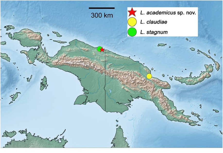
Distribution. Indonesia: Papua Province ( Fig. 7 View Figure 7 ).
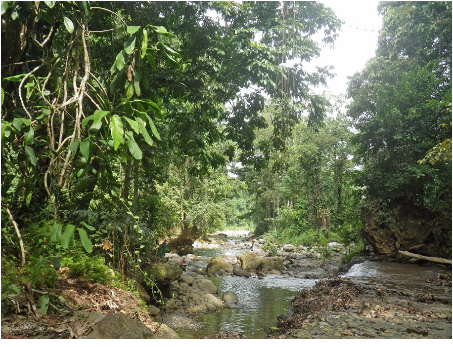
Biological aspects. The specimens were collected at an altitude of 160 m in a medium sized, shallow, moderately flowing river with stones and mud on the bottom, with few vegetation ( Fig. 8 View Figure 8 ). The new species was collected together with 12 other mayfly species: four other, yet undescribed species of Labiobaetis Novikova & Kluge, 1987 ;
Mystaxiops venatoris McCafferty & Sun, 2005 ; two species of Papuanatula LugoJOrtiz & McCafferty, 1999; two species of Centroptella Braasch & Soldán, 1980 ; one Cloeon sp. ; one Caenis sp. and Nonnullidens reductus Kluge, 2013 .
TypeJmaterial. Holotype. INDONESIA • larva; Papua Province, Jayapura, Waena , Kamp Wolker ; near UNCEN campus; 02°34'07"S, 140°38'51"E; 160 m; 26.v.2019; leg. Surbakti, Kellis & Sumoked; (PAP080); on slide; GBIFCH00592382 ; MZB GoogleMaps . Paratypes. INDONESIA • 17 larvae; same data as holotype; 3 on slides; GenBank MW041241 View Materials , MW041242 View Materials ; GBIFCH00673069 , GBIFCH00673081 , GBIFCH00592381 ; KSP, MZB; 14 in alcohol; GBIFCH00515503 ; KSP, MZB GoogleMaps • 4 ♂ and 2 ♀ imagos with individually associated larval and subimaginal exuviae, 1 ♀ subimago with its larval exuviae, 1 ♀ imago, 11 ♂ and 8 ♀ larvae ready to molt to subimagoes, 22 larvae; same locality as holotype; 9–13.viii.2012; leg. N. Kluge & L. Sheyko; SaintJPetersburg State University. GoogleMaps
| MZB |
MZB |
No known copyright restrictions apply. See Agosti, D., Egloff, W., 2009. Taxonomic information exchange and copyright: the Plazi approach. BMC Research Notes 2009, 2:53 for further explanation.
|
Kingdom |
|
|
Phylum |
|
|
Class |
|
|
Order |
|
|
Family |
|
|
Genus |