
Pterocerdale, Hoese, Douglass F. & Motomura, Hiroyuki, 2009
|
publication ID |
https://doi.org/ 10.5281/zenodo.191894 |
|
publication LSID |
lsid:zoobank.org:pub:4374E408-BA5D-4031-8B96-DCBC156EBC61 |
|
DOI |
https://doi.org/10.5281/zenodo.5625182 |
|
persistent identifier |
https://treatment.plazi.org/id/03AD87E2-FFA7-FFE3-FF35-FE6C8E39FB7C |
|
treatment provided by |
Plazi |
|
scientific name |
Pterocerdale |
| status |
gen. nov. |
Pterocerdale View in CoL gen. nov.
Type species: Pterocerdale insolita sp. nov.
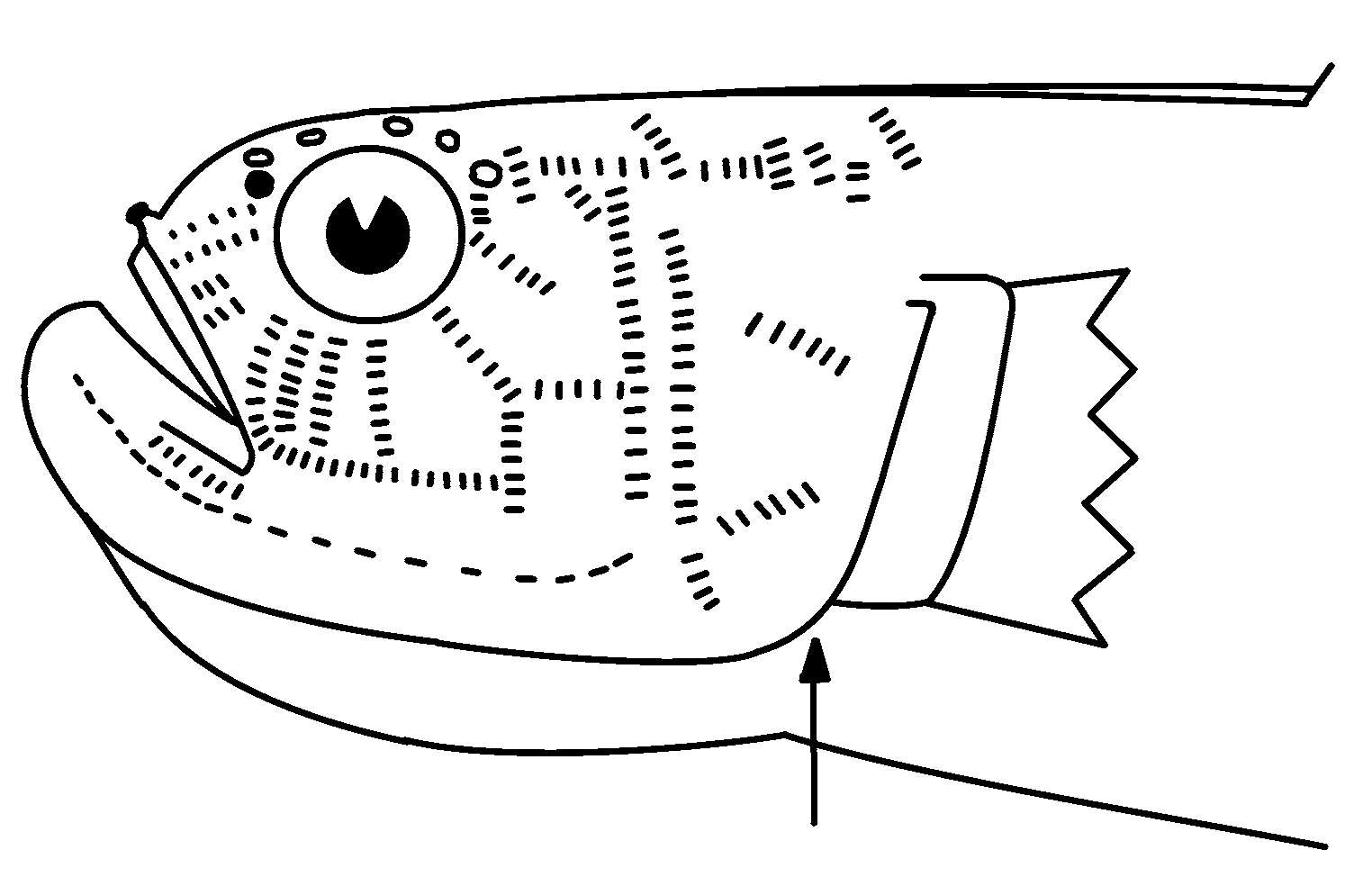
Diagnosis. Lower lip with free ventral margin posteriorly only; head and body compressed; body elongate; nape, cheek, preoperculum and operculum almost completely covered with scales; body covered with cycloid scales, imbricate, in 105 vertical rows; mouth terminal, only slightly protractile, forming an angle of about 60° to longitudinal axis of body; maxilla reaching posteriorly to below front margin of eye; head pores all paired laterally, with 5 pores around dorsal margin of each eye; snout relatively short, rounded, its length subequal to eye diameter; anterior nostril at end of short tube; posterior nostril a simple pore; no teeth on vomer, tongue or palatines; teeth conical, slightly curved; upper jaw with two rows of small, loosely attached teeth anteriorly, teeth in outer row slightly larger than those in inner row, widely spaced; lower jaw with single row of loosely attached, small teeth directed dorsally, no enlarged curved canines visible in holotype; along outer edge of dentary a series of blunt bony dorsoventrally flattened projections with rounded tip directed more or less horizontally (widely-spaced projections appear to be bony projections of dentary, not true teeth, but probably tooth sockets), space between projections about equal to width of projections; tongue tip broadly rounded; head papillae in transverse pattern; median nuchal crest, formed by low fold of skin from first dorsal spine onto head, low; gill opening vertical, extending ventrally from pectoral-fin base below upper margin to point just below lower pectoral base, below operculum ( Figure 4 View FIGURE 4 ); fleshy interorbital subequal to diameter of eye; 2 dorsal fins, first dorsal fin VI, second dorsal fin I,19; anal fin I,18; pectoral-fin rays 19; segmented caudal rays 9+8; branched caudal rays 8+7; pelvic fins separate 1,4; vertebrae 12+14.
Etymology. An arbitrary combination of letters from Ptereleotrinae + Cerdale , a genus of microdesmine gobioid, relating to the placement in the Ptereleotrinae and similarity to the microdesmine genus Cerdale .
Relationships. Placement of this genus is the Ptereleotrinae is provisional, because the genus displays features in common with both the Ptereleotrinae and the Microdesminae . Hoese (1984) noted that the ptereleotrines have a single pterygiophore preceding the first hemal arch, a feature shared with sicydiines, amblyopines, oxudercines and the eleotrids Thalasseleotris and Grahamichthys . However, Pterocerdale lacks that feature and its placement with the ptereleotrines is based on overall similarity, head pore pattern, reduced number of pelvic rays, elongate body and dorsal-fin and anal-fin ray counts. Pterocerdale is unique within the Ptereleotrinae in having 12 precaudal vertebrae, the upper lip with a free ventral margin posteriorly only, and cup-shaped bony projections from the dentary. In other ptereleotrines the first two anal pterygiophores lock into the first haemal spine (one before and one behind spine), but in Pterocerdal e the first 3 pterygiophores precede the first haemal spine, probably because of the increase of precaudal vertebrae from 10 to 12. In Gunnellichthys , Microdesmus and Cerdale three pterygiophores also precede the first haemal arch. The character was not recorded for other microdesmine genera. Pterocerdale also differs from other ptereleotrines, except Parioglossus in having well developed anterior zygapophyses on all vertebrae except the urostyle and a restricted gill opening. In Parioglossus the zygapophyses are developed on all precaudal and varying numbers of caudal vertebrae, but generally becoming smaller on posterior caudal vertebrae ( Rennis & Hoese, 1985). The zygapophyses and head pores of Pterocerdale suggest a close relationship to Parioglossus ; however, prominent anterior zygapophyses are also present on all vertebrae except the urostyle in microdesmines. Microdesmines are more similar to Pterocerdale in not showing any reduction in the size of the zygapophyses on the posterior vertebrae. The bony projections on the lower jaw of Pterocerdale are probably tooth sockets. These projections are unlike any other ptereleotrines, which have a smooth edge to the outer edge of the dentary. In microdesmines, there are large teeth in the outer row of the lower jaw, with the tooth sockets projecting slightly on the outer margin of the dentary. The projections in microdesmines are much smaller than those found in Pterocerdale . Pterocerdale also has a slender and slightly elongate skull, an intermediate condition between Parioglossus and the microdesmines. In Gunnellichthys , the ventral margin of the lower lip is free for about two-thirds of the length of the lower jaw, while in Microdesmus and Cerdale the ventral margin of the lower lip is free along the posterior quarter of the lower jaw, similar to the condition found in Pterocerdale . Paragunnellichthys has no free ventral margin of the lower lip. Other ptereleotrine genera have the ventral margin of the lower lip free or fused only to the chin. Thacker (2000) also noted that microdesmines have an elongate ventral (extended) dentary process at the anterior tip of the dentary, which is better developed than in other gobioid fishes. In Parioglossus the process is very short, but it is clearly long in Pterocerdale , although slightly shorter than that observed in radiographs of microdesmines. Thacker (2000) indicated that Hoese (1984) reported an elongate sphenotic in the ptereleotrinae ; however, Hoese in fact indicated “the expanded dorsal flange of the sphenotic reaching to the supraoccipital”, a condition identical to that in microdesmines.
Consequently, either Pterocerdale is strongly convergent with microdesmines or represents a lineage positioned between Parioglossus and microdesmines. If the latter proves to be correct then microdesmines would be more closely related to Parioglossus than other ptereleotrines and could not be its sister group without removing the latter genus from the ptereleotrines and including it within the microdesmines. Molecular and morphological studies comparing the two groups ( Thacker, 2000, 2009) have unfortunately not included Parioglossus . Other osteological features Thacker (2000) listed as distinctive for the microdesmines could not be observed from the radiographs. The relationship cannot be fully resolved until additional material becomes available for genetic and morphological studies.
Remarks. The genus is described here from a single specimen. Despite attempts to collect additional material and searches of museums in the U.S., Japan, Australia and Europe, no additional material has been found. It is described here because it is different from other gobioids in the several features mentioned above and because it raises the question of potential relationships between the ptereleotrines and microdesmines.
No known copyright restrictions apply. See Agosti, D., Egloff, W., 2009. Taxonomic information exchange and copyright: the Plazi approach. BMC Research Notes 2009, 2:53 for further explanation.