
Anopheles kleini Rueda
|
publication ID |
https://doi.org/10.5281/zenodo.190892 |
|
DOI |
https://doi.org/10.5281/zenodo.6219343 |
|
persistent identifier |
https://treatment.plazi.org/id/03ADA94B-FFC8-FFB1-FBCC-A293FA131D5B |
|
treatment provided by |
Plazi |
|
scientific name |
Anopheles kleini Rueda |
| status |
|
1. Anopheles kleini Rueda View in CoL
( Figs. 3 View FIGURE 3 A, 4A, 5A, 6A)
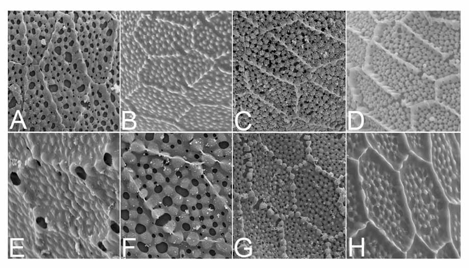
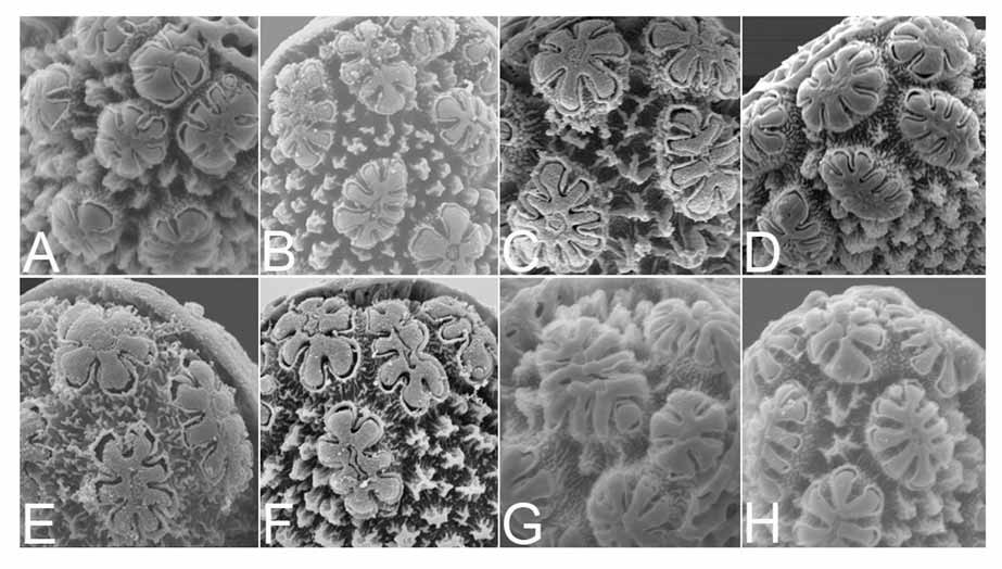
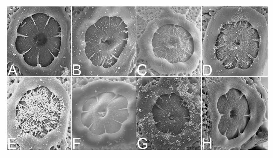
Size: Length 349.66–525.23 um (mean 476.34 + 73.38 um, n = 5); width 98.12–121.36 (mean 115.81 + 9.96 um, n = 5) (Table 1). Color: Black. Overall appearance: Boat-shaped in both ventral and dorsal views, anterior end blunt, posterior end blunt, sometimes pointed. Ventral surface concave, dorsal surface curved, float relatively short and wide in dorso-ventral plane, length 189.53–310.07 um (mean 243.44 + 47.23 um, n = 5); width 41.44–60.80 um (mean 53.90 + 8.74 um, n = 5). Dorsal and lateral surfaces: All surfaces uniformly covered with mostly pentagonal and hexagonal outer chorionic cells or plastron-type cells ( Hinton 1968) ( Fig. 3 View FIGURE 3 A), each longer than wide, long dimension oriented in long axis of egg. Interior of each cell with fine rounded structures, surrounded by an elevated, palisade-like outer chorionic reticulum. Cell area 206.78– 360.12 um (mean 293.32 + 49.92, n = 16) (Table 1). Float fairly short, about 0.51 length of egg; ratio of float length and width, and length in proportion to egg length and number of ribs as in Table 1. Ribs (= ridges, Harbach and Knight 1980) towards both ends of float wider than those at middle part, rarely striated on dorsal sides; number of ribs per float 20–25 (mean 23.20 + 1.30, n = 5). Ventral surface. Deck continuous, narrows in mid-line near center of float, degree of narrowing usually variable; anterior part of deck usually wider than posterior part; entire deck covered uniformly with fine tubercles ( Fig. 4 View FIGURE 4 A). Frill continuous, shallow along narrowed portion of deck. Lobed ventral tubercles at anterior end of the deck, 6–7 (mean 3.67 + 0.58, n = 3), and at posterior end, 8 (n = 3) (Table 1, Fig. 5 View FIGURE 5 A). Lobed ventral tubercles usually round, occasionally oval or oblong. Lobes of each anterior ventral tubercle, 6–9 (mean 7.31 + 0.85, n = 13); lobes of each posterior ventral tubercle, 3 (n = 3). Lobes clearly separated, often swollen at ends, outer walls often smooth. Lobes in slightly elevated, tuberculoid structures. Anterior end, micropyle. Anterior end slightly more blunt than posterior end. Micropylar collar irregular in outline, with smooth surface, inner edge uniformly and deeply excavated, peaks between excavations tapering to form radial ridges extending about half way across micropylar disc, dividing disc into sectors ( Fig. 6 View FIGURE 6 A). Number of sectors (or ridges) 6–8 (mean 7.25 +96, n = 4). Area of micropylar disc 33.98–146.76 um (mean 89.53 + 51.45 um, n = 4), usually with striated surface.

No known copyright restrictions apply. See Agosti, D., Egloff, W., 2009. Taxonomic information exchange and copyright: the Plazi approach. BMC Research Notes 2009, 2:53 for further explanation.