
Odontophotopsis tenuiptera
|
publication ID |
https://doi.org/ 10.5281/zenodo.179151 |
|
DOI |
https://doi.org/10.5281/zenodo.6242207 |
|
persistent identifier |
https://treatment.plazi.org/id/03AE2B55-FFEF-FFFF-1B9E-234BFBFA8E3C |
|
treatment provided by |
Plazi |
|
scientific name |
Odontophotopsis tenuiptera |
| status |
|
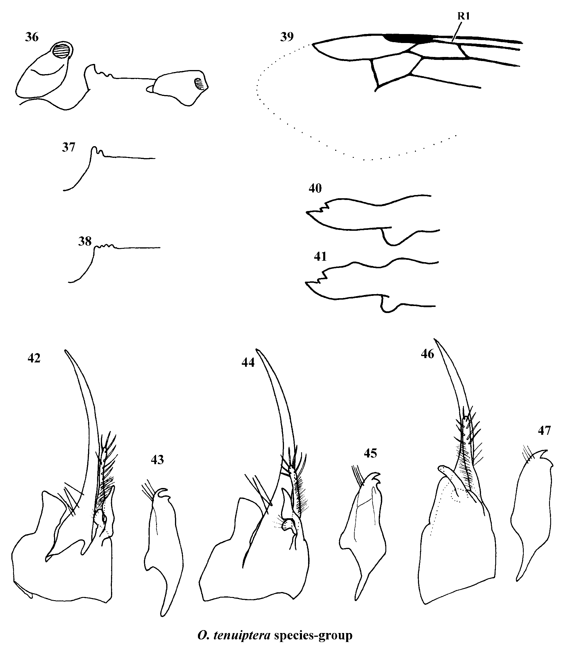
8. Odontophotopsis tenuiptera species-group
( Figs. 36–47 View FIGURES 36 – 47 )
Diagnosis. This species-group is easily diagnosed by the complete notauli; by the elongate marginal cell that has its length along costa subequal to twice the length of stigma ( Fig. 39 View FIGURES 36 – 47 ); by the elongate first metasomal segment that is 2–2.5X as long as its apical width in dorsal view; and by the unique mesosternal processes of the included species ( Figs. 36–38 View FIGURES 36 – 47 ). Also of use in identification are that the mandible is tridentate with a ventrobasal tooth ( Figs. 40, 41 View FIGURES 36 – 47 ), the clypeus is weakly tuberculate, the antennal scrobe is prominently carinate dorsally, the metasoma is not concolorous with head and mesosoma, the pygidium is glabrous medially, and the hypopygium has the apical margin dentate medially.
Male. Coloration and setal pattern. Head, mesosoma and first metasomal segment testaceous to ferruginous; antenna slightly paler. Metasomal segments 2–7 darker, dark ferruginous to piceous. Body clothed with sparse, erect, decumbent, pale yellow setae. Only few plumose setae on mesosoma. T1 without plumose fringe at distal margin. T2 with thin fringe of pale plumose setae. T3-7 and S1-7 lacking fringe of pale plumose setae. Apical margin of T2 and T3-7 with fine punctures bearing short decumbent setae. Pygidium with dense apical fringe of simple, yellow brown setae. Tibial spurs and legs concolorous with head and mesosoma.
Head. Rounded posteriorly. Mandible excised ventrally, angle of excision rounded ( Figs. 40, 41 View FIGURES 36 – 47 ). Dorsal carina strong, medially lamelliform, terminating at strong inner tooth ( Figs. 40, 41 View FIGURES 36 – 47 ). Subdistal inner tooth weak ( Figs. 40, 41 View FIGURES 36 – 47 ). Mandible varying from parallel to dilated beyond excision ( Figs. 40, 41 View FIGURES 36 – 47 ). Clypeus depressed below margin of mandibles, median area concave, posteriorly tuberculate. Median anterior margin of clypeus slightly narrower than length of F1, lateral angle tuberculate. Surface of clypeus polished, almost impunctate, with only few erect setae. Scape with single ventral carina. Antennal scrobes strongly dentate and carinate dorsally. Front, vertex, and gena with sparse, small, shallow punctures, immediately posterior to antennal insertion, becoming more separated on vertex and gena. Ocelli moderate in size.
Mesosoma. Sides and dorsum of pronotum coarsely punctate. Mesonotum with moderate, contiguous, shallow punctures. Notaulus distinct, complete. Scutellum coarsely, confluently punctate. Dorsum and posterior face of propodeum conspicuously, shallow reticulate, reticulations extending to sides of propodeum, but becoming coarse, punctate-reticulate. Metapleuron with moderate, close punctures on ventral 0.33, polished and impunctate on dorsal 0.66. Anterior 0.5X of mesopleuron impunctate, remainder of mesopleuron with shallow contiguous punctures. Mesosternal processes present, multidentate, subparallel, anteriorly placed ( Figs. 36–38 View FIGURES 36 – 47 ). Surface of mesosternum with shallow, depressed groove along midline, otherwise with moderate, close punctures. Metasternum bidentate. Mesocoxa, mesotrochanter and metatrochanter unarmed. Marginal cell elongate ( Fig. 39 View FIGURES 36 – 47 ). Legs with femur finely punctate. Trochanters and coxae unarmed. Tibial comb and tibial spurs straight.
Metasoma. First metasomal segment subnodose, approximately 2X –2.5X as long as apical width. T1 sparsely punctate, almost impunctate medially, polished between punctures; T2 polished, with fine, scattered punctures throughout, apical margin with small dense punctures, appearing shagreened, giving rise to decumbent setae; T3-6 with small dense punctures, appearing shagreened, giving rise to decumbent setae. Pygidial area basally with dense punctures, appearing shagreened, giving rise to decumbent setae; glabrous medially; punctate-granulate apically; elongate ovate; not defined laterally. S2 with small, shallow, well separated punctures. Sternal felt line ~0.5X length of tergal felt line. S3-5 with dense punctures, not as dense as tergites, appearing shagreened, giving rise to decumbent setae. Hypopygium with close, moderate punctures, apical margin dentate medially.
Genitalia. Parameres arcuate, stout at base, tapering toward apex, slightly outwardly and dorsally curved, asetose ( Figs. 42, 44, 46 View FIGURES 36 – 47 ). Cuspis elongate, at most reaching to midpoint of paramere, cylindrical, medially with longitudinal area of dense, fine setae, distal setae thicker and sparser ( Figs. 42, 44, 46 View FIGURES 36 – 47 ); basal accessory lobe present, knob-like, bearing several setae ( Figs. 42, 44, 46 View FIGURES 36 – 47 ). Digitus cylindrical to lobate, asetose. Penal valve bidentate ( Fig. 43, 45, 47 View FIGURES 36 – 47 ).
Remarks. This species-group is the same as defined by Schuster (1958) with the addition of the new species described here. Although this species-group is similar to the O. pudica species-group in having complete notauli, these two species-groups are probably not closely related. Given the unique combination of characters possessed by this species-group, it is only tentatively placed in Odontophotopsis .
No known copyright restrictions apply. See Agosti, D., Egloff, W., 2009. Taxonomic information exchange and copyright: the Plazi approach. BMC Research Notes 2009, 2:53 for further explanation.