
Odontophotopsis hexadonta
|
publication ID |
https://doi.org/ 10.5281/zenodo.179151 |
|
publication LSID |
lsid:zoobank.org:pub:BCC5C082-09ED-4DE6-B4EC-EDDC45216BFD |
|
DOI |
https://doi.org/10.5281/zenodo.6242185 |
|
persistent identifier |
https://treatment.plazi.org/id/03AE2B55-FFF6-FFE7-1B9E-25F2FDE08A0C |
|
treatment provided by |
Plazi |
|
scientific name |
Odontophotopsis hexadonta |
| status |
|
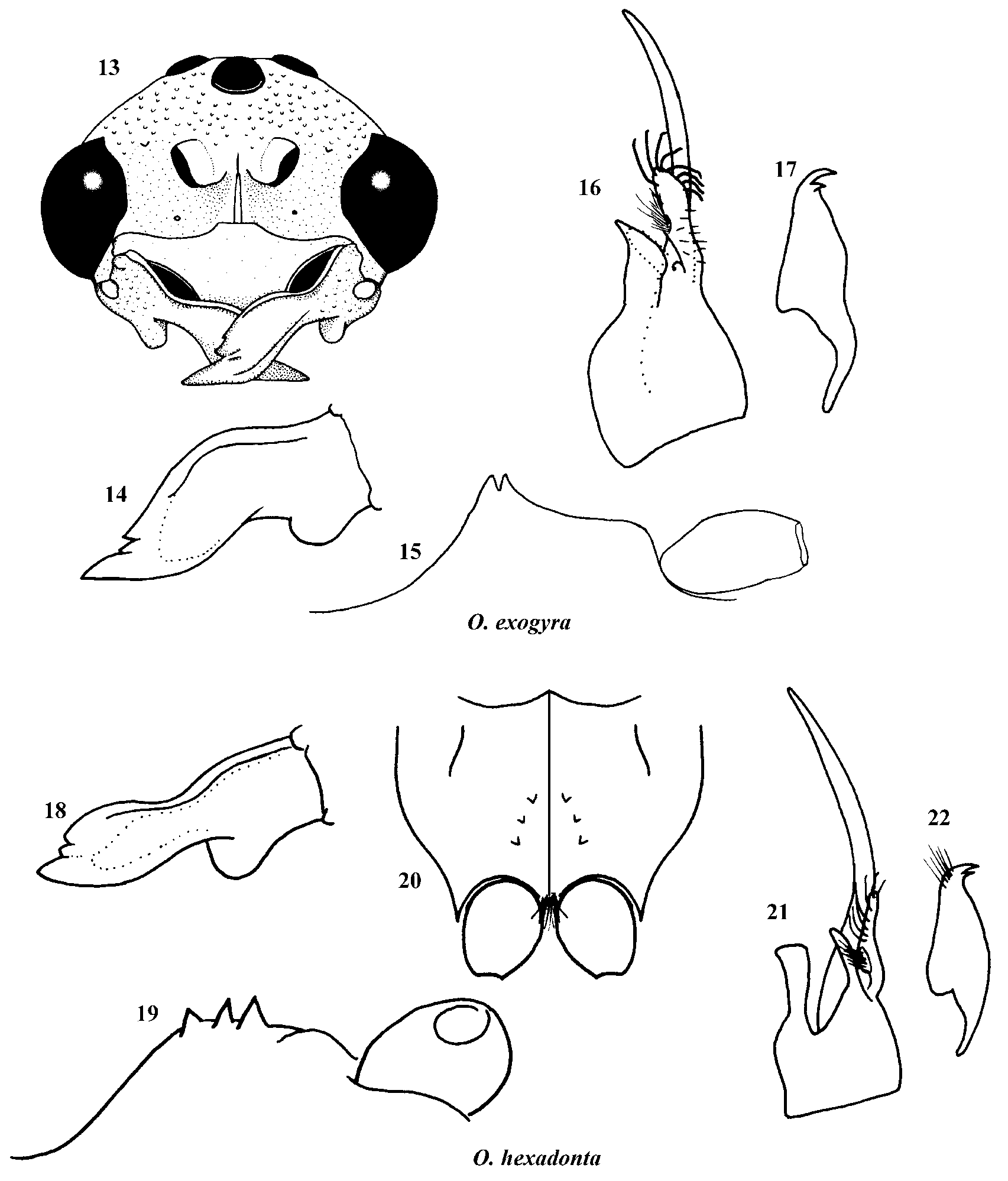
4. Odontophotopsis hexadonta species-group
( Figs. 18–22 View FIGURES 13 – 22 )
Odontophotopsis (Odontophotopsis) hexadonta Schuster, 1958 . Ent. Amer. (n. s.) 37: 51, male. Holotype: California, San Diego Co., Pine Valley, 27.Aug.1927, F.W. Kesley (UMSP).
Diagnosis. This species is easily diagnosed by the unique armature of the mesosternum with each side having a diverging row of three subequal teeth ( Figs. 19, 20 View FIGURES 13 – 22 ). Also, the mandible is tridentate and deeply excised ( Fig. 18 View FIGURES 13 – 22 ), the metasoma is subsessile, and the pygidium is polished, glabrous, and not defined laterally.
Male. Coloration and setal pattern. Body testaceous; antenna slightly darker, orange brown; head, mesosoma and metasoma uniform in color throughout; body clothed with sparse, erect, decumbent, pale yellow setae; only few plumose white setae on mesosoma; T1 with sparse plumose fringe at distal margin; T2 and S2 with thick fringe of pale plumose setae; T3-5 and S3-5 each with thinner, less conspicuous fringe of pale plumose setae. Tibial spurs and legs concolorous with body.
Head. Rounded posteriorly. Mandible deeply excised ventrally, angle of excision rounded; dorsal carina strong, medially lamelliform, terminating at strong inner tooth ( Fig. 18 View FIGURES 13 – 22 ); subdistal inner tooth weak ( Fig. 18 View FIGURES 13 – 22 ); mandible slightly dilated beyond excision ( Fig. 18 View FIGURES 13 – 22 ). Clypeus depressed below margin of mandibles, median area concave, lateral angle weakly tuberculate; surface of clypeus polished, almost impunctate, with only few erect setae; scape with single ventral carina. F1 ~0.75X length of F2. Front, vertex, and gena with moderate, shallow, close punctures, immediately posterior to antennal insertion, becoming separated and sparse on vertex and gena; ocellar area dark; ocelli moderate in size, ocellocular distance 2–2.5X greatest width of lateral ocellus.
Mesosoma. Sides and dorsum of pronotum coarsely punctate, dorsum with moderate, confluent, deep punctures, sides with somewhat larger, shallower punctures; mesonotum with moderate, contiguous, shallow punctures; notaulus weak, obsolete on anterior 0.5X of mesonotum; scutellum coarsely, confluently punctate; dorsum and posterior face of propodeum conspicuously, shallow reticulate, reticulations extending to sides of propodeum, but becoming coarse, punctate-reticulate; metapleuron with moderate, close punctures on ventral 0.33, polished and impunctate on dorsal 0.66; anterolateral area of mesopleuron with small, shallow, separated punctures; remainder of mesopleuron with deeper, contiguous to confluent punctures; mesosternum armed on each side with a diverging row of three subequal denticles ( Figs. 19, 20 View FIGURES 13 – 22 ); surface of mesosternum with shallow, depressed groove along midline, otherwise with moderate, close punctures. Metasternum bidentate. Marginal cell on costa ~1.5X length of stigma.
Metasoma. First metasomal segment subnodose; T1 weakly punctured at sides, almost impunctate medially; T2 polished, with fine, scattered punctures throughout; T3-5 weakly punctured, punctures most obvious at anterior and posterior margins; pygidium elongate ovate, 2X as long as wide, glabrous, polished, not defined laterally; S2 with small, shallow, well separated punctures, felt line 0.33X length of tergal felt line; S3-5 weakly punctured. Hypopygium with close, moderate punctures, rounded posteriorly. Genitalia as in Figs. 21 and 22 View FIGURES 13 – 22 .
Distribution. California.
Material examined. USA, California, Fresno Co., Coalinga, Mineral Springs Road, 10 males, 7.Apr.1987, 1 male, 29.May.1987, N.J. Smith ( FCDA, EMUS); Los Angeles Co., 1 male, Tanbark Flat, 10.Jul.1950, W.C. Bentinek ( EMUS); Riverside Co., Pinyon Flats Campground, 5 males, 4–5.Jul.2003, D. Yanega ( UCRC, EMUS); Riverside, 5 males, ( UCRC); Solano Co., Winters, 7 mi W, Stebbins Cold Canyon Res., 2 males, 13.Jul.2005, K.A. Williams ( EMUS); San Bernardino Co., Pinyon Hills, 12 males, 26.Jul.2003, G.R. Ballmer and D. Powell ( UCRC); San Diego Co., Pine Valley, 3 paratype males, 27.Aug.1927, F.W. Kesley ( UMSP; UAIC); Ranchita, 5 km W, 1 male, 1–5.Jun.2002, M.E. Irwin and F.D. Parker ( EMUS); San Ysidro, 1 male, 18–24.Jun.1924, Weedmark ( CDFA); Tulare Co., Ash Mt, Kaweah Power Station, 1 male, 26.Jun.1983, Halstead ( CDFA); Yolo Co., River Levee, 1 male, 22.Sep.1970, M.S. Wasbauer ( CDFA); Yuba Co., Shad Pad, S of Marysville, 1 male, 17.Jun.2006, K.A. Williams ( EMUS).
Remarks. This species-group is the same as defined by Schuster (1958). Besides the holotype, three paratypes were studied. The paratype labels were added to the specimens by Mickel in 1962, rather than by Schuster, even though these specimens were collected at the same time and locale as the holotype and were studied by Schuster (Mickel, unpub.).
No known copyright restrictions apply. See Agosti, D., Egloff, W., 2009. Taxonomic information exchange and copyright: the Plazi approach. BMC Research Notes 2009, 2:53 for further explanation.
|
Kingdom |
|
|
Phylum |
|
|
Class |
|
|
Order |
|
|
Family |
|
|
Genus |
Odontophotopsis hexadonta
| Pitts, James P. 2007 |
Odontophotopsis (Odontophotopsis) hexadonta
| Schuster 1958 |