
Alsophis antillensis ( Schlegel, 1837 )
|
publication ID |
https://doi.org/ 10.5252/geodiversitas2019v41a12 |
|
publication LSID |
urn:lsid:zoobank.org:pub:B0131324-1E7D-4D4E-97EA-AA812A5F7B94 |
|
DOI |
https://doi.org/10.5281/zenodo.3705026 |
|
persistent identifier |
https://treatment.plazi.org/id/03AE7E3B-FFE5-FFE0-FC37-F97EFAA7F4EB |
|
treatment provided by |
Valdenar |
|
scientific name |
Alsophis antillensis ( Schlegel, 1837 ) |
| status |
|
Alsophis antillensis ( Schlegel, 1837)
EXAMINED MATERIAL. — A total of 840 bones from all 20 sites and six islands are attributed to Alsophis antillensis ( Table 1 View TABLE ). Among these are 82 cervical and caudal vertebrae that could not be unambiguously identified because of their strong morphological variability. These elements, which are not described in this study, were associated with A. antillensis on the basis of their sizes, and the taxonomic composition of the snake material of the different sites.
DESCRIPTION
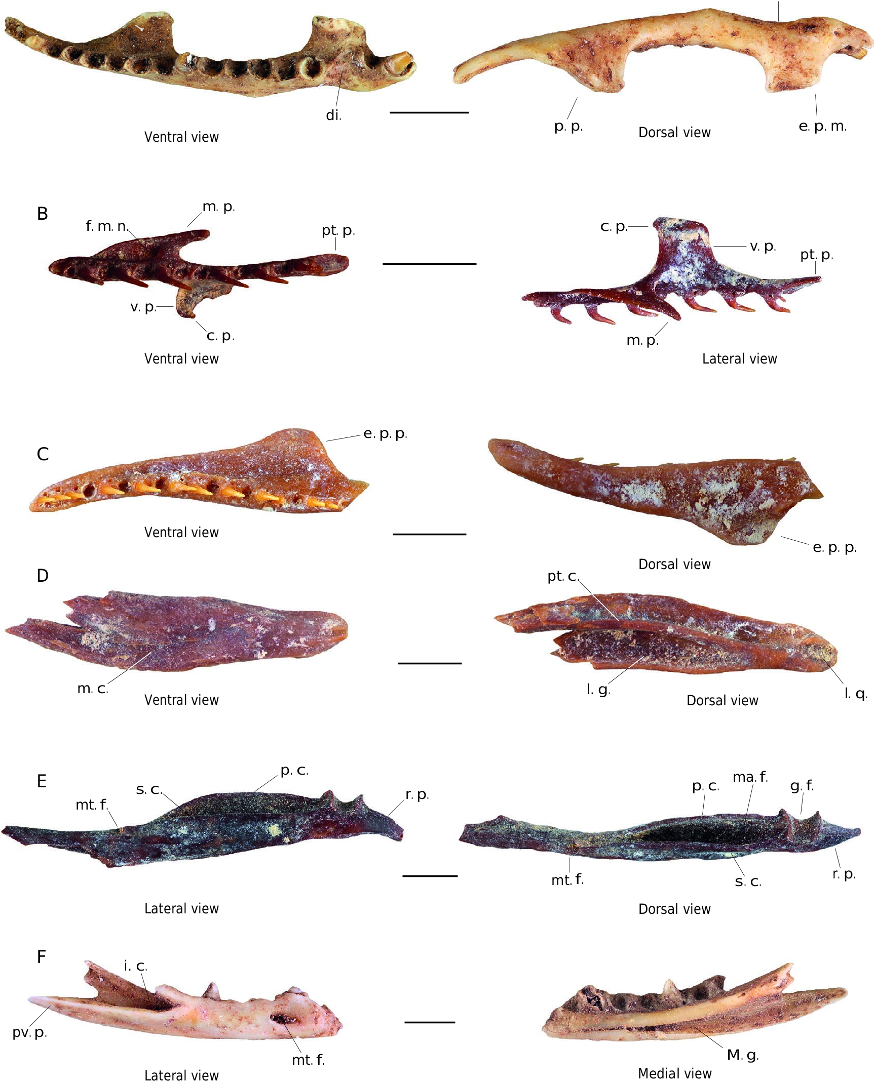
Maxilla (4 elements; Fig. 4A View FIG )
The most complete maxilla only lacks its anterior tip. It bears 16 tooth positions and measures 15.6 mm in maximum length. Maxillae are elongate, slightly incurved in dorsal and ventral views, and bear well-developed ectopterygoid and palatine processes. In ventral view, a short diastema is visible below the ectopterygoid process and separates the two last dental positions from the previous ones. The few teeth preserved and size of teeth alveolar niches show that the two last teeth are larger than the others (tendency to opisthomegadont dentition), no other observation can be made regarding the structure of maxillary teeth. In dorsal view, the ectopterygoid process is of sub-rectangular shape and occurs medial to the second and third last dental positions. The ectopterygoid process is wide, moderately long, and ventrally incurved with a rounded medial margin. The palatine process occurs medial to the fifth and ninth first dental positions. Still in dorsal view, this process is of sub-triangular shape with a longer anterior than posterior margin. The bone has a shallow dorsal notch and a weakly marked insertion area for the maxillary ramus of the ectopterygoid situated at the posterior base of the ectopterygoid process (broken on the bone depicted in Fig. 4A View FIG ).
Palatine (1 element; Fig. 4B View FIG )
The only available element is a complete right palatine discovered in Blanchard Cave. This bone measure 6.5 mm in maximum length and bears 14 tooth positions. In lateral view, the bone bears a long, sub-triangular, and ventro-posteriorly oriented maxillary process. The vomerine process (sensu Szyndlar 1984) is well-developed, long, and sub-rectangular shaped in lateral view. In dorsal view, the choanal process (sensu Szyndlar 1984) prolonges the vomerine process in medioventral direction and is slightly posteriorly incurved. The anterior and posterior tips of the bone are blunted and neither is bifurcated. In ventral view, a large foramen for the maxillary nerve (= palatine foramen sensu Cundall & Irish 2008) is visible at the base of the maxillary process. The pterygoid process is short and bears a small ventral articular facet for contact with pterygoid bone.
Pterygoid (3 elements; Fig. 4C, D View FIG )
Three fragments of pterygoids corresponding to anterior ( Fig. 4C View FIG ) and posterior ( Fig. 4D View FIG ) parts of the bone are present in the studied assemblage. This bone is nearly flat in lateral view and anteriorly incurved in ventral view. Still in ventral view, on the medial edge of its anterior portion, the bone bears a row of 19 small and posteriorly incurved teeth. The posterior fragment of this bone ( Fig. 4D View FIG ) shows that at least the posterior one-third of the bone lacks teeth. The ectopterygoid process is well developed, distinct,and lies in line with the 13 th to 19 th dental positions in ventral view. This process is subtriangular shaped with a blunt lateral margin in ventral view. The posterior part of the bone is elongate and its medial surface is concave in ventral view. In dorsal view, the posterior part of the bone bears a deep imprint of the mandibular condyle of the quadrate bone; this imprint is separated from the medial part of the bone by a high and thick pterygoid crest. The posterior tip of the bone is rounded and bears a small lateral notch that contacts the posterior process of the compound bone when the living animal closes its mouth ( Cundall & Irish 2008).
Compound bone (3 elements; Fig. 4E View FIG )
The two most complete compound bones are of two different sizes: the first one ( Fig. 4E View FIG ) is complete and measure 14 mm in maximum length, whereas the second one measure 28 mm long despite lacking its retroarticular process. The bone is elongate. The retroarticular process is well developed, longer than the glenoid fossa, of truncated conical shape, medially incurved in dorsal view, and perforated by a deep and large foramen near its anterior margin in medial view. In lateral view, the surangular crest is moderately developed with a nearly straight dorsal margin and a sharp ventral margin. The mental foramen is large, semi-circular, and opens anteriorly. In medial view, the development of the prearticular crest is variable, but this crest consistently is two times higher than the surangular crest on all specimens. In dorsal view, the mandibular fossa is long and deep, but its width varies. In medial view, the imprint of the angular is well marked and posteriorly pointed.
Dentary (1 element; Fig. 4F View FIG )
The only recovered dentary fragment corresponds to the posterior two-thirds of the bone. This fragment is moderately elongate and preserves seven tooth positions. In medial view, the Meckel groove is fully open with a narrower anterior opening that nearly reaches the anterior tip of the fragment. In lateral view, the bone has a large mental foramen situated in its median part. The bone bears a long posteroventral process with a blunted posterior apex. This process forms the ventral margin of a triangular insertion for the lateral process of the compound bone.
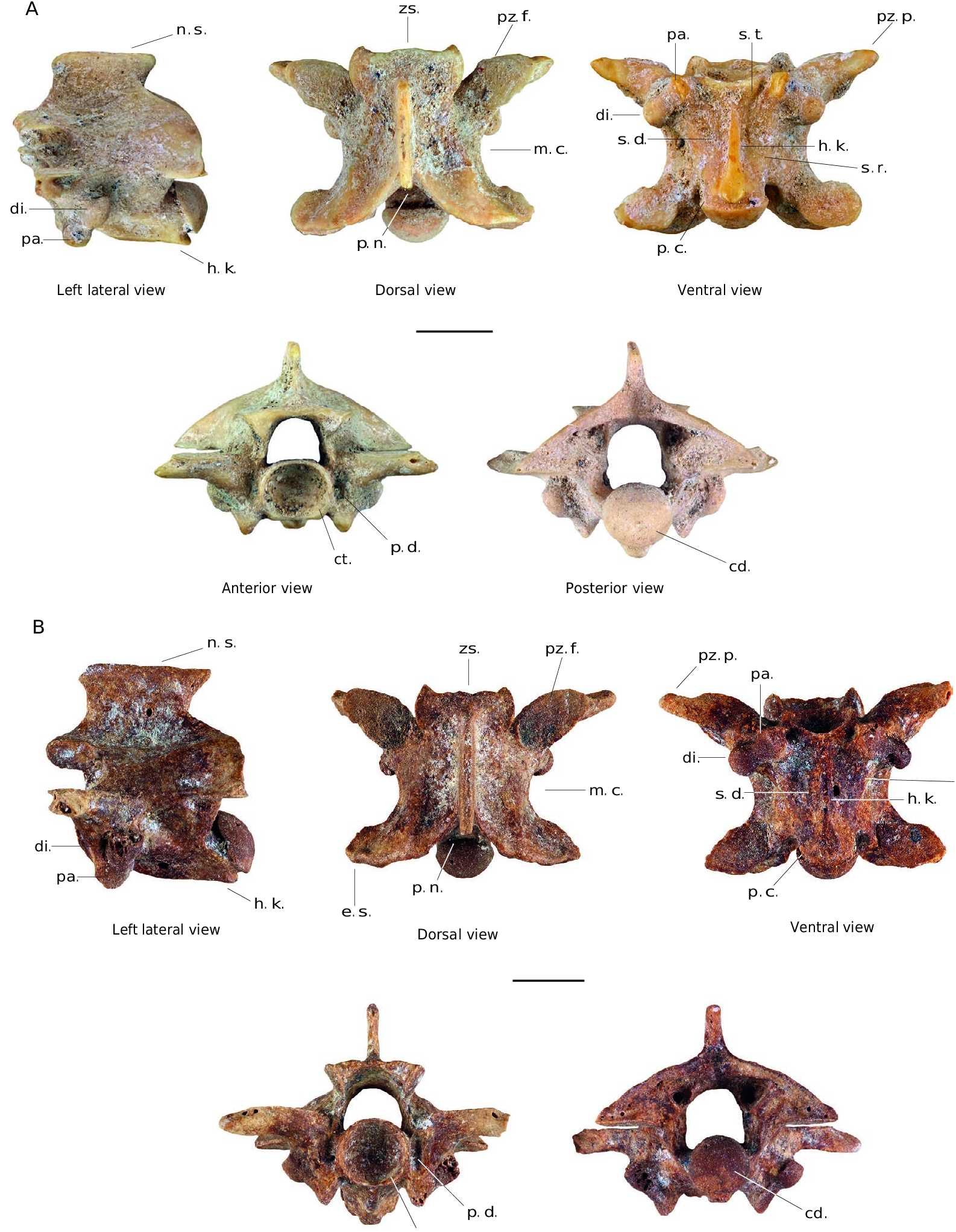
Trunk vertebrae (741 elements; Fig. 5A, B View FIG )
These vertebrae are weakly built and of variable size (centra lengths between 1 and 6.6 mm) corresponding to all ontogenetic stages from juvenile to adult individuals. In dorsal view, vertebrae are slightly wider than long to slightly longer than wide (ratio CL/WIC = 0.81-1.17) and exhibit a well-marked median constriction. The zygosphene is moderately wide and bears an anterior margin
A p. p. e. p. m. d. n.
di. p. p. e. p. m. Ventral view Dorsal view B m. p. f. m. n. pt. p. c. p. v. p. pt. p. v. p. c. p. Ventral view m. p. Lateral view e. p. p. C Ventral view e. p. p. Dorsal view D pt. c. m. c. l. g. l. q. Ventral view Dorsal view E mt. f. s. c. p. c. r. p. ma. f. p. c. g. f. mt. f. s. c. r. p. Lateral view Dorsal view F i. c. pv. p. mt. f. M. g. Lateral view Medial view
fossa; i. c., insertion area for the lateral process of the compound bone; i. q., imprint of the mandibular condyle of the quadrate bone; l. g., lateral groove; m. c., medial concave surface; M. g., Meckel groove; m. p., maxillary process; ma. f., mandibular fossa; mt. f., mental foramen; p. c., prearticular crest; p. p., palatine process; pt. c., pterygoid crest; pt. p., pterygoid process; pv. p., posteroventral process; r. p., retroarticular process; s. c., surangular crest, v. p., vomerine process. Scale bars: 3 mm.
whose shape varies from straight to slightly trilobate. The postero-medial notch of the neural arch is moderately deep and bracketed by flared lateral margins forming an angle of nearly 90°. On some specimens, a very reduced and slightly marked epizygapophyseal spine occurs, but this is absent on most of the vertebrae, including the two figured examples. Prezygapophyseal facets are rounded to ovoid in outline. The morphology of the prezygapophyseal processes is highly variable depending on the size of the specimen. These processes are anterolaterally oriented and conical shaped on small specimens, but tend to become shorter, wider, and blunter in large specimens although remaining elongate in some. In lateral view, the shape of these processes also varies from a transversally rounded in small specimens to a flattened shape in large specimens. Still in lateral view, the neural spine is well developed and longer than high. In most specimens the neural spine is two times longer than high, but this condition is variable in our material. The anterior margin of the spine is usually slightly incurved and a small dorsal anterior projection slightly overhangs the anterior edge of the spine whereas the posterior margin more strongly overhangs the posterior edge of the spine. The interzygapophyseal crest is straight in lateral view. Synapophysis bear moderately distinct diapophysis and parapophysis, the first being slightly larger and located more posteriorly. The hemal keel is well-visible in lateral view. The hemal keel is well-extended ventrally except along its anterior part where it shallows progressively until reaching the cotyle. In ventral view, the hemal keel is laterally well-delimited, thin, and exhibits a more or less spatulate shape (sensu Holman 2000) with a posterior enlargement of variable extend. However, rare specimens instead have a hemal keel shaped like a gladius (sensu Holman 2000). The hemal keel appears to exhibit important morphological variability in the material, but it is always laterally well-delimited. Still in ventral view, the centrum is triangular with well-marked lateral margins. It has subcentral depressions of variable extend and depth. A subcentral foramen occurs to either side of the hemal keel. In anterior view, the roof of the zygosphene is thin, ranging from slightly curved to straight. Still in anterior view, the cotyle is generally subcircular and slightly wider than high. However, the cotyle is round on some specimens. The cotyle is bordered by paracotylar foramina and deep paracotylar depressions of low extension. In ventral view, the cotyle also bears sub-cotylar tubercles on some specimens. In posterior view, the neural arch is moderately vaulted. In posterior outline, the condyle ranges from subcircular(often slightly dorsoventrally flattened) to round. It is separated from the centrum by a well-marked precondylar constriction best seen in ventral view.
REMARKS
The above described bones have been associated together on the basis of their size and of several morphological characters. They exhibit several characters occurring in “colubrid” (sensu lato) snakes: a slightly curved maxilla having well-developed palatine and ectopterygoid processes, with the number of teeth (16) between two and 36 ( Marx & Rabb 1972), and with the two most posterior teeth larger than the others ( Cundall & Irish 2008); the occurrence of distinct maxillary and choanal processes on the palatine bone ( Cundall & Irish 2008); vertebrae are weakly build, with a thin zygosphene and neural spine, differentiated paradiaopophyses, paracotylar foramina, and well-developed prezygapophyseal processes ( Rage 1984; Hsiou & Albino 2010). The fossils also exhibit a character occurring in xenodontine snakes: the occurrence of an extension (choanal process) of the vomerine process of the palatine in postero-ventral direction ( Cundall & Irish 2008). Among the Lesser-Antillean xenondontine snakes, these fossils exhibit several differences compared to the genus Erythrolamprus ( Erythrolamprus juliae ). In the later genus: the maxilla has more teeth (25-26) and its palatine process bears a distinct, pointed, and posteriorly oriented distal tip; the pterygoid has posterior lateral and medial grooves that are less deep and extended, its ectopterygoid process is weakly separated from the pterygoid flange; and the compound bone is more elongate and has a shorter mandibular fossa. Additional differences also occurr on the trunk vertebrae (see below in description of fossil Erythrolamprus ). By contrast, the fossil bones are entirely consistent with Alsophis antillensis , the second xenodontine species currently occurring on the Guadeloupe Islands. The fossils differ from other species of Alsophis we observed: on the compound bone the dorsal margin of the surangular crest is straight in medial view in A. antillensis and A. rijgersmaei , whereas it is slightly concave in A. rufiventris . The morphology of the vertebrae also is very different from A. rufiventris (see below in description of fossil Alsophis sp. 2) and also differs from A. rijgersmaei whose vertebrae bear a hemal keel consistently of gladius shape (sensu Holman 2000) with straight lateral margins and no posterior enlargement. In addition, gross size comparison with modern Alsophis specimens show that fossil vertebrae correspond to individuals between 25 and 150 cm in total length or between 18 and 112 cm in snout-vent length, a size matching that of modern A. antillensis (max. 93 cm of SVL see Henderson & Powell 2009), although some fossils appear to be larger than modern representatives. On the basis of these observations and the current occurrence of A. antillensis on the Guadeloupe Islands, we refer these remains to Alsophis antillensis . Compared with the snakes previously identified from fossils on the Guadeloupe Islands, our fossils aresimilar to the cf. Alsophis sp. and Colubroidea sp. 1 of Bochaton et al. (2015) in Cadet 2 cave (Marie-Galante Island), to the cf. Alsophis and Colubroidea of Bailon et al. (2015) in Blanchard cave (Marie-Galante Island), and to the Alsophis sp. of Boudadi-Maligne et al. (2016) from the site of Pointe Gros Rempart 6 (La Désirade Island). Our observations of ontogenetic variability of Alsophis antillensis and of Erythrolamprus vertebral morphology suggest that small fossil dipsadid vertebrae previously left unidentified can be confidently attributed to A. antillensis .
No known copyright restrictions apply. See Agosti, D., Egloff, W., 2009. Taxonomic information exchange and copyright: the Plazi approach. BMC Research Notes 2009, 2:53 for further explanation.